Figure 1.
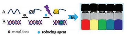
Schematic illustration of synthesis strategies and fluorescence properties of DNA-mediated metal nanoclusters: (A) ssDNA (B) dsDNA.

Development of metal nanomaterials has become a hot topic recently due to their distinctive properties such as easy synthesis of different sizes and shapes, high surface-to-volume ratio, stable optical properties, and good biocompatibility [1-3]. So far, several preparation methods of metal nanomaterials have been reported, including templatemethod[4], chemicalreduction[5], field assisted synthesis method [6, 7], seed-mediated method [8] and lithography method. Chemical reduction is the most common method for the synthesis ofmetal nanomaterials. Chemical reduction issimplewith low cost, but it is greatly affected by the experimental environment. Moreover, it is difficult to remove excess reagents during synthesis. Field assisted synthesis method refers to the chemical process driven by external physical fields such as ultrasonic field and radiation field that induce the formation of metal nanomaterials. Since external physical field can be applied or removed instantly, field assisted synthesis method is conducive for controlling the reaction process and the reaction time can be shortened to several hours or even minutes.However, this method has poor repeatability and is not easy to be controlled with severe side reactions.Seed-mediatedmethodis one of the most common methods for the preparation of nanorods and core-shell nanomaterials. The formation process includes the formation of crystal seeds and the epitaxial growth of shells. Seedmediated method can be operated under mild reaction conditions and can effectively control the morphology and size of nanomaterials. But there are many factors affecting the seed-mediated reactionprocess. Lithography method can accurately control the size and shape of metal nanostructures with high reproducibility, butthis method is costly, time consuming, and not suitable for large-scale applications.
Template method is easily controlled under mild reaction conditions and has been widely used for the synthesis of metal nanomaterials. It is essential to select suitable template molecules to achieve the successful synthesis of metal nanomaterials by template method. According to the different structures of template used in the synthesis process, the template is generally divided into hard template and soft template. Common hard templates include silicon dioxide [9], aluminum oxide [10], carbon nanotubes [11], and soft templates include synthetic polymers [12], surfactants [13], nucleic acids [14], etc. The hard template has limited sources and is prone to collapse during the synthesis of metal nanomaterials, which affects the properties of products and limits the wide application of hard template method. Besides, synthetic polymers and surfactants have the defects of complex structure and low mechanical strength. As a kind of biomacromolecules, DNA has the advantages of low toxicity, specific structure and high stability, and it has great potential as a template for the synthesis of metal nanomaterials.
DNA is a long-chain polymer with precise spatial configuration and controllable length composed of four bases called A, G, C and T, and can regulate the nanomaterials' morphology and structure which control the properties and characteristics of metal nanomaterials [15]. Besides, DNA can form other structures including G-quadruplexes, i-motifs, and high-level structures in different dimensions [16-21]. These structures not only increase the diversity of DNA microstructure, but also improve the thermal and chemical stability of DNA molecules. Therefore, DNA has been considered as a kind of efficient template molecules to mediate the precise synthesis of metal nanomaterials. DNA molecules have special nitrogenous bases with specific length which can stably coordinate with metal ions and precisely regulate the growth and development of metal nanometials with specific size and morphology. And the carbon skeletons of DNA molecules can prevent the aggregation of metal nanomaterials. Also, metal ions can be regularly accumulated on the negatively charged phosphate backbone through electrostatic interaction and are converted into metal nanomaterials during the reduction process.
DNA-mediated metal nanomaterials can be easily synthesized with tunable size, shape and morphology by controlling the length and rigidity of template DNAs, which could achieve the precise control of their surface plasmon resonance (SPR) and further the unique and excellent analytical performance such as molecule-like energy gaps, specific optical and electrochemical response [22, 23]. Moreover, artificial DNA strands as templates can be well designed and modified with various functional groups, which would make DNA-mediated metal nanomaterial excellent recognition and biocompatibility. Besides, DNA attached to the surface of the nanomaterial can protect the nanomaterial from aggregation and form specific active sites. Therefore, DNA-mediated metal nanomaterials have been widely used as ultrasensitive and selective probes or micro-sensors for in situ rapid analysis of trace targets in complicated biological and environmental samples. Compared with conventional analytical methods such as chromatography and mass spectrometry [24], DNA-mediated metal nanomaterials based methods are more selective and sensitive. Especially, when facing real samples with ultra-small volumes, new analytical methods based on DNAmediated metal nanomaterials show great priority to achieve the real-time quantitative information of trace targets in environmental and biological analysis.
This paper summarizes the recent progress on synthesis, morphology, and properties of DNA-mediated metal nanomaterials including nanoparticles and nanoclusters with various DNA strands as regulation templates. Moreover, the review introduces the applications of DNA-mediated metal nanomaterials based analytical methods to the ultrasensitive and selective analysis of environmental and biological samples. Finally, the developing prospect of DNA-mediated metal nanomaterials for environmental and biological analysis is discussed and proposed.
Nanoparticles are colloidal particles with particle size in the range of 1-100 nm. Metal nanoparticles have remarkable electrical and optical properties due to their unique surface and size effects. Moreover, since metal nanoparticles have good dispersibility, low biological toxicity and can be easily modified [25], they have been widely applied to the fields of catalysis, biomedicine and chemical sensing in recent years [26-28]. According to the types of elements that make up metal nanoparticles, they can be divided into single metal nanoparticles and multi-metal nanoparticles.
Commonly reported single metal nanoparticles mainly include gold nanoparticles (AuNPs) and silver nanoparticles (AgNPs). Single metal nanospheres have simple chemical composition and stable property which make them quite suitable as stable seeds for consequent fabrication of DNAs and deposition of homologous metal nanoparticles. The loading amounts of DNA strands on the surface of single metal nanospheres can be precisely controlled by adding the different amount of DNAs and adjusting related synthesis conditions. Thus, the size and morphology of DNA mediated single metal nanoparticles can be precisely controlled as designed with excellent analytical properties and good biocompatibility. Moreover, the further modification of probe molecules on DNA mediated single metal nanoparticles is relatively easy which would enhance the analytical selectivity for real samples.
Recently, researchers have used different structures of DNA strands as templates to synthesize many special DNA mediated single metal nanoparticles with specific shapes and morphologies. In 2010, Lu research group [29] in University of Illinois for the first time demonstrated that DNA could be used as templates to control the morphology of gold nanoparticles. They used three different single DNA strands, including poly A, poly C and poly Tas templates to synthesize three different DNA mediated metal nanoparticles in one pot. The results showed that poly A and poly C could induce the formation of gold nanoflowers, while poly T could not form flowerlike nanostructures. They also showed that DNA mediated gold nanoflowers could be readily uptaken by cells and visualized under dark-field microscopy, and these nanoflowers showed great potential in biological analysis. Later, they reported [30] that when gold nanoprisms were used as seeds, the single-stranded DNA sequences composed of different bases could regulate the formation of gold nanoparticles with complex shapes and rough surfaces. When different deoxyribonucleotides formed a singlestranded DNA together, they had competitive or synergistic effect. Subsequently, Lu group [31] and Yang group [32] in Xiamen University respectively explored the influence of different oligonucleotides on the morphology of sliver nanoparticles using silver nanocubes and silver nanospheres as seeds. The results showed that different sliver seeds also affected the final morphology of DNA mediated sliver nanoparticles. Due to the surface area, size and localized surface plasmon resonance (LSPR) are different, the antibacterial ability, biocompatibility and SERS properties of DNA mediated silver nanoparticles with different morphologies are significantly different, which broaden the application of DNA mediated single metal nanoparticles in environmental and biological analysis. Recently, Lu and his collaborators have systematically analyzed the formation of DNA-regulated gold nanoparticles through UV-vis, SEM, TEM, zeta potential, and cyclic voltammetry [33]. It was found that the formation process of nanoparticle could be divided into two stages, namely seed nucleation and deposition. DNA molecule regulated the nucleation process by influencing the diffusion process of the precursor to the seed, while the density and the rate of desorption and migration of DNA on the seed surface affected the deposition process of precursors, thus forming the nanoparticles with different morphologies.
Compared with nanospheres, anisotropic nanorods or nanogap particles have tunable surface plasmon resonance, stronger and more stable spectral characteristics. Therefore, synthesizing nanoparticles with stronger optical properties and higher stability has great significance in analytical chemistry. In recent years, there have been many reports on the subtle regulation of nanorods and core-shell nanogap structures using DNA strands as templates. Liang's and Yi Lu's group collaborated to adjust the geometric and plasmonic properties of gold nanoparticles by changing the template DNA sequences [22]. The results of the kinetic study indicated that gold nanorods eventually evolved into dumbbellshaped nanostructures due to the promotion of the longitudinal growth by A20, while the lateral growth was caused by G20, resulting in the octahedral shape of gold nanorods. By using different ratios of bases, it was further verified that DNA could regulate the SPR peaks of gold nanoparticles in the range of 550-1010nm. At the same time, Lim group [34] studied the influence of reaction conditions including pH and salt concentration on the structure and SERS properties of gold nanoparticles, and four kinds of gold nanoparticles with tunable intra-nanogap distance were obtained. They provided new insights into the design of plasmonic nanostructures and SERS-based biosensing applications by using DNA mediated gold nanoparticles.
Although many DNA mediated single metal nanoparticles with different surface morphologies and characteristics have been synthesized, it still remains a big challenge to precisely and predictably regulate the morphologies during synthesis. Despite many discoveries have shown that DNA can serve as programmable template to precisely regulate the morphologies of single metal nanoparticles, the mechanism of how the morphology can be well tuned by the template DNA still need further studied.
Compared with single metal nanoparticles or homologous metal nanostructures, due to the destruction of symmetry and the synergistic effect of different constituent elements, bimetallic nanoparticles exhibit more distinct surface plasmon resonance properties. Besides, bimetallic nanoparticles provide more ways to control nanoparticle configuration and surface properties, which would greatly affect their analytical performance and offer more diverse applications. Moreover, DNA mediated bimetallic nanoparticles can be assembled to form various aligned and unconventional nanostructures, which not only enrich the morphology and configuration of nanoparticles, but also have advantages in analytical application [35, 36].
In recentyears, several researches utilized DNAs as templates to design and synthesize bimetallic nanoparticles, including coreshell structures [37, 38] and Janus nanostructures [39]. Lu's group used palladium nanocubes as the seeds and four different DNA sequences T10, A10, C10, and G10 as templates to guide the formation of four different morphologies of bimetallic nanostructures [39]. Through detailed kinetic studies, it was found that different DNA sequences had different effects on the surface energy of palladium nanocubes and influenced the diffusion and deposition of gold precursors on the palladium surface, thus resulting in the formation of different palladium binary nanostructures. This research offered a fundamental understanding of how different DNA sequences controlled the morphology of bimetallic nanoparticles. The ability of DNA to precisely tune the morphology of bimetallic nanoparticles has great potential for thesynthesis ofmanymetal nanomaterials, which can be exploited for biomedical or analytical applications. Lee et al. [40] used DNAAuNPs as the seed to efficiently synthesize snowman-like Au/Ag nanoparticles by adjusting salt concentration, and proposed a formation mechanism for the asymmetric nanostructures. In addition, they assembled these snowman-like nanoparticles to form a variety of nanostructures with directionality, and provideda new way for the synthesis of anisotropic nanostructures that possessed special optical properties and higher chemical affinity, which provided wider and more diverse applications in analytical chemistry. Fan group [41] combined the nano-gap structure and the SERS enhancement effect of silver nanoparticle itself and synthesized mushroom like gold-silver nanoparticles with strong and stable SERS response. It was found that sodium chloride and ROX-labeled single-stranded DNA could effectively regulate the size of the nano-gap between gold and silver, finally affecting the size and SERS response of these DNA-mediated bimetallic nanoparticles.
Although there has been several reports on synthesizing DNA mediated bimetallic nanoparticles, their growth mechanism need further study. Compared with single metal nanoparticles, the synthesis process of bimetallic nanoparticles is more complex, and their final morphologies are also affected by more factors such as salt concentration and reducing agent, apart from the composition and length of DNA templates used. The precise control of the desired morphology and property of bimetallic nanoparticles would be a hot topic in this field in the next few years.
Metal nanoclusters are composed of hundreds of metal atoms with significant fluorescent characteristics, and their sizes are about 2nm [42]. Since the sizes of metal nanoclusters are close to the Fermi wavelength of the electron with the occurrence of band splitting, metal nanoclusters exhibit different optical and chemical properties from atoms or general nanoparticles. Compared with organic dyes, nanoclusters have higher fluorescent stability, and they are smaller, less toxic, and less prone to glare than quantum dots [43], so they have more advantages for the analysis and biosensing of heavy metal ions [44, 45].
As the template for synthesizing metal nanoclusters, DNA can well tune the size and fluorescent emission wavelength of metal nanoclusters by controlling the composition and length of DNA. Besides, the template DNA can protect metal nanoclusters from precipitation and oxidation, which is particularly important in complex sample analysis. In addition, DNA has the recognition function and is easy to be modified with specific groups, which increases the accuracy and sensitivity during real sample analysis [46]. The synthesis strategies and fluorescence properties of DNA mediated metal nanoclusters are showed in Fig. 1. In the following two sections, the synthesis and characteristics of metal nanoclusters that mediated by different structures of DNAs will be described.
Single-stranded DNA (ssDNA) is the most commonly used template for synthesizing metal nanoclusters because of its flexible design and simple configuration. Petty et al. [47] reported the synthesis of sliver nanoclusters (AgNCs) using ssDNA as the template for the first time. They found that Ag+ are more likely to bind with base C to form nanoclusters and different ssDNA sequences could result in different wavelength and intensity of fluorescence emission of AgNCs. Subsequent published works showed that the fluorescence emission characteristics of AgNCs were depended on the length and composition of template ssDNA. Via controlling the chain length, base composition and spatial structure of template ssDNA, the fluorescence emission wavelength of AgNCs could be adjusted from the visible to the nearinfrared range [48, 49].
Apart from the common primary structure, ssDNA can also form other secondary structures under different environmental conditions, including i-motif, G-quadruplex and hairpin DNA [50]. In addition to the sequence, the secondary structure of template DNA would also affect the fluorescence emission of metal nanoclusters. Gwinn et al. [51] found that ssDNA rich in base C or G could mediate the formation of fluorescent AgNCs. However, when ssDNA was thermally annealed and formed a double-stranded DNA, fluorescent AgNCs could not be mediated. Moreover, they also studied the effect of hairpin DNA on the fluorescence signal of AgNCs, and found that the fluorescence responses of AgNCs formed by the Gloop and C-loop were equivalent. However, the A-loop mediated AgNCs possessed weak fluorescence signal and the T-loop mediated AgNCs had no fluorescence signal. Zhang's group [52] used a series of guanine or cytosine rich DNA as templates to synthesize AgNCs with different fluorescent properties. The thermodynamic results showed that the order of affinity between silver ions and the DNA was as follows: coiled C-rich strand > imotif > duplex > G-quadruplex. This affinity could not only affect the fluorescence characteristics of AgNCs, but also decide the stability of AgNCs.
Compared with silver nanoclusters (AgNCs), gold nanoclusters (AuNCs) are highly stable, less toxicity and easy to combine with other materials for special functions. Thus, they have unique advantages as optical probe or detection platform for environmental or biological analysis. Cheng et al. [53] synthesized antibiotic-coated gold nanoclusters, combined with magnetic enrichment for the quantitative detection of bacteria in the range of 32-108 cfu/mL, and the detection limit was 16 cfu/mL. Compared with noble metals, copper was lower in cost and the preparation and application of copper nanoclusters (CuNCs) have also been reported recently [54, 55].
Apart from pure metal nanoclusters, bimetallic nanoclusters have also been reported in recent years [56, 57]. For example, Li et al. [58] used ssDNA as the template to efficiently synthesize gold/ silver nanoclusters for the detection of iodide ions. The good linear range was achieved in range of 0-10 μmol/L with the detection limit of 0.3 μmol/L. Current studies showed that fluorescence emission characteristics of metal nanoclusters can be well tuned via controlling the composition, length and spatial structure of template DNA, but there is no systematical studies focusing on specifying the regulation for precisely controlling the emission wavelength of DNA-mediated metal nanoclusters.
Double-stranded DNA (dsDNA) is more rigid and conformational than ssDNA. However, fully complementary dsDNAs have not enough space to bind to metal precursors and thus cannot form fluorescent metal nanoclusters. Therefore, dsDNAs need defects such as mismatch and abasic site to be used as template for the synthesis of metal nanoclusters with fluorescence signals [59]. Dadmehr et al. [60] used dsDNA with an inserted C6 loop as the template for the synthesis of fluorescent AgNCs, which were utilized as microprobes for the detection of methylated DNA. The good linearity was achieved between the increasing concentration of methylation DNA and the decreasing fluorescence intensity of AgNCs with a low detection limit of 9.4 ×10⁻¹⁰ mol/L. Yang et al. [61] produced massive DNA sequences that could be captured on the electrode via target-assisted polymerization nicking reaction. Then, the sequences triggered the hybridization chain reaction amplification to form dsDNA with numerous C-rich loops, which were used as template for the synthesis of AgNCs. By use of this preparation strategy the amount of AgNCs was significantly increased, leading to the highly sensitive electrochemical detection of miRNA, and the detection limit was as low as 0.64 fmol/L.
As a class of newly emerging fluorescent metal nanomaterials, CuNCs can be excited at the wavelength below 340 nm and exhibit fluorescence emission in the range of 570-600 nm, so the Stokes shifting would benefit the elimination of the interference of background signals from complex samples [62]. Therefore, many microprobes based on dsDNA mediated CuNCs have been synthesized and reported due to their unique properties in recent years. Wang et al. [63] developed an ultra-sensitive and label-free fluorescent method based on dsDNA mediated CuNCs for the detection of trypsin. The fluorescence intensity of CuNCs could be enhanced with the addition of protamine, due to the formation of protamine/DNA complexes between CuNCs and protamine. When trypsin was added, the fluorescence intensity of CuNCs would be quenched because of the protamine hydrolysis, which allowed the sensitive and selective detection of trypsin. The linear range achieved was 0.1-1000 ng/mL with the detection limit of 0.048 ng/mL. Recently, Chen et al. [64] designed a dumbbellshaped DNA molecule which consisted of two cytosine hairpin loops and an adenine-thymine-rich double-helical stem that was closed by the loops. This DNA molecule with special structure was used as template for the formation of AgNCs or CuNCs, which were developed for the sensitive detection of ATP. Through the comparison of detection limits of ATP based on AgNCs and CuNCs, it was found that the strategy based on CuNCs possessed lower detection limit of 81 pmol/L, which suggested CuNCs based strategy was more sensitive than AgNCs based one. Apart from being used as probes, dsDNA mediated CuNCs also have other potential applications. For example, Zhu et al. [65] developed a green method for staining DNA based on the in situ synthesis of dsDNA mediated CuNCs, and the skin toxicity experiments were carried out. Due to the difficult synthesis of dsDNA, especially the defective DNA, there are fewer reports on the synthesis of metal nanoclusters by using dsDNAs as templates.
DNA-mediated metal nanomaterials have attracted much attention due to their unique physical and chemical properties. As optical probes and chemical sensors, DNA mediated metal nanomaterials have higher sensitivity and selectivity because of their unique morphology, good stability and easy modification. Besides, analytical methods or platforms based on DNA-mediated metal nanomaterials are simple and fast, and they can realize realtime in situ analysis for ultra-micro samples, especially for environmental and biological analysis.
Heavy metal ions are environmental contaminants widely distributed in water and soils, and they are great harm to the ecological environment and human health. The US Environmental Protection Agency (EPA) stipulates that the maximum allowable concentrations of heavy metals in various environmental samples. For example, the maximum allowable concentrations of Hg2+, Pb2+ and Cu2+ in drinking water are 10 nmol/L, 72 nmol/L and 21 μmol/L, respectively. Traditional detection methods such as inductively coupled plasma mass spectroscopy (ICP-MS) and atomic absorption spectrophotometry (AAS) have good analytical performance for environmental samples, but these methods usually require complicated sample preparation process and high cost with large-scale analytical instrumentation and are not suitable for on-site or in situ analysis.
In recent years, DNA-mediated metal nanomaterials, especially nanoclusters, have been tentatively applied for environmental analysis due to their ultra-small size, strong fluorescent response and good stability. Besides, DNA-mediated metal nanomaterials based analytical methods are usually simple to operate and can realize rapid on-site detection with high sensitivity and selectivity. And the principles of DNA-mediated metal nanomaterials for environmental analysis are showed in Fig. 2. In recent years, many sensors based on DNA mediated metal nanomaterials have been developed for the colorimetric detection of heavy metal ions. The basic principle is based on the color change of nanomaterials dispersion system with the addition of heavy metal ions. Kanayama et al. [66] designed and synthesized a colorimetric sensor for the detection of Hg2+ using dsDNA as template. Without Hg2+ in the aqueous solution, the complex of dsDNA and gold was red. When Hg2+ existed, dsDNA would combine with Hg2+ via base T, thereby inducing the polymerization of gold nanoparticles and further the change of solution color from red to purple within 1 min. A satisfactory detection limit of this method was 0.5 μmol/L.
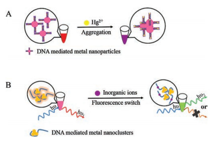
Another way for detecting metal ions is based on thefluorescence switching of nanomaterials. Lee et al. [67] used cytosine 12-mer as the template to synthesize AgNCs which showed red fluorescence. The redfluorescence can be switched into greenfluorescence by the addition of Ag+. This method had a minimum detection concentration for Ag+ as low as 10 nmol/L. Similar methods were also used for the analysis of Pb2+ [68] and Cu2+ [69]. Recently, Xu et al. [70] prepared a new fluorescence enhanced Hg2+ sensor using hairpin DNA as the template and the Exo Ⅲ was utilized for signal amplification, which greatly increased the detection sensitivity. And this method was applied to the detection of Hg2+ in real samples such as spiked tap water and the detection limit was 24 pmol/L.
Apart from cations, DNA mediated metal nanoclusters were also applied to the analysis of inorganic anions. Li et al. [58] developed a dual signal platform for the detection of I- based on ssDNA mediated gold/silver nanoclusters. When iodine ions were added, the detection system appeared fluorescence quenching and meanwhile the color changed from colorless to purple red. This method was simple and selective for the detection of I- with a good linear range from 0 to 10 μmol/L. Chen et al. [71] used ssDNA as the template to synthesize fluorescent gold/silver nanoclusters which were utilized as selective and sensitive probes for the detection of S2-. Due to the interaction between S2- and Au/Ag atoms/ions, the fluorescence of gold/silver nanoclusters was quenched with the addition of S2-. Moreover, the conformation of template DNA changed from packed hairpin to random coil structure. They had also demonstrated the practicality of this method for detection of S2- in hot spring and seawater samples.
DNA mediated metal nanomaterials usually have good biocompatibility, low toxicity and easy modification, so they have been widely used as sensitive and selective probes or sensors for the rapid analysis of small biomolecules, protein, nucleic acid, etc. And the principles of DNA-mediated metal nanomaterials for biological analysis are showed in Fig. 3. Some typical works in recent years have been summarized in Table 1 [72-74, 48, 49, 75-84].
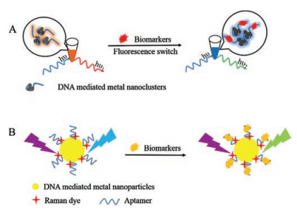
Imaging analysis has important practical significance in the field of biological and medical science, because the detailed information can be easily and intuitively observed in in vitro or in vivo biological samples. DNA-mediated metal nanomaterials have the advantages of easy labeling, easy modification, good optical stability and low biological toxicity, so they have good recognition function and can achieve the in situ analysis of trace targets, when they are used as bioprobes for imaging analysis. Many bioprobes based on DNA-mediated metal nanomaterials with high specificity and sensitivity such as AgNCs [85] have been designed and synthesized for bioimaging analysis coupled with suitable bioimaging technologies. Currently the most widely used bioimaging technologies include Raman imaging [86, 87], fluorescence imaging [88, 89] and multimodal imaging [90, 91]. Raman imaging analysis possesses highly sensitivity with narrow emission spectrum and excellent anti-water interference. Kang et al. [92] used DNA that had spacer sequences as the template to synthesize gold nanoparticles with highly narrow intra-nanogap, which could generate strong and stable Raman signals. Then, a variety of Raman molecules that could respond to near-infrared lasers were modified in the nanogap, and the target sites in hematopoietic stem cells were imaged rapidly. In addition, the change of the cell morphology during the death of induced cells could be well monitored. Fluorescence imaging technology is another widely used bioimaging technology with high imaging speed and wide application range. Xu et al. [93] designed ssDNAs with three different domains: the DNA scaffold for synthesis of fluorescent AgNCs, the recognition sequence to the target, and a quencher attached sequence. They used this multifunctional ssDNA as the template to synthesize an activatable silver nanoclusters beacon, which was utilized as versatile platform for detection of multiplex DNAs and small molecule. Via targetresponsive structure transformation of activatable silver nanoclusters beacon, target recognition induced the conformational transition and lit up the fluorescent signal of AgNCs. It was found that this platform had potentiality of ATP targeted fluorescence imaging in MCF-7 cancer cells, which revealed its promising application in complicated biosamples. Xu et al. [94] used Cy5 labeled DNA as the template to form a pyramid-like biological probe, which was used to detect telomerase activity in cells. In the presence of the target, the template DNA strand was replaced, resulting in the reduction of SERS signal and the recovery of fluorescence signal simultaneously, thereby realizing in situ quantification and imaging analysis of intracellular telomerase activity.
In this review the recent research progress on DNA-mediated metal nanomaterials including nanoparticles and nanoclusters were summarized from their synthesis strategies, morphologies, and special properties to their applications to environmental and biological analysis. Because the physical and chemical properties of metal nanomaterials are controlled by their morphology, so it is necessary to develop effective methods that can regulate the morphology of metal nanomaterials. DNA template based synthesis method is simple, time-saving, and can accurately regulate the optical and chemical properties of metal nanomaterials. Besides, DNA-mediated metal nanomaterials have the advantages of good stability and biocompatibility, and can be used as ultrasensitive and selective probes for in situ rapid analysis of targets in complicated biological and environmental samples. Therefore, they have more extensive applicability and practical significance in analytical chemistry.
Although many DNA-mediated metal nanomaterials with different morphologies and structures have been synthesized and reported with their tentative application to environmental and biological analysis, there are still many challenges in this field needing conquer in future. Firstly, the formation mechanism of DNA-mediated metal nanomaterials should be studied in detail to further reveal the role and function of DNA in the regulation process of corresponding nanomaterials. Secondly, the stability, anti-interference capability and biocompatibility of DNA-mediated metal nanomaterials need to be improved to meet the practical requirements of real sample analysis. Thirdly, the application range of DNA-mediated metal nanomaterials needs to be broadened in analytical chemistry. It is highly expected that more DNAmediated metal nanomaterials with different morphologies, structures and functions will be synthesized for broadening their practical applications in analytical chemistry.
This work was supported by the National Natural Science Foundation of China (Nos. 21475154, 21475153 and 21675178), the Special Funds for Public Welfare Research and Capacity Building in Guangdong Province of China (No. 2015A030401036), and the Guangzhou Science and Technology Program of China (No. 201604020165), respectively.
P. Miao, Y. Tang, L. Wang, ACS Appl. Mater. Inter. 9(2017) 3940-3947. doi: 10.1021/acsami.6b14247
Z. Wu, N. Xu, W. Li, J. Lin, Chin. Chem. Lett. 30(2019) 95-98. doi: 10.1016/j.cclet.2018.01.048
J. Yan, C. Hu, P. Wang, et al., Angew. Chem. Int. Ed. 54(2015) 2431-2435. doi: 10.1002/anie.201408247
X. Wang, J. Feng, Y. Bai, Q. Zhang, Y. Yin, Chem. Rev. 116(2016) 10983-11060. doi: 10.1021/acs.chemrev.5b00731
X. Liu, R. Huang, J. Zhu, Chem. Mater. 20(2015) 192-197.
Q. Zhang, J. Ge, T. Pham, et al., Angew. Chem. Int. Ed. 48(2009) 3516-3519. doi: 10.1002/anie.v48:19
H. Chen, E. Kern, C. Ziegler, A. Eychmueller, J. Phys. Chem. C 113(2009) 19258-19262. doi: 10.1021/jp906236q
Y. Huang, L. Dai, L. Song, et al., ACS Appl. Mater. Inter. 8(2016) 27949-27955. doi: 10.1021/acsami.6b05258
Y. Kanno, T. Suzuki, Y. Yamauchi, K. Kuroda, J. Phys. Chem. C 116(2012) 24672-24680. doi: 10.1021/jp308772b
A.M.M. Jani, D. Losic, N.H. Voelcker, Prog. Mater. Sci. 58(2013) 636-704. doi: 10.1016/j.pmatsci.2013.01.002
S. Kumar, V.C. Srivastava, G.K. Mandal, S.K. Pattanayek, K.L. Sahoo, J. Phys. Chem. C 121(2017) 20468-20480. doi: 10.1021/acs.jpcc.7b05973
Y. Umeda, C. Kojima, A. Harada, H. Horinaka, K. Kono, Bioconjugate Chem. 21(2010) 1559-1564. doi: 10.1021/bc1001399
B. Li, D.C. Higgins, S. Zhu, et al., Catal. Commun. 18(2012) 51-54. doi: 10.1016/j.catcom.2011.11.018
A. Kundu, S. Nandi, A.K. Nandi, Prog. Mater. Sci. 88(2017) 136-185. doi: 10.1016/j.pmatsci.2017.04.001
L.H. Tan, H. Xing, Y. Lu, Accounts Chem. Res. 47(2014) 1881-1890. doi: 10.1021/ar500081k
L.A. Yatsunyk, O. Mendoza, J. Mergny, Acc. Chem. Res. 47(2014) 1836-1844. doi: 10.1021/ar500063x
N.C. Seeman, Nature 421(2003) 427-431. doi: 10.1038/nature01406
P. Rothemund, Nature 440(2006) 297-302. doi: 10.1038/nature04586
Y. He, T. Ye, M. Su, et al., Nature 452(2008) 141-198.
J. Fu, M. Liu, Y. Liu, H. Yan, Acc. Chem. Res. 45(2012) 1215-1226. doi: 10.1021/ar200295q
Y. Ke, L.L. Ong, W.M. Shih, P. Yin, Science 338(2012) 1177-1183. doi: 10.1126/science.1227268
T. Song, L. Tang, L.H. Tan, et al., Angew. Chem. Int. Ed. 54(2015) 8114-8118. doi: 10.1002/anie.201500838
D. Lim, K. Jeon, J. Hwang, et al., Nat. Nanotechnol. 6(2011) 452-460. doi: 10.1038/nnano.2011.79
M. Jie, S. Mao, H. Li, J. Lin, Chin. Chem. Lett. 28(2017) 1625-1630. doi: 10.1016/j.cclet.2017.05.024
J. Li, Y. Zhang, S. Ding, R. Panneerselvam, Z. Tian, Chem. Rev. 117(2017) 5002-5069. doi: 10.1021/acs.chemrev.6b00596
Z. Dong, X. Le, C. Dong, et al., Appl. Catal. B Environ. 162(2015) 372-380. doi: 10.1016/j.apcatb.2014.07.009
G. Chen, I. Roy, C. Yang, P.N. Prasad, Chem. Rev. 116(2016) 2826-2885. doi: 10.1021/acs.chemrev.5b00148
A. Li, L. Tang, D. Song, et al., Nanoscale 8(2016) 1873-1878. doi: 10.1039/C5NR08372A
Z. Wang, J. Zhang, J.M. Ekman, P.J.A. Kenis, Y. Lu, Nano Lett. 10(2010) 1886-1891. doi: 10.1021/nl100675p
Z. Wang, L. Tang, L.H. Tan, J. Li, Y. Lu, Angew. Chem. Int. Ed. 51(2012) 9078-9082. doi: 10.1002/anie.201203716
J. Wu, L.H. Tan, K. Hwang, et al., J. Am. Chem. Soc. 136(2014) 15195-15202. doi: 10.1021/ja506150s
J. Li, Z. Zhu, F. Liu, et al., Small 12(2016) 5449-5487. doi: 10.1002/smll.v12.39
L.H. Tan, Y. Yue, N.S.R. Satyavolu, et al., J. Am. Chem. Soc. 137(2015) 14456-14464. doi: 10.1021/jacs.5b09567
H. Lee, S.H. Nam, Y.J. Jung, et al., J. Mater. Chem. C 3(2015) 10728-10733. doi: 10.1039/C5TC01915J
L. Xu, G. Wang, J. Shen, et al., Nanoscale 8(2016) 9337-9342. doi: 10.1039/C6NR00193A
J. Lee, M. You, G. Kim, J. Nam, Nano Lett. 14(2014) 6217-6225. doi: 10.1021/nl502541u
X. Lan, Q. Wang, ACS Appl. Mater. Inter. 8(2016) 34598-34602. doi: 10.1021/acsami.6b13280
D. Lim, I. Kim, J. Nam, Chem. Commun. (2008) 5312-5314. http://www.wanfangdata.com.cn/details/detail.do?_type=perio&id=CAS201303040000644931
N.S.R. Satyavolu, L.H. Tan, Y. Lu, J. Am. Chem. Soc. 138(2016) 16542-16548. doi: 10.1021/jacs.6b10983
J. Lee, G. Kim, J. Nam, J. Am. Chem. Soc. 134(2012) 5456-5459. doi: 10.1021/ja2121525
J. Shen, J. Su, J. Yan, et al., Nano Res. 8(2015) 731-742. doi: 10.1007/s12274-014-0556-2
Y. Tao, M. Li, J. Ren, X. Qu, Chem. Soc. Rev. 44(2015) 8636-8663. doi: 10.1039/C5CS00607D
S.Y. New, S.T. Lee, X.D. Su, Nanoscale 8(2016) 17729-17746. doi: 10.1039/C6NR05872H
G. Jie, L. Tan, Y. Zhao, X. Wang, Biosens. Bioelectron. 94(2017) 243-249. doi: 10.1016/j.bios.2017.03.015
N. Phuong-Diem, T.C. Vu, C. Baek, J. Min, Biosens. Bioelectron. 89(2017) 666-672. doi: 10.1016/j.bios.2015.12.031
C. Li, C. Wei, Sensor. Actuat. B Chem. 242(2017) 563-568. doi: 10.1016/j.snb.2016.11.091
J.T. Petty, J. Zheng, N.V. Hud, R.M. Dickson, J. Am. Chem. Soc. 126(2004) 5207-5212. doi: 10.1021/ja031931o
Q. Cao, Y. Teng, X. Yang, J. Wang, E. Wang, Biosens. Bioelectron. 74(2015) 318-321. doi: 10.1016/j.bios.2015.06.044
Y. Zhang, C. Zhu, L. Zhang, et al., Small 11(2015) 1385-1389. doi: 10.1002/smll.v11.12
L. Berti, G.A. Burley, Nat. Nanotechnol. 3(2008) 81-87. doi: 10.1038/nnano.2007.460
E.G. Gwinn, P. O'Neill, A.J. Guerrero, D. Bouwmeester, D.K. Fygenson, Adv. Mater. 20(2008) 279-283. https://www.mendeley.com/catalogue/sequencedependent-fluorescence-dnahosted-silver-nanoclusters/
W. Li, L. Liu, Y. Fu, et al., Photochem. Photobiol. Sci. 12(2013) 1864-1872. doi: 10.1039/c3pp50026h
D. Cheng, M. Yu, F. Fu, et al., Anal. Chem. 88(2016) 820-825. doi: 10.1021/acs.analchem.5b03320
Q. Song, R. Wang, F. Sun, et al., Biosens. Bioelectron. 87(2017) 760-763. doi: 10.1016/j.bios.2016.09.029
L. Sha, X. Zhang, G. Wang, Biosens. Bioelectron. 82(2016) 85-92. doi: 10.1016/j.bios.2016.03.066
L. Tian, Y. Li, T. Ren, et al., Talanta 170(2017) 530-539. doi: 10.1016/j.talanta.2017.03.107
Y. Dou, X. Yang, Anal. Chim. Acta 784(2013) 53-58. doi: 10.1016/j.aca.2013.04.038
Z. Li, R. Liu, G. Xing, T. Wang, S. Liu, Biosens. Bioelectron. 96(2017) 44-48. doi: 10.1016/j.bios.2017.01.005
Z. Yuan, Y. Chen, H. Li, H. Chang, Chem. Commun. 50(2014) 9800-9815. doi: 10.1039/C4CC02981J
M. Dadmehr, M. Hosseini, S. Hosseinkhani, M.R. Ganjali, R. Sheikhnejad, Biosens. Bioelectron. 73(2015) 108-113. doi: 10.1016/j.bios.2015.05.062
C. Yang, K. Shi, B. Dou, et al., ACS Appl. Mater. Inter. 7(2015) 1188-1193. doi: 10.1021/am506933r
F. Xu, H. Shi, X. He, et al., Anal. Chem. 86(2014) 6976-6982. doi: 10.1021/ac500955r
L. Wang, F. Shi, Y. Li, X. Su, Sensor. Actuat. B Chem. 222(2016) 945-951. doi: 10.1016/j.snb.2015.09.024
J. Chen, X. Ji, P. Tinnefeld, Z. He, ACS Appl. Mater. Inter. 8(2016) 1786-1794. doi: 10.1021/acsami.5b09678
X. Zhu, H. Shi, Y. Shen, et al., Nano Res. 8(2015) 2714-2720. doi: 10.1007/s12274-015-0778-y
N. Kanayama, T. Takarada, M. Maeda, Chem. Commun. 47(2011) 2077-2079. doi: 10.1039/c0cc05171c
J. Lee, J. Park, H.H. Lee, et al., Biosens. Bioelectron. 68(2015) 642-647. doi: 10.1016/j.bios.2015.01.058
B. Zhang, C. Wei, Talanta 182(2018) 125-130. doi: 10.1016/j.talanta.2018.01.061
G. Lan, C. Huang, H. Chang, Chem. Commun. 46(2010) 1257-1259. doi: 10.1039/b920783j
M. Xu, Z. Gao, Q. Wei, et al., Biosens. Bioelectron. 79(2016) 411-415. doi: 10.1016/j.bios.2015.12.081
W. Chen, G. Lan, H. Chang, Anal. Chem. 83(2011) 9450-9455. doi: 10.1021/ac202162u
J. Xu, C. Wei, Biosens. Bioelectron. 87(2017) 422-427. doi: 10.1016/j.bios.2016.08.079
K. Zhang, K. Wang, X. Zhu, M. Xie, Biosens. Bioelectron. 78(2016) 154-159. doi: 10.1016/j.bios.2015.11.038
J. Zhang, C. Li, X. Zhi, et al., Anal. Chem. 88(2016) 1294-1302. doi: 10.1021/acs.analchem.5b03729
J.D. Lee, J. Cang, Y. Chen, et al., Biosens. Bioelectron. 58(2014) 266-271. doi: 10.1016/j.bios.2014.02.068
Y. Zhang, Y. Cai, Z. Qi, L. Lu, Y. Qian, Anal. Chem. 85(2013) 8455-8461. doi: 10.1021/ac401966d
Q. Song, M. Peng, W. Le, et al., Biosens. Bioelectron. 77(2016) 237-241. doi: 10.1016/j.bios.2015.09.008
Y. Ye, L. Xia, D. Xu, et al., Biosens. Bioelectron. 85(2016) 837-843. doi: 10.1016/j.bios.2016.06.001
Q. Song, R. Wang, F. Sun, et al., Biosens. Bioelectron. 87(2017) 760-763. doi: 10.1016/j.bios.2016.09.029
L. Sha, X. Zhang, G. Wang, Biosens. Bioelectron. 82(2016) 85-92. https://www.sciencedirect.com/science/article/pii/S0956566316302664
J. Su, D. Wang, L. Noerbel, et al., Anal. Chem. 89(2017) 2531-2538. doi: 10.1021/acs.analchem.6b04729
W. Zhou, Y. Tian, B. Yin, B. Ye, Anal. Chem. 89(2017) 6121-6129.
L. Qi, M. Xiao, X. Wang, et al., Anal. Chem. 89(2017) 9850-9856. doi: 10.1021/acs.analchem.7b01861
Z. Mao, Z. Qing, T. Qing, et al., Anal. Chem. 87(2015) 7454-7460. doi: 10.1021/acs.analchem.5b01700
J. Yin, X. He, K. Wang, et al., Anal. Chem. 85(2013) 12011-12019. doi: 10.1021/ac402989u
C. Hu, J. Shen, J. Yan, et al., Nanoscale 8(2016) 2090-2096. doi: 10.1039/C5NR06919J
J. Lee, J. Oh, S.H. Nam, et al., Small 12(2016) 4726-4734. doi: 10.1002/smll.v12.34
J. Li, X. Zhong, F. Cheng, et al., Anal. Chem. 84(2012) 4140-4146. doi: 10.1021/ac3003402
J. Zhu, L. Zhang, Y. Teng, et al., Nanoscale 7(2015) 13224-13229. doi: 10.1039/C5NR03092G
J. Li, J. You, Y. Dai, et al., Anal. Chem. 86(2014) 11306-11311. doi: 10.1021/ac503026d
J. Xu, L. Shang, Chin. Chem. Lett. 29(2018) 1436-1444. doi: 10.1016/j.cclet.2017.12.020
J.W. Kang, P.T.C. So, R.R. Dasari, D. Lim, Nano Lett. 15(2015) 1766-1772. doi: 10.1021/nl504444w
G. Liu, J. Lo, D. Feng, J. Zhu, W. Wang, Anal. Chem. 89(2017) 1002-1008. doi: 10.1021/acs.analchem.6b04362
L. Xu, S. Zhao, W. Ma, et al., Adv. Funct. Mater. 26(2016) 1602-1608. doi: 10.1002/adfm.v26.10

Figure 1 Schematic illustration of synthesis strategies and fluorescence properties of DNA-mediated metal nanoclusters: (A) ssDNA (B) dsDNA.

Figure 2 Schematic illustration of principles of DNA-mediated metal nanomaterials for environmental analysis: (A) colorimetric method (B) fluorescence method.

Figure 3 Schematic illustration of principles of DNA-mediated metal nanomaterials for biological analysis: (A) fluorescence method, (B) SERS method.

 扫一扫看文章

扫一扫关注我们
 DownLoad:

 下载:
下载: