

Scheme 1.
Synthesis route of DIBS


Recently organic solids that can respond to external stimuli (mechanical force, heat, pH, etc.) with luminescence change have attracted wide attention owing to their applications in data recording, information security, anti-counterfeiting, etc[1-4]. However, most of the reported materials are based on short-lived emissions, and those based on visible afterglow are extremely rare. Since the latter can avoid the interference of background luminescence, it is an important development direction of stimulus-response materials[5-6].
Generally, external stimuli can change the interaction between molecules or the conformation of molecules of stimulus-response materials, resulting in a luminescence response[7]. However, most of the materials exhibit a crystalline-to-amorphous transition under stimulation, especially under mechanical force[8]. This makes it difficult to determine the final structure, which is not conducive to exploring the response mechanism. In contrast, the materials that bear crystalline-to-crystalline transition under stimulation are beneficial to reveal the mechanism[9]. But the preparation of such materials remains a huge challenge so far.
Organic afterglow materials, including room temperature phosphorescence (RTP) and delayed fluorescence (DF) materials, have emission that can be observed by the naked eye after excitation stops and have broad application prospects in information storage, anti-counterfeiting, biological imaging, and sensing[10-11]. Since the spin-orbit coupling constant of pure organic compounds is small, which is not conducive to the intersystem crossing between singlet and triplet states, and the triplet exciton is also easy to inactivate, it is still an arduous task to develop organic afterglow materials with high efficiency[12].
Previous studies have shown that some organic compounds with carbazole, thiophene, and imidazole groups can be used to construct long-lived RTP materials with stimulus-response properties[13]. Herein a new compound 3-(5, 7-dioxoimidazo[4, 5-f]isoindol-6(1H, 5H, 7H)-yl) benzenesulfonic acid (DIBS) was synthesized. DIBS(10℃) and DIBS(60℃)crystals with different packing structures were obtained by crystallization at different temperatures. DIBS(10℃) had RTP with a lifetime of 721.6 ms, while DIBS(60℃) exhibited both DF (259.8 ms) and RTP (336.3 ms). Interestingly, the afterglow color of the former changed from green to yellow-green under grinding or thermal stimulation, which is due to the structural transformation from the former to the latter. The structures and luminescent properties are described in detail below.
All reagents are of analytical grade and used as received. IR spectra were recorded on a Nicolet-20DXB spectrometer as KBr pellets (650-4 000 cm-1). ESI-MS spectra were recorded using an LCQ-Tof MS mass spectrometer. NMR spectra were recorded on a Bruker Avance Ⅱ 400M spectrometer. Thermogravimetric analysis (TGA) was performed under an N2 atmosphere with a heating rate of 10 ℃·min-1 using a TA-Q50 thermogravimetric analyzer. Elemental analyses (C, H, N) were performed on a Vario EL Ⅲ Elemental Analyzer. Powder X-ray diffraction (PXRD) patterns were collected on a D/MAX-2400 X-ray Diffractometer with Cu Kα radiation (λ=0.154 060 nm) at a scan rate of 10 (°)·min-1 (voltage: 40 kV, current: 25 mA, scan range: 5°-50°). Luminescence spectra were acquired by a Hitachi F-7000 fluorescence spectrophotometer. Luminescence lifetime was tested on an Edinburgh FLS1000 spectrofluorimeter.
A mixture of benzimidazole-5, 6-dicarboxylic acid (4.120 g, 20 mmol) and m-aminobenzene sulfonic acid (3.464 g, 20 mmol) in 60 mL DMF was heated at 135 ℃ for 10 h, then filtrated. A white solid was obtained upon adding an equal amount of water. The product was filtered, washed with EtOH, and dried in air. Yield: 65.0%. Element analysis Calcd. for C15H9N3O5S(%): C, 52.48; H, 2.64; N, 12.24. Found(%): C 52.43, H 2.703, N 12.25. IR (cm-1): 3 410(m), 3 110(s), 2 930(m), 1 700(m), 1 630(w), 1 480(w), 1 380(vs), 1 190(vs), 1 120(m), 1 030(w), 809(w), 740(m), 614(s). Negative ESI-MS (m/z): 341.9 (DIBS-2H), 342.9 (DIBS-H). 1H NMR (DMSO-d6, 400 MHz): δ 9.18 (s, 1H), 8.27 (s, 1H), 7.70 (t, J=1.5 Hz, 1H), 7.51 (s, 2H), 7.44 (s, 2H).
DIBS (10.3 mg, 0.03 mmol) was dissolved in DMSO solution in a 20 mL scintillation vial. The vial was heated to 105 ℃ for 30 min, then stored at 10 ℃ for 10 h and finally heated to room temperature to obtain colorless flake crystals, which were collected by filtration, washed with DMSO and dried in air (40% yield). Element analysis Calcd. for DIBS·2DMSO (C19H21N3O7S3, %): C, 45.64; H, 4.20; N, 8.41. Found(%): C, 45.62; H, 4.15; N, 8.44. IR (cm-1): 3 105(w), 2 983(m), 2 556(w), 1 770(s), 1 710(vs), 1 626(m), 1 482(s), 1 394(vs), 1 229(vs), 1 183(s), 1 118(vs), 1 025(vs), 954(m), 741(s). 693(s).
DIBS (10.3 mg, 0.03 mmol) was dissolved in DMSO solution in a 20 mL scintillation vial, which was heated to 105 ℃ for 30 min. Then the solution was evaporated in the oven at 60 ℃. The colorless block crystals were collected by filtration, washed with DMSO, and dried in air (45% yield).
Intensity data from single crystals of DIBS(10℃) and DIBS(60℃) were measured at 120 K on a Bruker SMART APEX Ⅱ CCD area detector system with graphite-monochromated Mo Kα (λ= 0.071 073 nm) radiation. Data reduction and unit cell refinement were performed with Smart-CCD software. The structures were solved by direct methods using SHELXS-2014 and were refined by full-matrix least squares methods using SHELXS-2014. All non-hydrogen atoms were refined anisotropically. The hydrogen atoms related to C and N atoms were generated geometrically. A summary of crystal structure refinement data is given in Table S1 (Supporting information). Selected bond lengths and angles are given in Table S2 and S3.
Scheme 1 shows the synthesis route of DIBS, which is derived from the condensation reaction of benzimidazole-5, 6-dicarboxylic acid and m-aminobenzene sulfonic acid. 1H NMR, ESI-MS, and IR demonstrate the successful synthesis of the compound. Detailed characterizations are shown in Fig.S1-S3.
Colorless flake (DIBS(10℃)) and block (DIBS(60℃)) crystals with formulas of DIBS·2DMSO and DIBS were obtained by recrystallization of the compound in DMSO solution at 10 and 60 ℃, respectively. Single-crystal X-ray diffraction analyses were performed for the two phases. DIBS(10℃) belongs to the C2/c space group and has a 2D supramolecular structure formed by weak interactions (Fig. 1). The asymmetric unit has an independent DIBS and two DMSO molecules. The proton of the sulfonic acid group is transferred to the benzimidazole group. There is a 74.67° torsion angle between the benzene and benzimidazole rings of the DIBS molecule. The benzimidazole nitrogen atom (N2) of one DIBS molecule and the sulfonate oxygen atom (O1A) of the adjacent molecule are connected by hydrogen bonds (N2—H…O1A: N2…O1A 0.264 2 nm, ∠N2—H…O1A=172.34°), forming a 1D chain running along the b-axis. Neighboring chains are connected by two kinds of offset face-to-face π-π interaction to form a layer extending along the b- and c-axes. One is the interaction between benzimidazole rings from the adjacent chains and the corresponding ring-ring separation is ca. 0.389 nm. Another is the interaction between the benzene and benzimidazole rings with a ring-ring separation of 0.426 nm. The layers are stacked along the b-axis and separated by DMSO molecules. There is also a hydrogen bond between another benzimidazole nitrogen atom (N1) of DIBS and an oxygen atom (O7B) of a DMSO molecule (N1—H…O7B: N1…O7B 0.264 5 nm, ∠N1—H…O7B=158.064°).
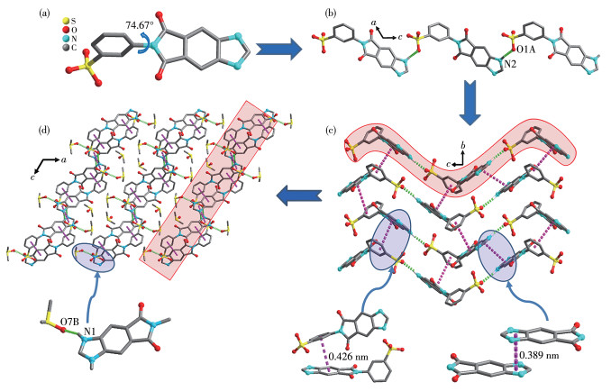
Hydrogen bond and π-π interactions are represented as green and pink dotted lines, respectively; H atoms are omitted for clarity except for those in hydrogen bonds; The red-shaded areas in c and d represent a chain and a layer respectively; Symmetry codes: A: x, 1-y, 1/2+z; B: 1/2-x, 1/2-y, 1-z.
DIBS(60℃) crystallizes in a P21/c space group. The asymmetric unit contains only one independent DIBS molecule (Fig. 2). The proton of the sulfonic acid group is also transferred to the benzimidazole group and a 43.69° torsion angle is observed for the molecule. Two DIBS molecules aggregate to form a dimmer through the dual offset face-to-face π-π interaction between benzimidazole rings. The corresponding ring-ring separation is about 0.384 nm. Then the dimmers are connected by the hydrogen bond between the benzimidazole nitrogen atom N1 and the sulfonate oxygen atom O3A (N1—H…O3A: N1…O3A 0.276 5 nm, ∠N1—H…O3A=172.00°), forming a 1D chain running along the a-axis. There is also offset face-to-face π-π interaction between the benzene rings from adjacent chains with ring-ring separation of 0.353 nm, resulting in a 2D layer extending along the a- and c-axes. Furthermore, a hydrogen bond is observed between the benzimidazole nitrogen atom N2 and sulfonate oxygen atom O3B (N2—H…O3B: N2…O3B 0.279 6 nm, ∠N2—H…O3B=163.37°) from adjacent layers. This finally results in a 3D supramolecular structure.
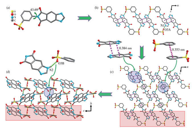
Hydrogen bond and π-π interactions are represented as green and pink dotted lines, respectively; H atoms are omitted for clarity except for those in hydrogen bonds; The red-shaded areas in c and d represent a chain and a layer respectively; Symmetry codes: A: 2-x, -y, 1-z; B: 1+x, 1/2-y, 1/2+z.
PXRD testing shows that the experimental patterns of the bulk samples were almost identical to the calculated patterns (Fig.S4a), indicating that the two phases have good purity. TGA (Fig.S4b) revealed that DIBS(10℃) was stable before 145 ℃ and then rapidly lost weight at 145-153 ℃, corresponding to two DMSO molecules (Obsd. 29.3%, Calcd. 31.2%). There was a platform region in a temperature range of 154-478 ℃. Then the compound decomposed upon further heating. The TGA curve of DIBS(60℃) showed that it was stable before 475 ℃, indicating that it has good thermal stability. Above 475 ℃, the compound decomposed quickly.
The photoluminescence (PL) properties of the two materials were further studied. DIBS(10℃) had blue fluorescence under a 365 nm UV lamp and presented green afterglow lasting about 6 s after the lamp was turned off (Fig. 3a). Under the same conditions, DIBS(60℃) had blue fluorescence and yellow-green afterglow that could last for about 3 s.

Strong emission peaks located at 450 and 510 nm were observed in the prompt and delayed spectra of DIBS(10℃). Time-resolved phosphorescence decay test (510 nm) showed it had a lifetime of 721.6 ms. In contrast, although DIBS(60℃) bore one emission peaked at 460 nm in the prompt spectrum, it had two emission peaks (460 and 520 nm) in the delayed spectrum (Fig. 3b). The 460 nm emission (τ=259.8 ms) has typical DF characteristic because its position overlaps with that of the fluorescence emission. The 520 nm emission had a lifetime of 363.3 ms (Fig.S5c), which can be attributed to RTP emission.
The temperature-dependent delayed spectra (Fig. 3c) of DIBS(60℃) were further gained. An emission peak at about 480 nm and a shoulder peak at about 460 nm can be observed in the spectrum at 77 K. With the increase in temperature, the intensity of the emission peak at 480 nm decreased gradually, accompanied by a redshift of the peak. When the temperature reached 240 K, the peak was located at 520 nm. With the further increase in temperature, the position of the peak remained unchanged but the intensity decreased gradually. When the temperature rose to 350 K, the peak could hardly be observed. In contrast, the shoulder peak at about 460 nm decreased gradually with the increase in temperature, but the position was unchanged. When the temperature rose to 240 K, the emission peak was almost invisible. The results preclude the possibility that the 460 nm peak is a thermally activated DF emission. The quantum yield of DIBS(10℃) and DIBS(60℃) was 3.34% and 4.05% respectively.
The sample DIBS(10℃)-G was obtained after grinding DIBS(10℃) in a mortar for 7 min (Fig. 4). The luminescence spectrum test (Fig. 3b) shows that its fluorescence peak shifted from 450 to 460 nm. In its delayed spectrum, a main peak at 520 nm and a shoulder peak at 460 nm could be observed. That is, the delay spectrum of DIBS(10℃)-G was the same as that of DIBS(60℃). PXRD characterization (Fig.S6a) proves that DIBS(10℃)-G still maintains good crystallinity. This is different from the commonly reported MCL molecules[14], which usually show the transition from crystalline to amorphous state under grinding. Further analysis shows that the pattern of DIBS(10℃)-G was the same as that of DIBS(60℃). The above results indicate that DIBS(10℃) can be transformed into DIBS(60℃) under grinding stimulation.
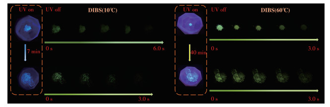
To observe the crystal structure changes of the compound during the grinding process, PXRD tests were conducted after grinding DIBS(10℃) samples for different times (labeled as DIBS(10℃)-X, X=30 s, 2 min, 4 min, and 7 min, respectively). As shown in Fig.S6a, with the increase of grinding time, the diffraction peak at 2θ=7.8°, 14.0°, 21.1°, and 27.3°of DIBS(10 ℃) gradually disappeared, At the same time, new peaks appeared at 2θ=10.9°, 18.7°, and 22.5°, and gradually increased. After grinding for 7 min, the PXRD pattern of the sample was almost consistent with that of DIBS(60℃), indicating that DIBS(10℃) has been successfully transformed into DIBS(60℃).
The spectra of DIBS(10℃) after grinding for different times were also measured (Fig.S6b). After 2 min of grinding, the prompt emission peak shifted to 460 nm and stabilized here. With the increase in grinding time, the strongest emission peak in delayed spectra (Fig.S6c) gradually redshifted, and reached 520 nm after 7 min of grinding. Interestingly, a small shoulder peak appeared at ca. 500 nm after grinding for 30 s. With the increase in grinding time, the peak position gradually blueshifted and stabilized at 460 nm after 4 min of grinding, while the emission intensity decreased gradually compared with that of the RTP peak. After 7 min of grinding, the shape of the delay spectrum was almost identical to that of DIBS(60℃).
The lifetimes of the 460 and 520 nm peaks of the DIBS(10℃)-G were 96.3 and 330.0 ms (Fig.S7) respectively, both shorter than that of DIBS(60℃). This may be because grinding leads to smaller crystal particles, thus exposing more surface areas to the air, and the quenching enhancement caused by oxygen in the air reduces the lifetimes. Unfortunately, many methods have been tried, but DIBS(10℃)-G can not return to the initial state. As a control, the MCL properties of DIBS(60℃) were also studied. Fig.S6d shows the PXRD patterns of the sample after grinding for different times. Interestingly, DIBS(60℃) maintained good crystallinity and the PXRD diffraction pattern did not change even after grinding for 40 min. PL spectrum (Fig.S6e, S6f) test of the sample showed that with the increase in grinding time, the emission peak in the prompt spectra had a slight red shift, while the peak in the delayed spectra was unchanged. The results indicate that DIBS(60℃) has a good ability to resist mechanical force.
TGA results show that heating will lead to the loss of DMSO in DIBS(10℃). So the influence of DMSO loss on its luminescence behavior was further studied. DIBS(10℃)-H was obtained by heating DIBS(10℃) at 170℃ for 20 min to remove the DMSO solvent molecules. Then the sample was submitted for characterization. The results show that it has both the same PL spectra(Fig.S8a) and diffraction pattern as that of DIBS(60℃) (Fig.S9).
The above results show that both heating and grinding stimulation can induce the transformation of DIBS(10℃) to DIBS(60℃). Similarly, various methods were tried, but DIBS(10℃)-H could not be restored to the initial state. After heating DIBS(60℃) for 20 min at 190℃, sample DIBS(60℃)-H was obtained. Its luminescence properties were consistent with that of DIBS(60℃) (Fig.S8b), indicating its afterglow has good thermal stability.
According to the literature[15], the different afterglow behavior of the two phases can be attributed to their different structures. The torsion angles of DIBS molecules in DIBS(10℃) (74.67°) and DIBS(60℃) (43.69°) are different. In addition, DIBS(10℃) has a loose stacking structure owing to the presence of DMSO solvent molecules. The density of DIBS(10℃) (1.480 g·cm-3) is smaller than that of DIBS(60℃) (1.643 g·cm-3), which also proves this point. Under external stimulation, the phase with large molecular distortion and loose stacking structure changes into the phase with small molecular distortion and close stacking structure. The irreversibility of the stimulus-response behavior may be attributed to the close stacking and highly stable crystal structure of DIBS(60℃), in which DMSO solvent molecules are extremely difficult to enter.
To sum up, a new benzimidazole derivative and its two solid phases, DIBS(10℃) and DIBS(60℃) were synthesized. Structure analyses show that DIBS molecules in the two phases have different conformations and stacking structures. DIBS(10℃) bears RTP emission, while DIBS(60℃) shows both RTP and DF emission. Remarkably, upon grinding or heating stimulation, DIBS(10℃) with a large molecular torsion angle and loose packing structure can be converted to DIBS(60℃) with a small molecular torsion angle and close packing structure. The work provides a good example of the preparation of organic materials with afterglow stimulation response behaviors.
刘进剑, 刘娜, 陆艺炜. 基于羧苄基紫精配体构筑的三种多重响应配合物[J]. 无机化学学报, 2022,38,(9): 1825-1833. LIU J J, LIU N, LU Y W. Three multiple-responsive complexes based on a carboxybenzyl viologen ligand[J]. Chinese J. Inorg. Chem., 2022, 38(9): 1825-1833.
Ekbote A, Mobin S M, Misra R. Stimuli-responsive phenothiazine-based donor-acceptor isomers: AIE, mechanochromism and polymorphism[J]. J. Mater. Chem. C, 2020, 8(10): 3589-3602. doi: 10.1039/C9TC05192A
Huang J X, Zhao H, Zhang J J, Zhang B L, Ni J, Song B, Li Y Q, Liu S Q, Duan C Y. Diemissive dye@CP composites with full-spectrum tunable mechanoluminescence[J]. J. Mater. Chem. C, 2021, 9(42): 15165-15174. doi: 10.1039/D1TC02841C
Fang M M, Yang J, Xie Y J, Dong Y Q, Peng Q, Li Z. Unexpected room-temperature phosphorescence from a non-aromatic, low molecular weight, pure organic molecule through the intermolecular hydrogen bond[J]. Mater. Chem. Front., 2018, 2(11): 2124-2129. doi: 10.1039/C8QM00396C
Gao M X, Tian Y, Yang J, Li X N, Ni J, Fang M M, Li Z. The same molecule but a different molecular conformation results in a different room temperature phosphorescence in phenothiazine derivatives[J]. J. Mater. Chem. C, 2021, 9(42): 15375-15380. doi: 10.1039/D1TC03460J
Zhang P P, Zhou H J, Ni A Y, Zhao H, Zhang J J, Wei H H, Liu S Q, Ni J, Duan C Y. Luminescent coordination polymer with its multistimuli-responsive sensitivity enabled and boosted by its dual emission[J]. Cryst. Growth Des., 2022, 22(8): 4845-4853. doi: 10.1021/acs.cgd.2c00338
王申宇, 陈典, 刘晓莉, 王硕文, 苑亚南, 王振平, 杨春. 基于(E)-N, N-二甲基-4-(2-(吡啶-4-基)乙烯基)苯胺的锌/镉有机-无机杂化金属卤化物的结构和发光性质[J]. 无机化学学报, 2021,37,(9): 1659-1664. WANG S Y, CHEN D, LIU X L, WANG S W, YUAN Y N, WANG Z P, YANG C. Structures and photoluminescence properties of zinc/cadmium-based organic-inorganic hybrid metal halides derived from (E)-N, N-dimethyl-4-(2-(pyridin-4-yl)vinyl)aniline[J]. Chinese J. Inorg. Chem., 2021, 37(9): 1659-1664.
Sudhakar P, Radhakrishnan T P. Stimuli responsive and reversible crystalline—Amorphous transformation in a molecular solid: Fluorescence switching and enhanced phosphorescence in the amorphous state[J]. J. Mater. Chem. C, 2019, 7(23): 7083-7089. doi: 10.1039/C9TC01600G
Liu Y J, Li A S, Xu S P, Xu W Q, Liu Y, Tian W J, Xu B. Reversible luminescent switching in an organic cocrystal: Multi-stimuli-induced crystal-to-crystal phase transformation[J]. Angew. Chem. Int. Ed., 2020, 59(35): 15098-15103. doi: 10.1002/anie.202002220
Shen Z Y, Yan X L, Cui S Q, Bo C C, Liu J Y, Wang B W, Li Y, Chen L G. Color-tunable, time-dependent, temperature and humidity-responsive afterglow from hyaluronic acid-based films[J]. Dyes Pigment., 2023, 212: 111113. doi: 10.1016/j.dyepig.2023.111113
Liu Y W, Li Y Z, Wen Y, Su X L, Xu M, Feng W, Liu Q, Li F Y. Significantly enhanced afterglow brightness via intramolecular energy transfer[J]. ACS Mater. Lett., 2021, 3(6): 713-720. doi: 10.1021/acsmaterialslett.1c00183
Nandi R P, Kalluvettukuzhy N K, Pagidi S, Thilagar P. Molecular persistent room-temperature phosphorescence from tetraarylaminoboranes[J]. Inorg. Chem., 2023, 62(3): 1122-1134. doi: 10.1021/acs.inorgchem.2c03386
Liu Y, Ma Z M, Liu J W, Chen M X, Ma Z Y, Jia X R. Robust white-light emitting and multi-responsive luminescence of a dual-mode phosphorescence molecule[J]. Adv. Opt. Mater., 2020, 9(2): 2001685.
Sun Y P, Qu H M, Zhang J C, Duan X Y, Zhang X K. Room temperature phosphorescence, thermally activated delayed fluorescence and multicolor mechanochromic luminescence of emitters through molecular interaction and conformational modulations[J]. Tetrahedron, 2022, 110: 132692. doi: 10.1016/j.tet.2022.132692
Zhang Y W, Ma H L, Wang S P, Li Z Q, Ye K Q, Zhang J Y, Liu Y, Peng Q, Wang Y. Supramolecular structure-dependent thermally-activated delayed fluorescence (TADF) properties of organic polymorphs[J]. J. Phys. Chem. C, 2016, 120: 19759-19767. doi: 10.1021/acs.jpcc.6b05537

Figure 1 Crystal structure of DIBS(10℃): (a) structure of DIBS; (b) 1D hydrogen-bonded chain; (c) 2D layer formed via connecting the 1D chains through π-π interactions; (d) packing of the layers viewed along the b-axis
Hydrogen bond and π-π interactions are represented as green and pink dotted lines, respectively; H atoms are omitted for clarity except for those in hydrogen bonds; The red-shaded areas in c and d represent a chain and a layer respectively; Symmetry codes: A: x, 1-y, 1/2+z; B: 1/2-x, 1/2-y, 1-z.

Figure 2 Crystal structure of DIBS(60℃): (a) structure of DIBS; (b) 1D chain based on hydrogen bond and π-π interaction; (c) 2D layer formed via connecting the 1D chains through π-π interaction; (d) 3D structure formed via connecting the 2D layers through hydrogen bonds
Hydrogen bond and π-π interactions are represented as green and pink dotted lines, respectively; H atoms are omitted for clarity except for those in hydrogen bonds; The red-shaded areas in c and d represent a chain and a layer respectively; Symmetry codes: A: 2-x, -y, 1-z; B: 1+x, 1/2-y, 1/2+z.

Figure 3 PL properties of the two phases: (a) photographs before and after turning off a 365 nm UV lamp at RT; (b) normalized luminescence spectra at RT (λex=365 nm); (c) temperature-dependent (77-400 K) luminescence spectra of DIBS(60℃) after turning off the excitation light source for 5 ms

 扫一扫看文章


扫一扫关注我们
 下载:
下载:

 下载:
下载:
