Scheme 1.
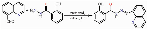
Synthetic route of HL
Sn(Ⅳ)/Ag(Ⅰ) Complexes of N-((Quinolin-8-yl)methylene)salicylhydrazide: Syntheses, Crystal Structures and DNA Interaction
Bei-Bei WANG , Hong-Xin CAI , Xue-Ao MEI , Xiu-Xiu ZHANG , Wei-Na WU , Yuan WANG , Zhong CHEN

Schiff bases are an important class of ligands in coordination chemistry and have been found extensive application in many different fields[1-4]. The Schiff base derivatives of acylhydrazone have attracted more attention due to their variable bonding modes towards metal ions and wide range of biological properties, such as antioxidant, anti-inflammatory, antibacterial and antitumor activities[5]. As one of the most important heterocycles, quinoline is frequently employed for the construction of transition metal complexes with significant pharmacological activities[6-7]. Our group has reported the structures of several Zn(Ⅱ)/Cd(Ⅱ) complexes with quinoline-containing acylhydrazone[8]. Never-theless, the investigation on Ag(Ⅰ) and Sn(Ⅳ) complexes with such type of ligands are relatively scarce. In fact, Ag(Ⅰ)-based complexes have been known for centuries, especially with respect to medical properties[9-10], and Sn(Ⅳ) compounds are widely used in catalysis, pesti-cides, polyvinyl chloride stabilizer, preservative, sterilization, anticancer drug and so on[11-13]. Herein, we report the crystal structures and DNA binding pro-perties of Sn(Ⅳ) and Ag(Ⅰ)complexes with N-((quinolin-8-yl)methylene)salicylhydrazide (HL, Scheme 1).
Solvents and starting materials for synthesis were purchased commercially and used as received. Elemental analysis was carried out on an Elemental Vario EL analyzer. The IR spectra (ν=4 000~400 cm-1) were determined by the KBr pressed disc method on a Bruker V70 FT-IR spectrophotometer. 1H NMR spectra of HL was acquired with Bruker AV400 NMR instrument in DMSO-d6 solution with TMS as internal standard. The UV spectra were recorded on a Purkinje General TU-1800 spectrophotometer. The interactions between three compounds and EB-DNA are measured using literature method[14] via emission spectra on a Varian CARY Eclipse spectrophotometer with the pass width of emission and excitation being 5 nm.
As shown in Scheme 1, the ligand HL was prepared by condensation of quinoline-8-carbaldehyde (1.57 g, 10 mmol) and 2-hydroxylbenzoyl hydrazine (1.52 g, 10 mmol) in methanol solution (100 mL) under refluxing for 1 h. The white solid was filtered and washed three times by methanol. Yield: 2.01 g (69%). Elemental analysis Calcd. for C17H13N3O2(%): C 70.09, H 4.50, N 14.42; Found(%): C 70.26, H 4.29, N 14.55. FT-IR (cm-1): ν(N-H) 3 259, ν(O=C) 1 651, ν(C=N) 1 604, ν(quinoline C=N) 1 575. 1H NMR (400 MHz, DMSO-d6): δ 12.13 (1H, s, NH-C=O), 12.01 (1H, s, OH), 9.73 (1H, s, CH=N), 8.96~8.98 (1H)/8.36~8.44 (2H)/7.94~8.09 (2H)/7.59~7.72 (2H)/7.40~7.42 (1H)/6.91~6.95 (2H) for Ar-H.
The complexes 1 and 2 were generated by reaction of the ligand HL (5 mmol) with equimolar of Ph2SnCl2 and AgNO3 in methanol solution (10 mL), respectively. Crystals suitable for X-ray diffraction analysis were obtained by evaporating the corresponding reaction solutions at room temperature.
1: Yellow blocks. Anal. Calcd. for C29.25H23ClN3O2.25Sn(%): C 57.97, H 3.79, N 6.92; Found(%): C 57.76, H 3.82, N 6.75. FT-IR (cm-1): ν(N=C-O) 1 624, ν(C=N) 1 589, ν(C=N quinoline) 1 575.
2: Colorless blocks. Anal. Calcd. for C18H17N4O6Ag(%): C 43.83, H 3.47, N 11.36; Found(%): C 43.62, H 3.77, N 11.16. FT-IR (cm-1): ν(N-H) 3 446, ν(N=C-O) 1 615, ν(C=N) 1 586, ν(C=N quinoline) 1 528, ν(NO3) 1 478 and 1 302.
The X-ray diffraction measurement for HL, complexes 1 and 2 were performed on a Bruker SMART APEX Ⅱ CCD diffractometer equipped with a graphite monochromatized Mo Kα radiation (λ=0.071 073 nm) by using φ-ω scan mode. Semi-empirical absorption correction was applied to the intensity data using the SADABS program[15]. The structures were solved by direct methods and refined by full matrix least-square on F2 using the SHELXTL-97 program[16]. All non-hydrogen atoms were refined anisotropically. All the other H atoms were positioned geometrically and refined using a riding model. Details of the crystal parameters, data collection and refinements for HL, complexes 1 and 2 are summarized in Table 1.
 下载:
导出CSV
下载:
导出CSV
| HL | 1 | 2 | |
| Empirical formula | C17H13N3O2 | C29.25H23CIN3O2.25Sn | C18H17N4O6Ag |
| Formula weight | 291.30 | 606.65 | 493.22 |
| T/ K | 296 | 296 | 296 |
| Crystal system | Orthorhombic | Monoclinic | Orthorhombic |
| Space group | Pca21 | P21/c | Pna21 |
| a / nm | 1.312 5(2) | 1.596 05(12) | 2.100 2(13) |
| b / nm | 0.647 38(12) | 1.205 79(8) | 0.500 5(3) |
| c / nm | 1.661 4(3) | 2.804 7(2) | 1.793 4(12) |
| V / nm3 | 1.411 6(4) | 5.205 4(6) | 1.855(2) |
| Z | 4 | 8 | 4 |
| Dc / (Mg·m-3) | 1.371 | 1.548 | 1.738 |
| Unique reflection | 2 189 | 9 172 | 3 070 |
| Rint | 0.051 | 0.036 | 0.071 |
| GOF | 1.021 | 1.067 | 1.016 |
| Final R indices [I>2σ(I)] | R1=0.043 8, wR2=0.088 7 | R1=0.032 1, wR2=0.066 1 | R1=0.049 2, wR2=0.095 5 |
| R indices (all data) | R1=0.071 6, wR2=0.102 4 | R1=0.051 1, wR2=0.073 9 | R1=0.116 6, wR2=0.115 7 |
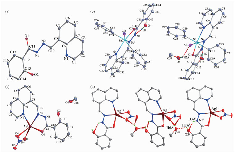
A diamond drawing of HL, complexes 1 and 2 are shown in Fig. 1a~c. Selected bond distances and angles are listed in Table 2. The bond length of carbonyl group in HL is 0.122 8(4) nm, which is similar as that in complex 2 (0.124 9(15) nm), while obviously shorter than that in complex 1 (0.128 1(4) and 0.128 9(4) nm). This fact shows that HL is anionic and neutral in complexes 1 and 2, respectively.
Symmetry codes: ⅰ 1.5-x, 0.5+y, -0.5+z; ⅱ 1.5-x, -0.5+y, 0.5+z
下载:
导出CSV
| HL | |||||
| C11-O1 | 0.122 8(4) | C10-N2 | 0.128 6(5) | C13-O2 | 0.136 4(5) |
| 1 | |||||
| Sn1-O1 | 0.213 2(2) | Sn1-C24 | 0.214 4(3) | Sn1-C18 | 0.214 7(3) |
| Sn1-N2 | 0.228 2(3) | Sn1-N1 | 0.236 9(3) | Sn1-Cl1 | 0.251 47(9) |
| Sn2-O3 | 0.208 4(2) | Sn2-C47 | 0.213 6(3) | Sn2-C53 | 0.213 7(3) |
| Sn2-N5 | 0.232 0(3) | Sn2-Cl2 | 0.250 34(9) | Sn2-N4 | 0.252 9(3) |
| 01-Sn1-C24 | 89.461(10) | 01-Sn1-C18 | 90.77(11) | C24-Sn1-C18 | 178.89(13) |
| 01-Sn1-N2 | 72.86(9) | C24-Sn1-N2 | 93.34(11) | C18-Sn1-N2 | 87.76(10) |
| 01-Sn1-N1 | 151.62(9) | C24-Sn1-N1 | 88.24(11) | C18-Sn1-N1 | 92.12(12) |
| N2-Sn1-N1 | 79.05(10) | 01-Sn1-Cl1 | 104.76(7) | C24-Sn1-Cl1 | 92.29(10) |
| C18-Sn1-Cl1 | 86.60(8) | N2-Sn1-Cl1 | 173.86(7) | N1-Sn1-Cl1 | 103.59(7) |
| 03-Sn2-C47 | 102.63(11) | 03-Sn2-C53 | 98.77(11) | C47-Sn2-C53 | 158.47(12) |
| 03-Sn2-N5 | 72.34(9) | C47-Sn2-N5 | 91.34(11) | C53-Sn2-N5 | 92.94(11) |
| 03-Sn2-Cl2 | 88.66(7) | C47-Sn2-Cl2 | 90.53(9) | C53-Sn2-Cl2 | 92.27(9) |
| N5-Sn2-Cl2 | 160.86(7) | 03-Sn2-N4 | 147.97(9) | C47-Sn2-N4 | 78.55(10) |
| C53-Sn2-N4 | 82.11(10) | N5-Sn2-N4 | 75.64(9) | Cl2-Sn2-N4 | 123.36(7) |
| 2 | |||||
| Ag1-N2 | 0.233 0(9) | Ag1-N1 | 0.238 0(10) | Ag1-O1 | 0.243 5(9) |
| Ag1-O3 | 0.237 4(12) | Ag1-O4 | 0.266 3(11) | ||
| N2-Agl-O4 | 142.9(4) | 01-Agl-O4 | 108.6(4) | 03-Agl-Nl | 103.5(4) |
| 03-Agl-O4 | 48.7(4) | 03-Agl-O1 | 107.1(4) | N2-Agl-O1 | 69.5(4) |
| Nl-Agl-O4 | 98.6(4) | N2-Agl-O3 | 168.2(4) | N2-Agl-Nl | 78.6(3) |
| Nl-Agl-O1 | 147.9(4) | ||||
As shown in Fig. 1a, the asymmetric unit of complex 1 contains two independent complexes molecules and a half of lattice water molecule. Each six-coordinated Sn(Ⅳ) ion is surrounded by two nitrogen atoms coming from quinoline and imine groups, one chloride ion, one oxygen atom coming from carbonyl group and two carbon atoms coming from two phenyl groups, exhibiting a distorted octahedral coordination geometry. There exist intramolecular O-H…N hydrogen bonds (O2-H2…N3, O4-H4…N6 and O5-H5A…Cl1, with D…A distance being 0.257 8, 0.259 1 and 0.341 0 nm, D-H…A angle being 145°, 144° and 147°, respectively) in the structure.
In complex 2, the Ag(Ⅰ) ion is surrounded by one tridentate hydrazone ligand with ON2 donor set and one bidentate nitrate anions, possessing a distorted square pyramid coordination geometry (τ=0.338). Furthermore, in the solid structure of 2, the complex molecules are linked by lattice methanol molecules into chains along c axis via various of intermolecular O-H…O hydrogen bonds(O2-H2A…O6, with D…A distance being 0.272 5(18) nm, D-H…A angle being 122.2°; O6-H6A…O4ⅰ, with D…A distance being 0.301 1(17) nm, D-H…A angle being 161.1°; O6-H6A…O5ⅰ, with D…A distance being 0.296 4(18) nm, D-H…A angle being 132.4°, Symmetry codes: ⅰ 1.5-x, 0.5+y, -0.5+z). The intramolecular O-H…O hydrogen bonds between amine N atoms and hydroxyl O atoms (N3-H3A…O2, with D…A distance being 0.259 8(15) nm, D-H…A angle being 130.7°) are also present.
The FT-IR spectral region for both complexes are more or less similar due to the similar coordination modes of the ligands. The ν(C=O) of the free ligand was 1 651 cm-1, and it disappeared in the spectra of comp-lexes. Meanwhile, new N=C-O stretching vibration absorption was observed at 1 624 and 1 615 cm-1 in complexes 1 and 2, respectively, revealing that the C=O in O=C-N moiety has enolizated and the oxygen atom coordinates to the metal ion in both complexes[8]. The ν(C=N) bands of the imine group, quinoline ring in the hydrazone ligand shifted to lower frequency values in the complexes, indicating that the N atoms of such two units take part in the coordination[17-18]. In addition, the general pattern of the IR spectroscopy for complex 2 supports the normal coordination of bidentate nitrate group[19]. It is in accordance with the crystal structure study.
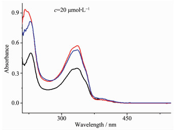
The UV spectra of HL, complexes 1 and 2 were measured in CH3OH solution (c=20 μmol·L-1) at room temperature (Fig. 2). The spectra of HL featured two bands located at 229 nm (ε=50 168 L·mol-1·cm-1) and 336 nm (ε=34 974 L·mol-1·cm-1), which could be assigned to characteristic π-π* transition centered on quinolone ring and the imine moiety[20], respectively. Both bands exhibited significant hyperchromicity in the spectra of both complexes (ε=93 738 and 57 310 L·mol-1·cm-1 for complex 1; ε=81 974 and 53 125 L·mol-1·cm-1 for complex 2), indicating that the ligand HL takes part in the coordination[21].
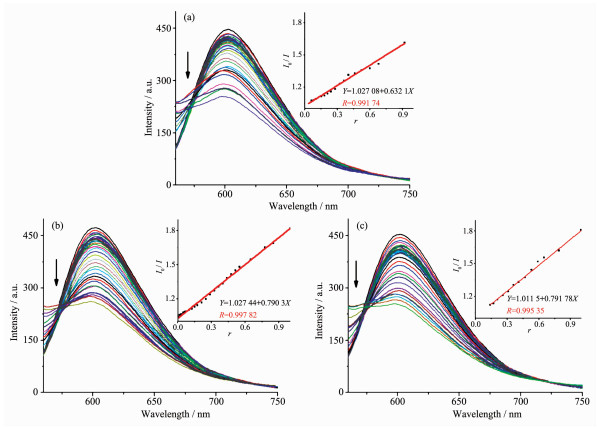
It is well known that EB can intercalate nonspecifically into DNA, which causes it to fluoresce strongly. Competitive binding of other drugs to DNA and EB will result in displacement of bound EB and a decrease in the fluorescence intensity[22-23]. The effects of the ligand and complexes on the fluorescence spectra of EB-DNA system are presented in Fig. 3. The fluorescence intensities of EB bound to ct-DNA at about 600 nm showed remarkable decreasing trends with the increasing concentration of the tested compounds, indicating that some EB molecules are released into solution after the exchange with the compounds. The quenching of EB bound to DNA by the compounds is in agreement with the linear Stern-Volmer equation: I0/I=1+Ksqr[22], where I0 and I represent the fluorescence intensities in the absence and presence of quencher, respectively; Ksq is the linear Stern-Volmer quenching constant; r is the ratio of the concentration of quencher and DNA. In the quenching plots of I0/I versus r, Ksq values are given by the slopes. The Ksq values were 0.632 1, 0.790 3 and 0.791 9 for the ligand HL, complexes 1 and 2, respectively. The results indicate that interactions of the complexes with DNA are stronger than that of the ligand HL, because the complexes have higher rigidity to bind the base pairs along DNA, thus increasing their binding abilities[14].
Beyene B B, Das K, Kerayu B A, et al. Catal. Commun., 2019, 119:111-114 doi: 10.1016/j.catcom.2018.10.027
Saghatforoush L, Moeini K, Hosseini-Yazdi S A, et al. RSC Adv., 2018, 62:35625-35639
Yousif E, Ahmed D S, El-Hiti G A, et al. Polymers, 2018:103-105
Bourosh P N, Revenko M D, Stratulat E F, et al. Russ. J. Inorg. Chem., 2014, 59:545-557 doi: 10.1134/S0036023614060059
Bahmudov K T, Kopylovich M N, Pombeiro A J L. Coord. Chem. Rev., 2013, 257:1244-1281 doi: 10.1016/j.ccr.2012.12.016
Kasiotis, K M, Fokialakis N, Haroutounian S A. Synthesis, 2006, 11:1791-1802 https://www.researchgate.net/publication/239729265_Synthesis_of_Novel_Conformationally_Constrained_Pyrazolo43-_c_quinoline_Derivatives_as_Potential_Ligands_for_the_Estrogen_Receptor
Stringer T, Taylor D, Guzgay H, et al. Dalton. Trans., 2015, 33:14906-14917 https://www.ncbi.nlm.nih.gov/pubmed/26226082
许志红, 吴伟娜, 刘树阳, 等.无机化学学报, 2018, 34(2):375-380 http://www.wjhxxb.cn/wjhxxbcn/ch/reader/view_abstract.aspx?flag=1&file_no=20180222&journal_id=wjhxxbcnXU Zhi-Hong, WU Wei-Na, LIU Shu-Yang, et al. Chinese J. Inorg. Chem., 2018, 34(2):375-380 http://www.wjhxxb.cn/wjhxxbcn/ch/reader/view_abstract.aspx?flag=1&file_no=20180222&journal_id=wjhxxbcn
Movahedi E, Rezavani A R. J. Mol. Struct., 2017, 1139:407-417 doi: 10.1016/j.molstruc.2017.03.042
Písařík P, Jelínek M, Remsa J, et al. Mater. Sci. Eng. C, 2017, 77:955-962 doi: 10.1016/j.msec.2017.04.005
Aamal AK, Zhu H B, Harb M, et al. Catal. Sci. Technol., 2017, 7:581-586 doi: 10.1039/C6CY02187E
Grzeskiewicz A M, Owczarzak A, Kucinska M, et al. J. Coord. Chem., 2017, 70:1776-1789 doi: 10.1080/00958972.2017.1316841
Attanzio A, Ippolito M, Girasolo M A, et al. J. Inorg. Biochem., 2017, 7:36056-36071
沈伟, 胡未极, 吴小勇, 等.无机化学学报, 2016, 32(6):1101-1110 http://www.wjhxxb.cn/wjhxxbcn/ch/reader/view_abstract.aspx?flag=1&file_no=20160623&journal_id=wjhxxbcnSHEN Wei, HU Wei-Ji, WU Xiao-Yong, et al. Chinese J. Inorg. Chem., 2016, 32(6):1101-1110 http://www.wjhxxb.cn/wjhxxbcn/ch/reader/view_abstract.aspx?flag=1&file_no=20160623&journal_id=wjhxxbcn
Sheldrick G M. SADABS, University of Göttingen, Germany, 1996.
Sheldrick G M. SHELX-97, Program for the Solution and the Refinement of Crystal Structures, University of Göttingen, Germany, 1997.
Huang Y Q, Zhao W, Chen J G, et al. Z. Anorg. Allg. Chem., 2012, 638:679-682 doi: 10.1002/zaac.v638.3/4
Huang Y Q, Wan Y, Chen H Y, et al. New J. Chem., 2016, 40:7587-7595 doi: 10.1039/C6NJ01231K
王娜, 贺新前, 林秋月, 等.中国稀土学报, 2010, 28(2):141-146 http://www.cnki.com.cn/Article/CJFDTotal-XTXB201002001.htmWANG Na, HE Xin-Qian, LIN Qiu-Yue, et al. Journal of the Chinese Rare Earth Society, 2010, 28(2):141-146 http://www.cnki.com.cn/Article/CJFDTotal-XTXB201002001.htm
Song X Q, Zeng Z P, Liu W S, et al. J. Solid State Chem., 2009, 182:841-848 doi: 10.1016/j.jssc.2008.12.025
李晓静, 吴伟娜, 徐周庆, 等.无机化学学报, 2015, 31(11):2265-2271 http://www.wjhxxb.cn/wjhxxbcn/ch/reader/view_abstract.aspx?flag=1&file_no=20151123&journal_id=wjhxxbcnLI Xiao-Jing, WU Wei-Na, XU Chou-Qing, et al. Chinese J. Inorg. Chem., 2015, 31(11):2265-2271 http://www.wjhxxb.cn/wjhxxbcn/ch/reader/view_abstract.aspx?flag=1&file_no=20151123&journal_id=wjhxxbcn
Yang Z Y, Wang Y, Wang Y. Bioorg. Med. Chem. Lett., 2007, 17:2096-2101 doi: 10.1016/j.bmcl.2006.10.049
Joseph M, Suni V, Kurup M R P, et al. Polyhedron, 2004, 23:3069-3080 doi: 10.1016/j.poly.2004.09.026

Figure 1 Diamond drawing of HL (a), and complexes 1 (b) and 2 (c) with 10% thermal ellipsoids; Chain-like structures in complex 2 formed by hydrogen bonds shown in dashed line (d)
Symmetry codes: ⅰ 1.5-x, 0.5+y, -0.5+z; ⅱ 1.5-x, -0.5+y, 0.5+z

Figure 2 UV spectra of the ligand HL, complexes 1 and 2in CH3OH solution at room temperature

Figure 3 Emission spectra of EB-DNA system in the presence of HL (a) and complexes 1 (b) and 2 (c), respectively
Arrow shows the fluorescence intensities change of EB-DNA system upon increasing tested compound concentration; Inset: plot of I0/I versus r, r=ccompound/cDNA
Table 1. Selected crystallographic data for HL, complexes 1 and 2
| HL | 1 | 2 | |
| Empirical formula | C17H13N3O2 | C29.25H23CIN3O2.25Sn | C18H17N4O6Ag |
| Formula weight | 291.30 | 606.65 | 493.22 |
| T/ K | 296 | 296 | 296 |
| Crystal system | Orthorhombic | Monoclinic | Orthorhombic |
| Space group | Pca21 | P21/c | Pna21 |
| a / nm | 1.312 5(2) | 1.596 05(12) | 2.100 2(13) |
| b / nm | 0.647 38(12) | 1.205 79(8) | 0.500 5(3) |
| c / nm | 1.661 4(3) | 2.804 7(2) | 1.793 4(12) |
| V / nm3 | 1.411 6(4) | 5.205 4(6) | 1.855(2) |
| Z | 4 | 8 | 4 |
| Dc / (Mg·m-3) | 1.371 | 1.548 | 1.738 |
| Unique reflection | 2 189 | 9 172 | 3 070 |
| Rint | 0.051 | 0.036 | 0.071 |
| GOF | 1.021 | 1.067 | 1.016 |
| Final R indices [I>2σ(I)] | R1=0.043 8, wR2=0.088 7 | R1=0.032 1, wR2=0.066 1 | R1=0.049 2, wR2=0.095 5 |
| R indices (all data) | R1=0.071 6, wR2=0.102 4 | R1=0.051 1, wR2=0.073 9 | R1=0.116 6, wR2=0.115 7 |
 下载: 导出CSV
Table 2. Selected bond lengths (nm) and angles (°) in HL, complexes 1 and 2
| HL | |||||
| C11-O1 | 0.122 8(4) | C10-N2 | 0.128 6(5) | C13-O2 | 0.136 4(5) |
| 1 | |||||
| Sn1-O1 | 0.213 2(2) | Sn1-C24 | 0.214 4(3) | Sn1-C18 | 0.214 7(3) |
| Sn1-N2 | 0.228 2(3) | Sn1-N1 | 0.236 9(3) | Sn1-Cl1 | 0.251 47(9) |
| Sn2-O3 | 0.208 4(2) | Sn2-C47 | 0.213 6(3) | Sn2-C53 | 0.213 7(3) |
| Sn2-N5 | 0.232 0(3) | Sn2-Cl2 | 0.250 34(9) | Sn2-N4 | 0.252 9(3) |
| 01-Sn1-C24 | 89.461(10) | 01-Sn1-C18 | 90.77(11) | C24-Sn1-C18 | 178.89(13) |
| 01-Sn1-N2 | 72.86(9) | C24-Sn1-N2 | 93.34(11) | C18-Sn1-N2 | 87.76(10) |
| 01-Sn1-N1 | 151.62(9) | C24-Sn1-N1 | 88.24(11) | C18-Sn1-N1 | 92.12(12) |
| N2-Sn1-N1 | 79.05(10) | 01-Sn1-Cl1 | 104.76(7) | C24-Sn1-Cl1 | 92.29(10) |
| C18-Sn1-Cl1 | 86.60(8) | N2-Sn1-Cl1 | 173.86(7) | N1-Sn1-Cl1 | 103.59(7) |
| 03-Sn2-C47 | 102.63(11) | 03-Sn2-C53 | 98.77(11) | C47-Sn2-C53 | 158.47(12) |
| 03-Sn2-N5 | 72.34(9) | C47-Sn2-N5 | 91.34(11) | C53-Sn2-N5 | 92.94(11) |
| 03-Sn2-Cl2 | 88.66(7) | C47-Sn2-Cl2 | 90.53(9) | C53-Sn2-Cl2 | 92.27(9) |
| N5-Sn2-Cl2 | 160.86(7) | 03-Sn2-N4 | 147.97(9) | C47-Sn2-N4 | 78.55(10) |
| C53-Sn2-N4 | 82.11(10) | N5-Sn2-N4 | 75.64(9) | Cl2-Sn2-N4 | 123.36(7) |
| 2 | |||||
| Ag1-N2 | 0.233 0(9) | Ag1-N1 | 0.238 0(10) | Ag1-O1 | 0.243 5(9) |
| Ag1-O3 | 0.237 4(12) | Ag1-O4 | 0.266 3(11) | ||
| N2-Agl-O4 | 142.9(4) | 01-Agl-O4 | 108.6(4) | 03-Agl-Nl | 103.5(4) |
| 03-Agl-O4 | 48.7(4) | 03-Agl-O1 | 107.1(4) | N2-Agl-O1 | 69.5(4) |
| Nl-Agl-O4 | 98.6(4) | N2-Agl-O3 | 168.2(4) | N2-Agl-Nl | 78.6(3) |
| Nl-Agl-O1 | 147.9(4) | ||||
下载: 导出CSV

 扫一扫看文章

扫一扫关注我们