
Figure 1.
The classification of nanoparticles. By Figdraw.
Tumor microenvironment-sensitive polymeric nanoparticles for synergetic chemo-photo therapy
Tingting Hu , Chao Shen , Xueyan Wang , Fengbo Wu , Zhiyao He

Despite the remarkable achievements in the understanding of cancer and the great advances in the treatment of cancer, cancer remains a huge threat to human health and life, leading to more than 9.9 million deaths worldwide in 2020 [1]. There are three main strategies for systemic treatment of cancer, including chemotherapy, targeted therapy, and cancer immunotherapy. Chemotherapy is still the predominantly used modality for almost all types of cancers at different stages [2]. However, challenges including poor tumor targeting, systemic serious adverse reactions, chemotherapy resistance, and the limited therapeutic efficacy of monotherapy modality have limited its further development [3-5]. To address these challenges, multimodal combination cancer therapy has emerged and has demonstrated significant potential in enhancing the effectiveness and security of chemotherapeutic strategies [6,7].
With the rapid advancement of cancer treatment technology, the emerging treatment modalities such as gene therapy, phototherapy (mainly refers photothermal therapy (PTT) and photodynamic therapy (PDT), and sonodynamic therapy are gradually applied to clinical cancer treatment [8]. Specially, light-mediated phototherapy is a hopeful "green" physical therapy [9,10]. PTT involves converting the light (typically within the near-infrared region) into locally high temperatures with photothermal conversion agents, subsequently ablating tumor cells by inducing cell membrane lysis and rupture through local hyperthermia [11]. PDT, on the other hand, typically requires three main components: light, photosensitizer (PS), and oxygen, which together undergo photodynamic reaction and generate cytotoxic reactive oxygen species (ROS) to trigger damage of tumor cell membranes and induction of cellular necrosis and apoptosis [11]. In recent years, phototherapy has garnered significant interest due to its advantages of non-invasiveness, spatiotemporal control, low off-target toxicities, and negligible drug resistance [12,13], and has become an effective complementary therapy modality for cancer treatment [14]. On the cellular level, both PTT and PDT have been proved to surmount chemotherapy resistance and block compensatory signaling pathways [15]. On the tumor microenvironment (TME) level, PDT and PTT can enhance tumor perfusion, vascular, and extracellular matrix permeability, thereby facilitating anticancer agent penetration deep into the tumor site [16-18]. Nevertheless, the penetration depth of light in tumor tissue is limited, and the treatment duration is short, which generally fails to completely kill tumor cells [12]. Consequently, the combination of chemotherapy and phototherapy is a good attempt for cancer therapy, which can merge the advantages of both treatment modalities, surmount the shortcomings of the two methods, and enhance the efficacy of cancer treatment while reducing adverse reactions.
To achieve the efficient synergetic anticancer effects of chemotherapy and phototherapy, it is necessary to deliver chemotherapeutic agents and phototherapeutic agents simultaneously to the tumor site and then specifically release them within the tumor cells. With the development of nanotechnology, a wide variety of nanocarriers (Fig. 1), including polymer-based, lipid-based, carbon-based, and metal-based nanoparticles, have been developed as ideal platforms for the co-delivery and controlled release of multiple anticancer agents [19-22]. Among the aforementioned candidates, polymer-based nanoparticles that are self-assembled from amphiphilic blocks or grafted polymers, are considered to be a superior option due to their low toxicity, favorable biocompatibility, good biodegradability, reduced immunogenicity, comparative stability, increased water solubility, ease of synthesis, flexible surface modification, tailored design, and abilities to load both hydrophilic and hydrophobic agents [23-25]. Specially, owing to the exceptional safety and biocompatibility of polymer materials, the Food and Drug Administration of the United States (FDA) has authorized a list of pharmaceutical polymer carrier materials, including polylactic acid-glycolic acid (PLGA), polylactic acid (PLA), polyglycolic acid (PGA), polymethyl methacrylate (PMMA), polyprolactam (PCL), etc. [26]. Recent advances in modern synthetic polymer chemistry, along with other formulation techniques, have broadened the range of polymeric materials synthesis with complex structures and advanced polymer nanoformulation, facilitating the advancement of polymer-based nanomedicines [27-30]. In particular, the application of stimuli-sensitive polymeric materials capable of responding to endogenous stimuli like weak acidity, hypoxia and tumor specific enzymes in the TME, or ectogenous stimuli like X-rays, ultrasonic waves and heat, have become a trend in order to mediate on-demand and programmable rapid drug release at the desired sites [24,31,32]. Compared with external stimuli, TME-responsive polymeric nanoparticles have the advantages of safety and convenience, offer the opportunity to mimic the elegant mechanisms of natural biological systems, and have attracted more and more attention in synergistic chemo-photo therapy [33,34]. Given the paramount importance of TME-responsive polymeric nanoparticles, we present an overview of the most recent advancements in TME-responsive polymeric nanoparticles for synergetic chemo-photo therapy.
TME, as a soil for cancer cell culture, is mainly composed of blood vessels, stromal cells, fibroblasts, extracellular matrix (ECM), immune cells, and signaling molecules, which profoundly affects the initiation and progression of tumors [35]. Tumor neovascularization is crucial for the occurrence and development of tumors. Mature tumor vascular matrix structure is chaotic, leading to increased interstitial fluid pressure, inadequate blood perfusion, persistent hypoxia, and drug delivery difficulties, all of which contribute to cancer progression, metastasis, and drug resistance. The ECM is another important component of TME with multiple crucial roles, including mechanical support, TME modulation, and a source of signaling molecules that trigger tumor metastasis and further malignant transformation. Cytokines, enzymes and growth factors secreted by fibroblasts and immune cells in the tumor tissues make tumors more aggressive and invasive. In addition to the various components in the tumor region, the physiology of TME is also different from that of normal tissues. Unlike normal tissues, TME typically exhibits a more acidic microenvironment, a higher state of hypoxia, elevated interstitial fluid pressure and ATP, abundant ROS and glutathione (GSH), and overexpressed enzymes [36,37]. Unfortunately, these hostile features of TME are strongly associated with tumor angiogenesis, treatment resistance, and tumor progression [38,39]. Therefore, TME is an important marker in cancer treatment and has been widely utilized as a stimulus to stimulate size shrinkage, charge conversion, shell deshielding, or structure disintegration of stimuli-responsive polymeric nanoparticles to facilitate the programmable controlled release of anticancer agents (Fig. 2). In this section, we briefly describe the characteristics of TME related to "stimulus-responsive nanodrug strategies", rather than describe all the components of TME in detail.

Research suggests that the extracellular space of tumor tissue has a slightly acidic environment with a pH range of 6.5–6.8, which is due to a large number of metabolic abnormalities and increased lactate accumulation [40]. Unlike normal cells that generate energy primarily through mitochondrial oxidative phosphorylation, cancer cells prefer to meet their energy requirements through aerobic glycolysis. Through this altered energy metabolism, cancer cells are capable of fabricating large amounts of lactate, resulting in the acidification of TME [41]. Such mild acidity shows great potential as an internal stimulus for the construction of weak acid-sensitive polymeric nanoparticles. Typically, polymers containing tertiary amine structures possess pH-stimuli-cleavable performance owing to the protonation of tertiary amine groups in the acidic TME [42]. In addition, several acid-cleavable chemical groups such as cis-aconityl, maleimide, and benzoic-imine bond have been widely used to exploit pH-responsive polymeric nanoparticles [43].
Owing to the unrestrained proliferation of tumor cells, large amounts of nutrients and oxygen are required inside the tumor, which leads to the formation of abnormal tumor-associated angiogenesis, resulting in impaired microcirculation [44]. And, hypoxia is mainly caused by vascular defects and irregular microvessels in the deep regions of tumor [45,46]. In certain regions of tumor, the oxygen partial pressure can decrease to 0–2.5 mmHg, which is significantly lower than that in normal tissues of 30–40 mmHg [47]. Normal cells cannot endure hypoxic environment, while cancer cells can accommodate the hypoxic changes and survive by regulating the gene expression of hypoxia-inducible factor-1-alpha (HIF-1α), endothelin-1 (EDN-1), and nuclear factor kappa B (NF-κB) [48]. Due to the significant difference between tumor tissues and normal tissues, hypoxia is well widely acknowledged as a major target for cancer diagnosis and treatment [49,50]. Nitroaromatic, quinone, and azobenzene derivatives are the primary functional chemical groups sensitive to hypoxia, and have been extensively exploited to fabricate hypoxia-labile polymeric nanoparticles for tumor targeted therapy [51,52].
Cancer cells produce large amounts of ROS (mainly includes H2O2, HO•, 1O2 and O2•−) through the abnormally active nicotinamide adenine dinucleotide phosphate (NADPH) oxidase, increased cellular receptor signaling, and mitochondrial respiratory chain dysfunction [53,54]. In addition, mutations in genes and modified patterns of energy metabolism in cancer cells can additionally promote ROS production [55]. ROS, especially H2O2, plays an important role in a variety of physiological processes [56]. And, research confirms that ROS play a vital function in stabilizing HIFs, which is essential for the formation and progression of tumors [55]. It is estimated that the concentration of H2O2 in the TME is as high as 100 × 10−6 mol/L, which is approximately 100 times greater than the concentration found in normal tissues [57], making it another promising stimulus for the development of TME-activatable polymeric nanoparticles. In recent years, various ROS-cleavable linkers, including thioketal, thioether, phenylboronic esters, selenide, aminoacrylate, and arylboronic ester, have been widely used to fabricate ROS-sensitive polymeric nanomedicines [58].
In order to maintain the balance of the redox state in tumor cells, GSH, an antioxidant that helps eliminate excessive ROS, is invariably upregulated in the TME for avoiding irreversible damage to tumors [59,60]. According to reports, the GSH levels in tumor cells are typically 5–10 mmol/L, significantly exceeding the concentrations found in normal tissues (1–5 mmol/L) [61]. More importantly, the cytosol contains a concentration of GSH that is approximately 1000 times greater than the concentration in the extracellular space [62,63], making it an attractive stimulus for the design of endogenous stimuli-sensitive nanoparticles. The chemical groups that are sensitive to GSH are mainly disulfide and ditelluride, which have been widely incorporated into various polymers to develop GSH-cleavable polymer materials [63].
Researchers show that enzymes exhibit different expression levels in many disease-related microenvironments, including cancer [64]. Notably, TME shows excessive specific enzyme secretion compared with normal tissues, including hyaluronidase (HAdase), matrix metalloproteinases (MMPs), esterase, cathepsins, fibroblast activation protein-α (FAP-α), γ-glutamyl transpeptidase, and glucuronidase [64]. Compared with other abnormalities in the TME, enzyme overexpression in tumors possesses high spatial and functional specificity in a peculiar way. Therefore, more intelligent nanocarriers can be designed to facilitate on-demand drug release by using enzymes as specific stimuli. As with other polymers that respond to stimuli, the design of enzyme-sensitive polymers also involves incorporating particular substrates into the vector structures by chemical conjugation. Small peptides, peptide sequences, and gelatin are typically utilized as enzyme recognition substrates to synthesize enzyme-sensitive polymers [65-67].
Studies suggest that adenosine triphosphate (ATP) levels differ between cancer cells and normal cells, and even between the extracellular and intracellular spaces, which motivates the development of ATP-reliable or ATP-sensitive nanoparticles [68,69]. It is estimated that tumor cells have approximately 1.2 times more ATP than normal cells [69]. Consequently, the application of ATP as a stimulus to develop responsive nanoparticles for cancer therapy has become a research hotspot. A variety of chemical moieties can be utilized to construct ATP-response nanocarriers. Competitive binding of ATP to poly (amino acids) has been used to build theranostic nanoparticles [70]. Phenylboronic acids can be used to bind ATP by forming covalent esters [71,72]. In addition, ATP responsiveness can be achieved by introducing ATP-sensitive aptamers into polymeric nanoparticles [73,74].
Among the various kinds of endogenous stimuli, weak acidity is the most commonly used stimulus. The tumor regions exhibit a slightly lower pH of 6.5–6.8, which can trigger structural distortion, charge reversal, dimensional change, or disintegration of pH-responsive polymeric nanoparticles [75-77]. And, therapeutic agents are then released either through the direct dissociation of therapeutic compounds from polymers or by the decomposition of nanoparticles [78,79].
Yu and colleagues well-designed a doxorubicin (DOX)-grafted amphiphilic copolymer with pH-sensitive hydrazine bond and DIP moiety to co-deliver DOX and semiconducting polymers (SPs) [80]. In their study, the SP-loaded pH-sensitive polymeric prodrug micelles (PADD@SPs) with two pH-sensitive structures enabled the quick drug release of DOX in the acidic TME, and, the hyperthermia triggered by SP/near infrared (NIR) light not only directly ablated cancer cells but also effectively facilitated DOX penetration into the deep parts of tumor tissues to eradicate the cancer cells surviving from PTT. All in all, this combination of photothermal SPs and DOX using pH-sensitive polymeric micelles represents a viable strategy for synergistic photothermal-chemotherapy.
In another way, Husni and colleagues fabricated a novel type of chem-photothermal combination agent, wherein DOX and gold nanoclusters (GNCs) were simultaneously loaded into pH-responsive poly(ethylene glycol)-poly[(benzyl-L-aspartate)-co-(N-(3-aminopropyl)imidazole-L-aspartamide)] (PEG-PABI) micelles [81]. In their study, the dual drug-loaded micelles (Dox/GNC-Ms) exhibited excellent pH-reliable characteristics due to the protonation of the imidazole groups in PEG-PABI, thereby enabling rapid drug release when exposed to weakly acidic conditions in the TME. Moreover, PEG-PABI micelles surmounted the shortcomings of conventional GNCs by increasing their photothermal stability while boosting high heat generation efficiency. This advancement makes the Dox/GNC-M nanosystem a highly promising nanomedicine for chemo-photothermal combination therapy.
The combination of PTT and chemotherapy can greatly improve the efficacy of tumor treatment. However, due to the complexity of TME, excellent permeability of nanoparticles in tumors is still highly desired [82,83]. Among the strategies to enhance tumor penetration, charge reversal is a simple but effective method [84,85]. Nanoparticles, which circulate in the blood with a negative charge but can be reversed to a positive charge when they reach the tumor site, often have greater tumor penetration. Wang and colleagues constructed a smart core-shell structure polymeric nanocarrier (MPPD@IR825/DTX) with pH-sensitive charge-reversal property to co-deliver docetaxel (DTX) and IR825 for synergistic therapy [86]. The core was positively charged poly(ethylene imine)-poly(ε-caprolactone) block polymer (PEI-PCL) micelles loaded with DTX and IR825, and the shell was dimethylmaleic anhydride (DMMA)-modified PEGylated polymer with negatively charge at neutral conditions. The charge-reversal characteristics of MPPD@IR825/DTX were due to the re-exposure of core micelles following the rapid hydrolysis of DMMA under the weak acidic conditions, which significantly improved the efficiency of cellular uptake and tumor penetration. In the same time, MPPD@IR825/DTX showed excellent thermal conversion performance and increased the cytotoxicity of DTX, providing a simple but new approach for targeting and combinational chemo-photothermal therapy. In a similar way, Hao and colleagues successfully developed a pH-dependent charge-reversing drug delivery system (Fe3O4-DOX@PDA-GOx@PEOz, FDPGP) for collaborative multimodal cancer therapy [87]. To achieve the charge-reversal properties of multifunctional nanoparticles, PEG-grafted poly(2-ethyl-2-oxazoline) (PEOz) polymer was utilized to coat the DOX-glucose oxidase (GOx) dual-loaded magnetic polydopamine nanoparticles. PEOz is a polymer containing tertiary amine structure, which possesses the charge conversion capability from negative charge at physiological condition to positive charge in the acid TME [88]. In their study, excellent tumor accumulation, deep tumor penetration, and superior tumor ablation were achieved, thereby resulting in a combination of cascade-augmented chemotherapy/starvation therapy/chemodynamic therapy (CDT)/PTT. Overall, FDPGP provides a new perspective for the development of multimodal synergistic cancer treatment.
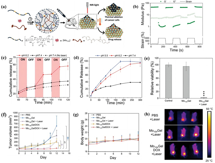
Although pH-responsive polymeric nanoparticles have been extensively studied in cancer treatment, their inadequate response rate and drug release regulation ability remain major obstacles affecting the therapeutic efficacy. Therefore, it is desirable to construct a smart nanocarrier that combines pH-sensitive polymers and other stimuli-responsive intelligent materials to efficiently regulate its functional drug release. Li and colleagues fabricated an intelligent NIR-/pH-triggered dual-responsive nanoplatform (MPDA-BTZ@CMCS-DOX) for the simultaneous administration of hydrophobic/hydrophilic therapeutic agents and photothermal synergetic tumor elimination [89]. First, hydrophobic bortezomib (BTZ) was encapsulated in mesoporous polydopamine nanospheres (MPDAs) through the pH-cleavable borate ester bond to realize sustained release of BTZ triggered by tumor acidity. Subsequently, pH-sensitive carboxymethyl chitosan (CMCS) coated on the surface of BTZ-loaded MPDAs was used to capture hydrophilic DOX while avoiding pre-leakage of BTZ. In their study, CMCS remained unchanged under physiological conditions but disintegrated under weak acid conditions to achieve pH-regulating DOX release. Moreover, when exposed to NIR radiation, the MPDA efficiently killed cancer cells through hyperthermia and further accelerated the drug release rate, thereby enabling NIR-/pH-triggered robust chemo-photothermal therapy. Guedes and colleagues successfully constructed an injectable and self-healing hydrogel (Mo154GelDOX) with pH/NIR dual-responsiveness consisting of benzylaldehyde functionalized polyethylene glycol (DF-PEG), poly(N-isopropylacrylamide) functionalized thermo-responsive chitosan (CS-g-PNIPAAm), Mo154, and DOX [90]. Briefly, the hydrogel was created through the dynamic imine bonds between the aldehydes from DF-PEG and the amines from CS-g-PNIPAAm, along with the dynamic electrostatic interactions between the negatively charged Mo154 and the positively charged CS-g-PNIPAAm. In their study, this double cross-linked dynamic network showed fast self-healing properties after injection. Interestingly, the imine bonds formed during the dynamic cross-linking process are pH-reliable, allowing accelerated and more complete drug release in the acidic TME. Taken together, this dual-stimulus-responsive dynamic hydrogel nanosystem holds great potential for the combined application of photothermal and chemotherapy (Fig. 3). Additionally, a summary of pH-responsive polymeric nanoparticles for synergetic chemo-photo therapy is displayed in Table S1 (Supporting information).
It is well known that intratumoral hypoxia is one of the obstacles in the treatment of solid tumors and is highly associated with tumor progression and poor prognosis [91]. According to the characteristics of hypoxia microenvironment, a variety of strategies have emerged to overcome/alleviate the hypoxia condition in the TME [92-94]. Interestingly, hypoxia is also closely associated with reducing species such as nitrogen reductase and nitroreductase [95]. Therefore, various hypoxia‐responsive prodrugs and drug delivery systems have been constructed using the trigger of hypoxia [96-99]. However, the heterogeneously distributed hypoxic regions within the tumor significantly weakens the sensitivity of hypoxic responsiveness and reduces the therapeutic efficiency of the hypoxia-responsive nanosystems. To overcome the limitations of the hypoxia-responsive nanocarriers, a noteworthy strategy is to combine them with PDT systems. During the process of converting oxygen into singlet oxygen (1O2) in PDT, the continuous consumption of oxygen and the resulting vascular damage can further intensify the hypoxia conditions within tumors [100-102]. Therefore, the combination of PDT with hypoxia‐sensitive nanocarriers may potentially strengthen the antitumor effect of conventional monotherapy.
Xu and colleagues fabricated a hypoxia-sensitive lipid-polymer drug delivery system (LN (DOX + ICG)) consisting of hypoxia-sensitive poly(nitroimidazole)25 (P-(Nis)25), glioma-targeting peptide angiopep-2 (A2), indocyanine green (ICG), and DOX for combinational multitherapy of chemotherapy, PDT, and PTT targeting glioma [103]. In their study, LN (DOX + ICG) successfully targeted the gliomas owing to its A2 moiety, and then realized rapid release of DOX and ICG in the hypoxic regions within the tumor. Simultaneously, the released ICG not only mediated local PDT and PTT but also created a more hypoxic environment by continuous oxygen consumption during ROS generation, thereby accelerating the release of residual DOX and ICG. This work shows that polymer nanoparticles based on hypoxia-responsive nitroimidazole linkers can rapidly release therapeutic agents after PDT and may enhance the synergistic anticancer effect.
In another study, Zhang and colleagues constructed a tumor acidity and hypoxia dual-responsive nanocarrier (DANPCT) to realize effective PDT, PDT-triggered tumor hypoxia aggravation, accelerated drug release, and enhanced photodynamic-chemotherapy [104]. Firstly, TAT-modified PEGylated polyphosphoesters (TAT-PEG-PHEP), 2-nitroimidazole-grafted PEGylated polyphosphoesters (PEG-b-P(AEP-g-NI)), tirapazamine (TPZ) and chlorin e6 (Ce6) were mixed and self-assembled to NPCT. And then, NPCT was reacted with 2,3-dimethylmaleic anhydride to shield the TAT ligands using the DA moiety. Upon reaching the tumor site, weak acid-induced DA deshielding exposed TAT to effectively facilitate cellular internalization. Laser-mediated PDT induced a large amount of ROS production, thereby contributing to tumor cell apoptosis, continuous oxygen consumption, and hypoxia aggravation. Then, hypoxia-responsive NI moiety led to the rapid disassembly of DANPCT, thus leading to accelerated TPZ release. Finally, TPZ was reduced to cytotoxic agents, resulting in chemotherapeutic cascade reactions.
In most cases, therapeutic agents or prodrugs are physically loaded into nanoparticles, and there is a risk of drug leakage in normal tissues before reaching the target sites. Conjugation of therapeutic agents on the block copolymer backbones can effectively avoid burst and uncontrollable release of drugs, which has attracted more and more attention due to its advantage of negligible drug leakage during delivery [105]. Dutta and colleagues prepared a multifunctional light-enhanced hypoxia-responsive block copolymer polymeric prodrug delivery system (ICG loaded PEG-b-P(CPTNBMA-co-PEMA, ICG@CPTNB) consisting of PEG, copolymerized segments of ortho-nitrobenzyl-linked camptothecin (CPT) methacrylate, and PEMA monomers to load ICG for combined photodynamic chemotherapy [106]. In their study, the charge transfer of PEMA units triggered by tumor acidity significantly promoted the cellular internalization of nanoparticles, and, ICG encapsulated in the nanoparticles not only produced local hyperthermia for PTT but also generated large amount of 1O2 to trigger cell apoptosis. At the same time, the continuous oxygen consumption during ROS generation aggravated hypoxia within the tumor, thereby amplifying the hypoxia-reliable CPT release. All in all, ICG@CPTNB can overcome the limitations of hypoxia-responsive prodrugs or PDT monotherapy and holds great potential for synergic treatment of solid tumors. Zhou and colleagues successfully constructed another hypoxia-responsive block copolymer polymeric prodrug delivery system (PEG-b-P(CPTNMA-co-TPPMA), PMMs) similar to ICG@CPTNB [107]. Both in vitro and in vivo studies have demonstrated that the well-defined PMMs with a PDT-enhanced hypoxia-triggered drug release ability offers another effective strategy to achieve complementary photodynamic-chemotherapy.
To achieve deeper intratumoral penetration, an ideal nanocarrier should have a relatively large size to enhance tumor accumulation by the EPR effect, and then release therapeutic ultra-small nanoparticles under TME stimuli after reaching the tumor site [108-110]. Yang and colleagues designed a versatile human serum albumin (HSA)-based nanoplatform (HCHOA) with size-changeable property triggered by hypoxia for co-achievement of deep penetration and enhanced photodynamic-chemotherapy [111]. Briefly, HCHOA was constructed by using a hypoxia-responsive azobenzene linker to covalently bridge Ce6-conjugated HSA (HC) and oxaliplatin prodrug-conjugated HSA (HO). In their study, HCHOA was stable when subjected to regular oxygen partial pressure and had a particle size of 100–150 nm, but in the hypoxic TME, HCHOA could rapidly disintegrated into ultrasmall HC and HO nanoparticles with a diameter less than 10 nm. Taken together, HCHOA provides a unique but simple strategy to enhance tumor penetration and improve collaborative chemo-photodynamic tumor ablation through hypoxia-induced size shrinkage. In another way, Xu and colleagues fabricated an "all-in-one" hypoxia-responsive copolymer polymeric delivery system to simultaneously realize hypoxia-responsiveness, size shrinkage, oxygen consumption, hypoxia aggravation, deep tumor penetration, and improved chemo-photodynamic tumor ablation [112]. Firstly, hypoxia-responsive azobenzene-centered copolymers (POEGMA-b-PCL-Azo-PCL-b-POEGMA) that could self-assemble into spherical micelles were successfully synthesized. Then, Ce6 and DOX were loaded inside the smart micelles. In their study, Ce6-mediated PDT further aggravated the hypoxic conditions in the tumor area by continuously consuming oxygen, thereby triggering the cleavage of azobenzene to formed smaller spherical micelles for deep penetration. In addition, Ce6 continuously converted oxygen to toxic ROS, synergizing with DOX, resulting in a roust synergistic effect of chemo-photodynamic therapy.
ROS-responsive polymeric nanocarriers have attracted much attention in recent years. In addition to the endogenous ROS within tumor cells as a stimulus, the exogenous ROS generated during PDT, CDT or sonodynamic therapy can also be utilized to endow intelligent properties to the nanocarriers [113-115]. Undoubtedly, the utilization of ROS-sensitive polymer-based nanocarriers to encapsulate/conjugate chemotherapy drugs, combined with the regulation of in situ ROS production by PSs, can activate multimodal ROS cascades, improve drug delivery, and realize better anticancer effects [116,117].
Kim and colleagues prepared a ROS-sensitive polymeric prodrug delivery system (PEG-TK-DOX/PhA) with ROS cascade actions to achieve spatiotemporally controlled, photo-boosted chemo-photodynamic therapy [118]. In their study, DOX was conjugated to PEG via an ROS-responsive thioketal (TK) linker, which could then self-assemble into micelles to encapsulate pheophorbide A (PhA). Upon internalization of PEG-TK-DOX/PhA by tumor cells, the release of DOX and PhA was temporally and spatially controlled in a cascade-like manner, initially by endogenous ROS and subsequently by exogenous ROS produced during PDT. Moreover, PDT-mediated by PhA not only directly induced cell apoptosis but also triggered an ROS cascade, which further accelerated the release of therapeutic cargos via the ROS-reliable structural disintegration of micelles, thereby resulting in intensive chemo-photodynamic therapy.
Diao and colleagues constructed an H2O2-sensitive self-amplified nanodrug delivery system (DPPa NP) to improve chemo-photodynamic therapy and extend the therapeutic time window of PDT [119]. Briefly, DPPa NP was constructed by encapsulating BSA-SOH-conjugatable PS DPPa with a H2O2-responsive polymer prodrug mPEG-TK-CL. In their study, the overexpressed endogenous H2O2 within tumor cells triggered the cleavage of mPEG-TK-CL and thereby induced the release of chlorambucil (CL) and DPPa. Then, the liberated CL only directly killed tumor cells but also up-regulated the ROS levels of tumor cells, thereby further accelerating the disassembly of DPPa NP via a self-amplification mechanism to achieve complete release of the therapeutic agents. More importantly, the released DPPa could further bind to BSA-SOH and then transfer DPPa from small molecule PSs into more efficient macromolecular PSs, thereby greatly prolonging their retention time in tumors and thus extending the therapeutic window of PDT. Collectively, this smart delivery system is of great significance for extending the therapeutic time window of PDT and enhancing the effectiveness of synergistic chemotherapy and PDT.
Extensive studies have been conducted to enhance the efficacy effectiveness of cancer treatment by combining chemotherapy and PDT. Nevertheless, the excessive GSH in tumor cells can scavenge 1O2 produced by PS, which generally leads to undesirable outcomes for PDT [120,121]. In order to overcome this challenge, Wang and colleagues successfully designed and synthesized an H2O2-reliable polymer prodrug with GSH scavenging capability to load Ce6 for enhanced synergistic tumor ablation [122]. The prodrug P(EG-a-CPBE) was synthesized by conjugating chlorambucil (CB) to the copolymer P(EG-a-PD) via an H2O2-activatable linker 4-(hydroxymethyl)phenylboronic acid (PBA). In their study, PBA was quickly cleaved by the H2O2 within tumor cells, resulting in the release of free CB and Ce6. Simultaneously, PBA was converted into quinine methide (QM) under endogenous H2O2, which effectively removed the intracellular GSH and prevented the consumption of 1O2 produced during PDT, thus resulting in cooperatively enhanced chemo-photodynamic therapy (Fig. 4). In another study, Tan and colleagues fabricated a novel type of GSH/ROS dual-responsive polymeric nanoparticles (HES-SeSe-DOX/Ce6) with GSH scavenging capability by using diselenide-bridged doxorubicin (DOX)-hydroxyethyl starch conjugate (HES-SeSe-DOX) to encapsulate Ce6 [123]. Once internalized by tumor cells, the disintegration of HES-SeSe-DOX/Ce6 was occurred in a two-step manner by either breaking or oxidizing the diselenide-bridged linkages in response to endogenous GSH, H2O2, and Ce6-induced exogenous 1O2, thereby resulting in effective GSH depletion, complete drug release, and enhanced ROS levels. More importantly, selenic acid generated by in situ oxidation of diselenide bonds further promoted the production of abundant ROS, resulting in the cyclic amplification of intracellular ROS. Overall, HES-SeSe-DOX/Ce6 p presents a compelling approach to strengthen the effect of chemo-photodynamic therapy.

Conductive polymers have attracted much attention in the fabrication of smart nanomedicines for chemo-photo therapy. Whereas, the clinical application of conductive polymers is restricted because their non-degradability raises concerns about their long-term biosafety in vivo. Liu and colleagues prepared a biodegradable H2O2-sensitive conductive polymer, polyacrylic acid stabilized poly(pyrrole-3-COOH) (PAA@PPyCOOH), to encapsulate DOX for safe and accurate chemo-photothermal tumor ablation [124]. In their study, PAA@PPyCOOH proved to be an outstanding photothermal nanodrug with excellent biocompatibility and remarkable photothermal performance, and completely released DOX in response to both pH and H2O2. Furthermore, PAA@PPyCOOH was further degraded by the over-expressed H2O2 within tumor cells, and subsequently excreted through urine and feces without any prolonged accumulation in the body. Collectively, this degradable pH/H2O2 dual-sensitive nanocarrier opens new horizons for developing biodegradable intelligent nanomedicines for future clinical cancer treatment. Additionally, a summary of ROS-responsive polymeric nanoparticles for synergetic chemo-photo therapy is displayed in Table S2 (Supporting information).
Tumor cells typically have higher GSH levels than the extracellular matrix, which has implications for the construction of GSH reduction-responsive polymeric nanoparticles [6,31]. On the one hand, GSH-responsive nanocarriers can achieve rapid release of encapsulated cargos in response to endogenous GSH within tumors, leading to decent treatment outcomes. On the other hand, GSH depletion due to the cleavage of GSH-responsive bonds can further provide efficient cancer therapy by disrupting the tumor GSH-defense system and boosting oxidative stress [125,126].
Luo and colleagues designed a disulfide bond-bridged amphiphilic and block polymer prodrug (polyHPMA-DOX), which could self-assemble into GSH-responsive micelles to load Ce6 for combined chemo-photodynamic therapy [127]. In their study, intracellular overexpressed GSH effectively disrupted the prodrug micelles, resulting in precise DOX release within tumor cells. When combined with PDT mediated by Ce6, the released DOX exhibited a significant anticancer effect with a TGI more than 70% (Fig. 5).

In another study, Ma and colleagues synthesized a carrier-free GSH-responsive heterotetrameric prodrug (THPP-(S-S-PTX)4) by conjugating PTX with THPP using disulfide bonds [128]. Subsequently, the self-assembled prodrug nanoparticles were coated by polymer DSPE-PEG-RGD, resulting in enhanced circulation stability and effective active tumor targeting. Upon internalization of THPP-(S-S-PTX)4/DSPE-PEG-RGD by tumor cells, PTX and THPP were rapidly liberated owing to the cleavage of disulfide bonds triggered by intracellular GSH and ROS. Furthermore, the drug release was amplified by exogenous ROS produced during PDT. Overall, THPP-(S-S-PTX)4 represents a novel nanodrug delivery system that can combine chemotherapy and PDT without extra carrier.
The dynamic protecting shell and adaptable size of nanoparticles are key solutions to increase blood circulation time, facilitate efficient endocytosis, and enhance tumor permeability. Taking together, Yan and colleagues fabricated a thermo- and GSH-responsive size-changeable polymer prodrug (PEG(-COOH)Fu/MI(-SS-)CPT) by conjugating hydrophilic Fu(-COOH)-PEG chains and hydrophobic disulfide-functionalized CPT prodrug blocks MI(-SS-)-CPT via a thermally reversible Diels-Alder reaction [129]. In their study, the IR780-loaded polymer prodrug could effectively detach the Fu(-COOH)-PEG protecting layer by a retro-D-A reaction when exposed to NIR radiation, and subsequently reduce the particle size to promote cellular internalization and tumor deep penetration. And, once internalized by tumor cells, GSH could trigger the cleavage of disulfide bonds to liberate active CPT for chemotherapy.
Studies have shown that both DOX and phototherapy can boost the immunogenicity of tumor cells, thereby activate the anti-tumor immune system [130-133]. Therefore, DOX combined with phototherapy is considered to be a simple but effective strategy to achieve a "1 + 1 > 2" treatment strategy. Herein, Han and colleagues constructed a GSH-responsive delivery system (Ag2S-PAsp-cRGD) combined with DOX to synergistically realize enhanced chemo-photothermal therapy and systemic antitumor immunotherapy [134]. In their study, DOX-loaded Ag2S-PAsp-cRGD actively targeted the tumor tissue and exhibited a dual response to GSH and NIR light. In addition, PTT mediated by Ag2S effectively killed tumor cells and also contributed to the release of DOX deep into the tumor, thereby enhancing the chemotherapy efficacy. More importantly, combined with the immunogenic cell death (ICD) effect induced by both PTT and DOX, this treatment further activated the systemic antitumor immune response, triggering photochemotherapy-enhanced immunotherapy.
Both chemotherapy and PDT have their own barriers. Tumor hypoxia, low penetration and elevated GSH are the main common problems. To realize enhanced chemo-photodynamic therapy and surmount these challenges at the same time, Han and colleagues constructed an intelligent core-shell nanocarrier (GC@MCS) that can be activated by hypoxia and GSH simultaneously [135]. Briefly, this nanocarrier consisted of hypoxia-sensitive hyaluronic acid-nitroimidazole (HA-NI) as the shell, MnO2 as the oxygen supply agent, and GSH-activatable poly(l-glutamic acid) derivatives (γ-PFGA) as the core to simultaneously deliver Ce6 and gambogic acid (GA). After enhanced endocytosis mediated by HA-CD44 receptor interaction, the hypoxia-sensitive HA-NI shell automatically fell off in response to intracellular hypoxia, resulting in complete release of MnO2, partial release of GA, and adequate exposure of GA/Ce6-loaded charge reversible nanocore. With the apoptosis of superficial tumor cells induced by the partially released GA, GA/Ce6-loaded nanocores gradually penetrated deep into the tumor and ultimately achieved intensive PDT under a hyperoxic condition facilitated by MnO2. More importantly, ROS produced during PDT in turn contributed to GA-mediated apoptosis by scavenging high level of intracellular GSH. Collectively, GC@MCS provides an interactive combination strategy that enables smart mutual promotion and outstanding chemo-photodynamic therapy. Additionally, a summary of GSH-responsive polymeric nanoparticles for synergetic chemo-photo therapy is displayed in Table S3 (Supporting information).
Tumor-specific enzymes play an important role in nanomedicines due to their excellent catalytic properties and biological recognition abilities. In this regard, enzyme-activatable materials with advantage of higher tumor selectivity represent a new generation of modules for the development of intelligent nanodrug delivery systems [31,64,136]. The extensively studied enzyme-stimuli cleavable nanocarriers for programmable drug release and tumor targeting are cataloged by the effector biomolecule, such as MMPs, cathepsin B, and HAdase.
Cong and colleagues constructed a hyaluronic acid (HA) stacked nanosystem, which allows HA- and folic acid (FA)-mediated active tumor targeting and intracellular conversions of particle size and surface charge to simultaneously improve tumor penetration, cellular endocytosis, drug release behavior, and cooperative chemo-photo therapy [137]. Firstly, the basic component of this nanocarrier was composed of positively charged hexadecapeptide modified with indocyanine green derivative (ICGD). Then, the obtained self-assembled small micelles (< 30 nm) were coated with FA- and DPA-grafted HA to develop negatively charged large nanoparticles (~130 nm), designated as hICP. After enhanced cellular internalization mediated by HA-CD44 receptor and FA-FA receptor interactions, hICP experienced an impressive size shrinkage and charge conversion as a result of the disassembly of the HA layer triggered by the overexpressed HAdase and low pH in the TME, thereby facilitating deep penetration and rapid release of SN38 within tumors. In addition, ICGD in the nanocarrier not only achieved effective PTT/PDT but also contributed to NIR-induced SN38 release, resulting in improved anticancer efficacy.
In another study, Hao and colleagues carefully fabricated a multifunctional delivery system (Fe3O4-DOX@PDA-GOx@HA, FDPGH) by ingeniously introducing HA onto a nanoreactor surface to realize improved CDT combined with chemotherapy, starvation therapy, PTT, and GSH consumption [138]. Firstly, DOX and Gox were encapsulated into a nanocarrier made of Fe3O4@PDA. Secondly, the obtained nanoreactor was coated with HA to improve tumor targeting and circulatory stability. Upon internalization by tumor cells, the HA and PDA shells rapidly disintegrated in the presence of acidic TME and overexpressed HAdase, respectively, facilitating the release of Fe3O4, GOx and DOX. Afterwards, intracellular GSH promoted the reduction of Fe3+ to Fe2+, resulting in GSH consumption and CDT activation. GOx oxidized glucose to generate gluconic acid and H2O2, enabling enhanced CDT through the Fenton reaction and tumor cell apoptosis by initiating starvation treatment. Additionally, the Fe3O4 core and PDA layer had outstanding photothermal performance, which in turn enabled PTT-enhanced CDT, and PTT-enhanced chemotherapy. This novel cascade nanoreactor provides an attractive strategy for multimodal treatment modalities by combining multiply enhanced CDT with chemotherapy, starvation therapy, and PTT.
Shen and colleagues genetically engineered histidine (His), HSA, matrix metalloproteinase reaction site, and RGD into a recombinant protein for co-loading DTX and Au nanoparticles to develop RHMH18@AuD [139]. Among them, DTX was enclosed within the self-assembled micellar fraction of His, while ultra-small Au nanoparticles were aggregated in the HSA fraction through biomimetic mineralization. Under physiological conditions, RHMH18@AuD nanoparticles were quite stable and able to maintain the same conformation as HSA. However, once arrived at the tumor site, RHMH18@AuD underwent structural separation into RGD-HSA@Au and His@DTX in response to MMP-2. Subsequently, RGD-HSA@Au achieved a positive photothermal performance in the extracellular matrix, while His@DTX delivered DTX into tumor cells, triggering a rapid release of DTX under the stimulation of tumor acidity, providing a feasible new modality for high-quality tumor therapy.
Tan and colleagues developed a novel stimuli-responsive polymeric delivery system (OEGMA)-PTX@Ce6, NPs@Ce6) composed of Ce6 and enzyme-sensitive polymer-PTX prodrug [140]. Among them, the enzyme-reactive polymeric prodrug was prepared by covalently attaching PTX to the polymer backbone through a cathepsin B-sensitive tetrapeptide, allowing it to self-assemble into nanostructures. With synergistic action between Ce6 and PTX, this cathepsin B-responsive delivery system effectively improved chemo-photodynamic tumor ablation with tumor growth inhibition greater than 98%. More interestingly, Tam and colleagues carefully constructed an MMP-2/cathepsin B dual-responsive photodynamic molecular beacon (PMB) for precise cancer treatment [141]. Briefly, PMB was designed and prepared by connecting distyryl boron dipyrromethene (DSBDP)-based PS and Black Hole Quencher 3 moiety through two peptide segments that were sensitive to MMP-2 and cathepsin B, respectively. Due to the effective Forster resonance energy transfer between the two components, PMB was completely quenched under physiological state. Only upon simultaneous interaction with MMP-2 and cathepsin B, both of the peptides were cleaved, followed by a complete decomposition of PMB, thereby resulting in the restoration of the photodynamic activity of the DSBDP fraction. Their research proved that this intelligent PMB that was designed to have a "double-lock" mechanism had the great potential to act as an enzymatic AND logic gate and improve the tumor specificity of PDT.
Quinone propionate, a highly specific quinone oxidoreductase-1 (NQO1) substrate, is a perfect stimulus for the design of NQO1-responsive drug delivery systems [142]. Liu and colleagues constructed an NQO1-cleavable self-assembling prodrug (ICG@IrQ) for co-delivery of Irinotecan and ICG to enhance the chemotherapeutic effect with phototherapy [143]. Among them, the prodrug was prepared by the simple insertion of quinone propionate into Irinotecan, which could self-assemble into nanoparticles. In their study, ICG@IrQ exhibited a highly specific sensitivity to the elevated NQO1 in the TME, thereby resulting in a rapid disassembly of nanostructures and efficient drug release. Additionally, ICG@IrQ was further coated with biomimetic cell membranes to form a biomimetic nanodrug (CM-ICG@IrQ) possessing two advantages: reduced clearance by the reticuloendothelial system and enhanced active tumor targeting. Their study provides a fresh perspective on the rational development of enzyme-reactive nanomedicines for synergetic chemo-photo therapy (Fig. 6).

Fibroblast activation protein alpha (FAPα), a transmembrane protease of the dipeptidyl peptidase (DPP) subfamily, is overexpressed in more than 90% of solid tumors but hardly expressed in normal tissues [144,145], making it another attractive stimulus for the construction of tumor enzyme-activatable delivery systems for accurate tumor targeting and highly-specific cancer treatment. Luo and colleagues rationally fabricated a multicomponent prodrug PCP@R848/DOX by co-loading DOX and R848 into a bifunctional PD-1/PD-L1 peptide antagonist containing FAPα-sensitive peptide [146]. Upon arriving at the tumor tissue, PCP@R848/DOX was specifically disintegrated by FAP-α in the stroma, followed by the complete release of DOX and R848 from the nanostructures, which together triggered a cascade of antitumor immune responses through a synergy mechanism of ICD induction, TME reversal, and T lymphocyte activation. Overall, this FAPα-responsive prodrug represents a triple combination strategy for cancer therapy. Additionally, a summary of enzyme-responsive polymeric nanoparticles for synergetic chemo-photo therapy is displayed in Table S4 (Supporting information).
In recent years, in addition to single stimulus-responsive nanoparticles, multiple stimulation-responsive nanoparticles have also received extensive attention due to their efficient, intelligent, and multistage drug delivery/release behavior [5,147,148]. Most importantly, the diversity and complexity of multi-stimulus responsive nanoparticles provide a practical way to precisely control the release of different cargos under different conditions. Given these advantages, multi-stimulus responsive nanoparticles have an important role in drug delivery applications.
Zhang and colleagues designed a ROS/pH dual-cleavable PEG-DOX conjugate (TPD) using PEG propiolate (PEGB), thioketal molecule terminating amino groups (TKL), and amine-containing DOX [149]. Because of the resulting ene-amines and thioketals in the backbone structures, the developed TPD exhibited not just a high drug loading rate to Ce6, but also demonstrated high sensitivity towards the acidic TME and elevated ROS. In their study, DOX and Ce6 were liberated gradually through a cascade reaction to realize cooperative photodynamic-chemotherapy.
In another study, Ni and colleagues constructed a ROS/pH dual-activatable core-shell tecto dendrimer (CSTD) by assembling PBA and mannose modified PAMAM dendrimers using phenylboronic ester bonds sensitive to both tumor acidity and H2O2 [150]. Then, copper ions and DSF were efficiently co-loaded into CSTD to achieve synergistic cuproptosis-boosted CDT and chemo-photodynamic therapy. Upon specifically internalized by tumor cells, the obtained CSTD-Cu(II)@DSF disintegrated to release drug molecules in response to the weak acidity and elevated intracellular H2O2 in the TME, which thereby exerted a highly effective antitumor effect by integrating chemotherapy, cuproptosis, and CDT.
Jia and colleagues successfully prepared a tumor acidity-dependent charge-variable and MMP-2-dependent shape-changing nanoplatform PEG-His@BPC for improved circulation time and enhanced tumor penetration and retention, ultimately amplifying chemo-photodynamic therapy [151]. In this system, Ce6, berberrubine (BBR) and MMP-2-cleavable peptide were conjugated to construct a linear triblock molecule called BPC, which could self-assemble into spherical nanostructures with positive charge. Then, BPC and PEG-His were mixed to fabricate negatively charged PEG-His@BPC to realize prolonged circulation time. Upon arriving at the tumor site, His was protonated, causing the detachment of the PEG shell from PEG-His@BPC, thereby ensuring adequate exposure of the positively charged BPC for effective deep penetration. Simultaneously, the overexpressed tumor enzyme cleaved the MMP-2 response peptide, leading to the transformation of the spherical nanoparticles into linear nanofibers, which further promoted the deep penetration and tumor retention of nanocarriers. Based on this dual-responsive nanoplatform with charge reversal and shape transformation, PEG-His@BPC achieved a significant synergistic antitumor effect (Fig. 7).

Studies have shown that intratumor bacteria can contribute to drug resistance in pancreatic ductal adenocarcinoma (PDAC) [152]. Therefore, delivery of anticancer drugs using antimicrobial polymers that can rapidly kill bacteria before drug release may be the optimal choice for overcoming drug inactivation caused by bacteria. In this regard, Kang and colleagues fabricated a HAdase/GSH dual-cascade responsive nanostructure (sNP@G/IR) with a so-called "offensive-defensive" strategy to promote the treatment of pancreatic cancer by triggering deep penetration, eliminating intratumor bacteria, and delivering drugs accurately in a sequential manner [153]. Firstly, a GSH-cleavable polymer with excellent antibacterial properties was used to encapsulate gemcitabine and IR1048. Then, the obtained nanoparticle core (NP@G/IR) was further coated with a HAdase-activatable HA shell. After specifically accumulated in the tumor site, the shell of sNP@G/IR was gradually degraded by HAdase, thereby facilitating deep tumor penetration of the resultant NP@G/IR. And then, the exposed guanidine on NP@G/IR effectively eliminated bacteria within tumor cells. Additionally, intracellular GSH triggered the dissociation of disulfides to liberate free Gem and IR1048. In this way, intratumor bacteria were eliminated and Gem was protected, which exhibited a remarkable antitumor effect when combination with IR1048-mediated PDT.
Interestingly, Shu and colleagues carefully engineered a pH/ROS/MMP-2 triple-responsive tumor-penetrating peptide (PEG-M-PPMT) as a novel nanomaterial for co-delivery of sorafenib (SRF) and Ce6, which showed an extraordinarily high antitumor efficiency [154]. In the case of the overexpressed MMP-2 in the TME, the PEG corona was partially detached form the PEG-M-PPMT, resulting in the formation of smaller particles with greater tumor penetration ability. After internalization of the resultant nanoparticles by tumor cells, the tumor acidity and elevated intracellular ROS triggered significant swelling of the nanoparticles, thus accelerating the drug release for rapid tumor cell apoptosis. Additionally, Ce6 activated with NIR amplified the level of intracellular ROS so as to further magnify the drug release efficiency. Collectively, SRF/Ce6-loaded PEG-M-PPMT exhibited great potential for chemo-photodynamic antitumor therapy, resulting in complete eradication of approximately 29% of the tumors.
Regenerated silk fibroin (RSF), a biocompatible polymer with intrinsic pH/H2O2/GSH-activatable capacities has been proved to be a promising drug-delivery material [155,156]. In this regard, Chen and colleagues fabricated a novel TME-responsive nanoplatform (PC-Mn@Dox-NPs) by co-loading DOX and Mn2+ into RSF-based nanoparticles, followed by the surface modification with phycocyanin (PC) [157]. In their study, PC-Mn@Dox-NPs exhibited accelerated drug release rates in a triple pH-, H2O2 and GSH-triggered manner. By combining DOX-based chemotherapy, Mn2+-mediated CDT, and PC-triggered PDT cascade, PC-Mn@Dox-NPs demonstrated a robust anticancer effect. Their research suggests that PC-Mn@Dox-NPs shows great potential as a nanococktail for cascade-mediated collaborative cancer therapy. Additionally, a summary of multiple stimuli-responsive polymeric nanoparticles for synergetic chemo-photo therapy is displayed in Table S5 (Supporting information).
Stimuli-responsive nanoparticles have emerged as a trend in the field of drug delivery because they have the ability to trigger programmable drug release at specific sites in response to environmental changes. Based on the unique characteristics of TME such as low pH, hypoxia, overexpressed GSH and ROS, and tumor-specific enzymes, this paper reviewed a series of TME-responsive polymeric nanoparticles for synergistic chemo-photo therapy (Fig. 8). Due to their ability to adapt their structures and functions in various physiological conditions, TME-responsive polymeric nanoparticles can extend their circulation time, enhance tumor accumulation, promote deep tumor penetration, facilitate tumor retention, and achieve controlled drug release, which is conducive to improving the efficiency of chemo-photo therapy and sheds new light on the development of the next generation of anticancer nanomedicine.

Despite the significant advancements of TME-responsive polymeric nanoparticles in the field of cancer diagnosis and treatment, the clinical translation of these intelligent nanoparticles still faces great challenges.
Firstly, the biological safety of nanomedicine is the prerequisite for its biomedical applications. Although the short-term biosafety of TME-sensitive polymeric nanoparticles has been demonstrated in animals, comprehensive evaluations of their long-term safety are still needed. In fact, multiple-stimulus combinations in nanoparticles have not been validated in clinical trials at all, and only a limited number of them have been used in commonly used mice models. We encourage the use of large animals (e.g., macaque models) and novel, multifunctional monitoring systems to monitor the pharmacokinetic processes and toxicological features of drug delivery systems, which will greatly promote the development of TME-responsive nanoparticles. Additionally, according to the FDA standards, therapeutics should be eliminated from the body once they have accomplished their therapeutic mission. In this regard, TME-responsive polymeric nanoparticles with inherent biodegradability can disintegrate into small fragments or molecules, allowing for rapid renal excretion, which provides an additional advantage for their possible future clinical transformation.
Secondly, due to the complicated pathophysiological conditions, the development of TME-stimuli sensitive building blocks remains a challenge. For example, the TME has only slightly acidic conditions compared to intracellular compartments or normal tissues. Thus, traditional acid-activatable building blocks in response to intracellular pH cannot be extended to construct TME-responsive nanocarriers. Another case in point concerns the self-balance of the redox state in tumor cells. In brief, the overexpressed ROS in tumor cells are generally scavenged by the upregulated GSH via self-adaptation mechanisms, thereby avoiding irreversible cellular damage. Despite previous explorations of ROS-based cancer therapy, the therapeutic efficacy remains restricted due to the elevated levels of GSH in tumor cells. Therefore, it is desirable to fabricated multifunctional TME-responsive polymeric nanodrug delivery systems that can simultaneously scavenge intracellular GSH to avoid ROS depletion.
Thirdly, although PTT and PDT have been used in clinical trials for several tumors, including colorectal cancer, in situ squamous cell carcinoma, and high-grade glioma. The poor tissue penetration of NIR I light (700–900 nm) limits their further application. In contrast, NIR II light (1000–1700 nm), particularly within 1000–1100 nm, possesses the advantages of higher maximum permissible exposure, deeper tissue penetration, less scattering, and lower photon absorption. Therefore, with the development and application of NIR II absorption small molecules/polymer materials, TME-reliable phototherapy will be further advanced to meet the needs of clinical transformation. In addition, for tumors located deep in the body, some medical devices (e.g., endoscopes and optical fibers) can be introduced to transmit light to those deep lesions, ensuring that all tumor masses can be fully exposed to light and maximizing the combined anti-tumor efficiency.
Finally, the complexity of formulation and the use of numerous nanoparticle components will largely hinder the high repeatability and large-scale production of TME-responsive polymeric nanoparticles. Therefore, the design of stimuli-responsive polymers may need to be closer to clinical translation, and it is very important for researchers to develop intelligent but simple nanomedicines to realize mass production and batch stability. One strategy to overcome these problems is to follow a "all-in-one" paradigm, where a single nanoparticle building block possesses multiple functions. The combination of tumor targeting, real-time imaging, stimulus-reliable drug release, and other features of high drug-loading TME-responsive polymeric nanomedicines with multiple functions but simple structures is expected to further improve the chemo-phototherapy effects without significantly increasing the system complexity.
Fortunately, with the tireless efforts of scientists, the breakthroughs in polymeric nanomedicines have led to clinical transformation of several formulations (Table S6 in Supporting information). The pH-responsive, epirubicin-loaded polymeric micelles have entered Phase II study (NCT03168061) for evaluating the highest tolerated dose and safety in patients with advanced solid tumors or soft tissue sarcoma. The docetaxel-loaded polymeric micelles have also entered Phase II study (NCT05254665) for dose confirmation and indication expansion. The triptorelin pamoate-loaded polymer prodrug nanoparticles have entered Phase III study (NCT05458856) to determine whether triptorelin administered subcutaneously every 6 months is effective and safe for the treatment of prostate cancer in adult men. In addition, several polymer-based nanomedicines have been approved by the FDA for cancer treatment, such as Abraxane, Eligard, and Oncaspar. All in all, stimuli-responsive polymeric nanomedicines have great potential for cancer therapy.
The authors declare that they have no known competing financial interests or personal relationships that could have appeared to influence the work reported in this paper.
This work was supported by National Key Clinical Specialties Construction Program, the National Natural Science Foundation of China (No. 81602699), and the Sichuan Science and Technology program (No. 2019YFG0266).
Supplementary material associated with this article can be found, in the online version, at doi:
C. Xia, X. Dong, H. Li, et al., Chin. Med. J. 135 (2022) 584–590. doi: 10.1097/CM9.0000000000002108
M. Ashrafizadeh, M. Delfi, A. Zarrabi, et al., J. Control. Release 351 (2022) 50–80. doi: 10.1016/j.jconrel.2022.08.001
Q. Wu, Z. Yang, Y. Nie, et al., Cancer Lett. 347 (2014) 159–166. doi: 10.1016/j.canlet.2014.03.013
Y. Wang, Y. Zhang, X. Zhang, et al., Pharmaceutics 14 (2022) 1735. doi: 10.3390/pharmaceutics14081735
H. Wang, L. Gao, T. Fan, et al., ACS Appl. Mater. Interfaces 13 (2021) 54621–54647. doi: 10.1021/acsami.1c13634
M. Yu, R. Cao, Z. Ma, M. Zhu, J. Mater. Chem. B 11 (2023) 1416–1433. doi: 10.1039/D2TB02248F
L. Menilli, C. Milani, E. Reddi, F. Moret, Cancers 14 (2022) 4462. doi: 10.3390/cancers14184462
W. Fan, B. Yung, P. Huang, X. Chen, Chem. Rev. 117 (2017) 13566–13638. doi: 10.1021/acs.chemrev.7b00258
M. Overchuk, R.A. Weersink, B.C. Wilson, G. Zheng, ACS Nano 17 (2023) 7979–8003. doi: 10.1021/acsnano.3c00891
J.P.J. Merlin, A. Crous, H. Abrahamse, Wiley Interdiscip. Rev. Nanomed. Nanobiotechnol. 16 (2023) e1930.
H. Luo, S. Gao, J. Control. Release 362 (2023) 425–445. doi: 10.1016/j.jconrel.2023.08.056
S. He, X. Jia, S. Feng, J. Hu, Small 19 (2023) e2300078. doi: 10.1002/smll.202300078
Y.T. Zhong, Y. Cen, L. Xu, et al., Adv. Healthc. Mater. 12 (2023) e2202307. doi: 10.1002/adhm.202202307
D. Meng, S. Yang, Y. Yang, et al., J. Control. Release 352 (2022) 146–162. doi: 10.1016/j.jconrel.2022.10.019
J. Cao, Z. Chen, J. Chi, et al., Artif. Cells Nanomed. Biotechnol. 46 (2018) 817–830.
L. Zhang, Y. Zhang, Y. Xue, et al., Adv. Mater. 31 (2019) e1805936. doi: 10.1002/adma.201805936
J. Beik, Z. Abed, F.S. Ghoreishi, et al., J. Control. Release 235 (2016) 205–221. doi: 10.1016/j.jconrel.2016.05.062
L. Wu, X. Cai, H. Zhu, et al., Adv. Funct. Mater. 28 (2018) 04324.
H. Gao, Z. Cao, H. Liu, et al., Theranostics 13 (2023) 1974–2014. doi: 10.7150/thno.80887
M.J. Mitchell, M.M. Billingsley, R.M. Haley, et al., Nat. Rev. Drug Discov. 20 (2021) 101–124. doi: 10.1038/s41573-020-0090-8
S. Fan, H. Han, Z. Yan, et al., Med. Rev. 3 (2023) 230–269. doi: 10.1515/mr-2023-0020
R. Liu, C. Luo, Z. Pang, et al., Chin. Chem. Lett. 34 (2023) 107518. doi: 10.1016/j.cclet.2022.05.032
R. De, M.K. Mahata, K.T. Kim, Adv. Sci. 9 (2022) e2105373. doi: 10.1002/advs.202105373
L. Mao, P. Ma, X. Luo, et al., ACS Nano 17 (2023) 9826–9849. doi: 10.1021/acsnano.3c02273
Y. Chen, Q. Zeng, B. Chu, et al., Chin. Chem. Lett. 34 (2023) 108133. doi: 10.1016/j.cclet.2023.108133
F. Hu, J. Qi, Y. Lu, et al., Chin. Chem. Lett. 34 (2023) 108250. doi: 10.1016/j.cclet.2023.108250
A.K. Tewari, S.C. Upadhyay, M. Kumar, et al., Polymers 14 (2022) 3545. doi: 10.3390/polym14173545
M. Wang, T. Wang, P. Cai, X. Chen, Small Methods 3 (2019) e1900025. doi: 10.1002/smtd.201900025
Z. Hong, X. Zan, T. Yu, et al., Chin. Chem. Lett. 34 (2023) 107603. doi: 10.1016/j.cclet.2022.06.026
M. Li, W. Zhang, J. Li, et al., Chin. Chem. Lett. 34 (2023) 108177. doi: 10.1016/j.cclet.2023.108177
S. Luo, Z. Lv, Q. Yang, et al., Pharmaceutics 15 (2023) 1928. doi: 10.3390/pharmaceutics15071928
J. Xu, Y. Lai, F. Wang, et al., Chin. Chem. Lett. 34 (2023) 108332. doi: 10.1016/j.cclet.2023.108332
Y. Li, X. Zhang, X. Liu, et al., Chem. Sci. 12 (2021) 3130–3145. doi: 10.1039/D0SC06557A
S. Peng, F. Xiao, M. Chen, H. Gao, Adv. Sci. 9 (2022) e2103836. doi: 10.1002/advs.202103836
M. Fane, A.T. Weeraratna, Nat. Rev. Cancer 20 (2020) 89–106. doi: 10.1038/s41568-019-0222-9
H. Park, G. Saravanakumar, J. Kim, et al., Adv. Healthc. Mater. 10 (2021) e2000834. doi: 10.1002/adhm.202000834
Y. Yang, H. Wu, B. Liu, Z. Liu, Adv. Drug Deliv. Rev. 179 (2021) 114004. doi: 10.1016/j.addr.2021.114004
F. Klemm, J.A. Joyce, Trends Cell Biol. 25 (2015) 198–213. doi: 10.1016/j.tcb.2014.11.006
H. Fang, Y.A. Declerck, Cancer Res. 73 (2013) 4965–4977.
M.G. Vander Heiden, L.C. Cantley, C.B. Thompson, Science 324 (2009) 1029–1033. doi: 10.1126/science.1160809
A. Anemone, L. Consolino, F. Arena, et al., Cancer Metastasis Rev. 38 (2019) 25–49. doi: 10.1007/s10555-019-09782-9
Y. Wang, K. Zhou, G. Huang, et al., Nat. Mater. 13 (2014) 204–212. doi: 10.1038/nmat3819
X. Pang, Y. Jiang, Q. Xiao, et al., J. Control. Release 222 (2016) 116–129. doi: 10.1016/j.jconrel.2015.12.024
Y. Li, K. Tang, X. Zhang, et al., Chem. Commun. 58 (2022) 8754–8765. doi: 10.1039/D2CC02759C
D.M. Gilkes, G.L. Semenza, D. Wirtz, Nat. Rev. Cancer 14 (2014) 430–439. doi: 10.1038/nrc3726
V. Petrova, M. Annicchiarico-Petruzzelli, G. Melino, I. Amelio, Oncogenesis 7 (2018) 10. doi: 10.1038/s41389-017-0011-9
P. Vaupel, K. Schlenger, C. Knoop, M. Hockel, Cancer Res. 51 (1991) 3316–3322.
G. Gruber, R.H. Greiner, R. Hlushchuk, et al., Breast Cancer Res. 6 (2004) R191–R198. doi: 10.1186/bcr775
H. Wang, J. Li, Y. Wang, et al., J. Control. Release 319 (2020) 25–45. doi: 10.1016/j.jconrel.2019.12.028
C.D. Phung, T.H. Tran, L.M. Pham, et al., J. Control. Release 324 (2020) 413–429. doi: 10.1016/j.jconrel.2020.05.029
J. Yin, H. Cao, H. Wang, et al., Acta Pharm. Sin. B 10 (2020) 2246–2257. doi: 10.1016/j.apsb.2020.06.004
Z. Li, P. Liu, W. Chen, et al., J. Nanobiotechnol. 21 (2023) 221. doi: 10.1186/s12951-023-01939-7
E. Panieri, M.M. Santoro, Cell Death Dis. 7 (2016) e2253. doi: 10.1038/cddis.2016.105
W.S. Wu, Cancer Metastasis Rev. 25 (2006) 695–705.
F. Weinberg, N. Ramnath, D. Nagrath, Cancers 11 (2019) 1191. doi: 10.3390/cancers11081191
M.T. Elnakish, H.H. Hassanain, P.M. Janssen, et al., J. Pathol. 231 (2013) 290–300. doi: 10.1002/path.4255
N. Yang, W. Xiao, X. Song, et al., Nano Micro Lett. 12 (2020) 15. doi: 10.1007/s40820-019-0347-0
Z. Cao, D. Li, J. Wang, X. Yang, Acta Biomater. 130 (2021) 17–31. doi: 10.1016/j.actbio.2021.05.023
G. Lian, J.R. Gnanaprakasam, T. Wang, et al., eLife 7 (2018) e36158. doi: 10.7554/eLife.36158
F. Gong, L. Cheng, N. Yang, et al., Nano Lett. 18 (2018) 6037–6044. doi: 10.1021/acs.nanolett.8b02933
Y. Liu, Y. Tian, Y. Tian, et al., Adv. Mater. 27 (2015) 7156–7160. doi: 10.1002/adma.201503662
S. Peng, Y. Men, R. Xie, et al., J. Colloid Interface Sci. 539 (2019) 19–29. doi: 10.1016/j.jcis.2018.12.035
Y. Yang, W. Sun, Nanoscale Adv. 4 (2022) 3504–3516. doi: 10.1039/D2NA00222A
M. Shahriari, M. Zahiri, K. Abnous, et al., J. Control. Release 308 (2019) 172–189. doi: 10.1016/j.jconrel.2019.07.004
D.S. Chu, R.N. Johnson, S.H. Pun, J. Control. Release 157 (2012) 445–454. doi: 10.1016/j.jconrel.2011.10.016
L. Luo, F. Xu, H. Peng, et al., J. Control. Release 318 (2020) 124–135. doi: 10.1016/j.jconrel.2019.12.017
X. Chen, H. Gao, Y. Deng, et al., ACS Nano 14 (2020) 5121–5134. doi: 10.1021/acsnano.0c02197
J. Deng, A. Walther, Adv. Mater. 32 (2020) e2002629. doi: 10.1002/adma.202002629
E. Sameiyan, E. Bagheri, S. Dehghani, et al., Acta Biomater. 123 (2021) 110–122. doi: 10.1016/j.actbio.2020.12.057
J. Lai, B.P. Shah, Y. Zhang, et al., ACS Nano 9 (2015) 5234–5245. doi: 10.1021/acsnano.5b00641
X. Zhang, K. Achazi, R. Haag, Adv. Healthc. Mater. 4 (2015) 585–592. doi: 10.1002/adhm.201400550
M. Naito, T. Ishii, A. Matsumoto, et al., Angew. Chem. Int. Ed. 51 (2012) 10751–10755. doi: 10.1002/anie.201203360
R. Mo, T. Jiang, R. DiSanto, et al., Nat. Commun. 5 (2014) 3364. doi: 10.1038/ncomms4364
Y. Zhang, Y. Lu, F. Wang, et al., Small 13 (2017) e1602494. doi: 10.1002/smll.201602494
G. Gao, Y.W. Jiang, W. Sun, et al., Small 15 (2019) e1900501. doi: 10.1002/smll.201900501
J. Li, Y. Wang, C. Xu, et al., Acta Biomater. 134 (2021) 546–558. doi: 10.1016/j.actbio.2021.04.022
M. Xu, C.Y. Zhang, J. Wu, et al., ACS Appl. Mater. Interfaces 11 (2019) 5701–5713. doi: 10.1021/acsami.8b13059
S. Xiong, Z. Wang, J. Liu, et al., Colloids Surf. B: Biointerfaces 173 (2019) 346–355. doi: 10.1016/j.colsurfb.2018.10.012
R. Yan, X. Liu, J. Xiong, et al., RSC Adv. 10 (2020) 13889–13899. doi: 10.1039/D0RA01241F
D. Yu, Y. Wang, J. Chen, et al., Acta Biomater. 137 (2022) 238–251. doi: 10.1016/j.actbio.2021.10.009
P. Husni, Y. Shin, H. Jeon, et al., J. Control. Release 359 (2023) 52–68. doi: 10.1016/j.jconrel.2023.05.025
Z. Li, X. Shan, Z. Chen, et al., Adv. Sci. 8 (2020) 2002589.
H. Yang, Z. Tong, S. Sun, Z. Mao, J. Control. Release 328 (2020) 28–44. doi: 10.1016/j.jconrel.2020.08.024
Z. Zhang, T. Wang, R. Yang, et al., ACS Appl. Mater. Interfaces 12 (2020) 38499–38511. doi: 10.1021/acsami.0c06872
L. Hu, C. Xiong, G. Wei, et al., J. Colloid Interface Sci. 608 (2022) 1882–1893. doi: 10.1016/j.jcis.2021.10.070
X. Wang, Y. Gu, Q. Li, et al., Colloids Surf. B: Biointerfaces 209 (2022) 112164. doi: 10.1016/j.colsurfb.2021.112164
S. Hao, J. Zuo, H. Huang, et al., Int. J. Biol. Macromol. 242 (2023) 124048. doi: 10.1016/j.ijbiomac.2023.124048
N. Jia, W. Li, D. Liu, et al., Mol. Pharm. 17 (2020) 1516–1526. doi: 10.1021/acs.molpharmaceut.9b01189
S. Li, Y. Gan, C. Lin, et al., ACS Appl. Bio Mater. 4 (2021) 1605–1615. doi: 10.1021/acsabm.0c01451
G. Guedes, S. Wang, F. Fontana, et al., Adv. Mater. 33 (2021) e2007761. doi: 10.1002/adma.202007761
Z. Chen, F. Han, Y. Du, et al., Signal Transduct. Target. Ther. 8 (2023) 70. doi: 10.1038/s41392-023-01332-8
L. Hou, X. Gong, J. Yang, et al., Adv. Mater. 34 (2022) e2200389. doi: 10.1002/adma.202200389
M. Liao, F. Chen, L. Chen, et al., Small 19 (2023) e2302744. doi: 10.1002/smll.202302744
M. Jiang, B. Qin, L. Luo, et al., J. Control. Release 335 (2021) 408–419. doi: 10.1016/j.jconrel.2021.06.001
Z. Li, M. Wu, H. Bai, et al., Chem. Commun. 54 (2018) 13127–13130. doi: 10.1039/C8CC08445A
R. Huang, D. Fan, H. Cheng, et al., Int. J. Nanomed. 18 (2023) 3359–3375. doi: 10.2147/IJN.S415139
D. Hao, Q. Meng, B. Jiang, et al., ACS Nano 16 (2022) 14693–14702. doi: 10.1021/acsnano.2c05341
K. Yang, L. Yue, G. Yu, et al., Biomaterials 275 (2021) 120822. doi: 10.1016/j.biomaterials.2021.120822
Y. Li, J. Jeon, J.H. Park, Cancer Lett. 490 (2020) 31–43. doi: 10.1016/j.canlet.2020.05.032
X. Li, N. Kwon, T. Guo, et al., Angew. Chem. Int. Ed. 57 (2018) 11522–11531. doi: 10.1002/anie.201805138
J. Yin, C. Wang, L. Zhao, et al., Biomaterials 296 (2023) 122094. doi: 10.1016/j.biomaterials.2023.122094
C. Song, W. Xu, Z. Wei, et al., J. Mater. Chem. B 8 (2020) 648–654. doi: 10.1039/C9TB02248A
H. Xu, Y. Han, G. Zhao, et al., ACS Appl. Mater. Interfaces 12 (2020) 52319–52328. doi: 10.1021/acsami.0c12971
Z. Zhang, J. Feng, T. Zhang, et al., Front. Bioeng. Biotechnol. 11 (2023) 1197404. doi: 10.3389/fbioe.2023.1197404
I. Ekladious, Y.L. Colson, M.W. Grinstaff, Nat. Rev. Drug Discov. 18 (2018) 273–294.
D. Dutta, Q. Zhou, J.F. Mukerabigwi, et al., Biomacromolecules 22 (2021) 4857–4870. doi: 10.1021/acs.biomac.1c01152
Q. Zhou, F. Mohammed, Y. Wang, et al., J. Control. Release 339 (2021) 130–142. doi: 10.1016/j.jconrel.2021.09.023
K. Zhang, X. Meng, Z. Yang, et al., Biomaterials 258 (2020) 120278. doi: 10.1016/j.biomaterials.2020.120278
W. Yu, C. Hu, H. Gao, A.C.S. Appl, Bio Mater. 3 (2020) 5455–5462.
J. Sun, J. Li, X. Li, et al., Chin. Chem. Lett. 34 (2023) 107891. doi: 10.1016/j.cclet.2022.107891
G. Yang, S.Z.F. Phua, W.Q. Lim, et al., Adv. Mater. 31 (2019) e1901513. doi: 10.1002/adma.201901513
Z. Xu, C. Pan, W. Yuan, Biomater. Sci. 8 (2020) 3348–3358. doi: 10.1039/D0BM00328J
M. Ding, Y. Fan, Y. Lv, et al., Acta Biomater. 149 (2022) 334–346. doi: 10.1016/j.actbio.2022.06.041
X. Zhang, H. Gao, D. Wei, et al., ACS Appl. Mater. Interfaces 15 (2023) 29827–29840. doi: 10.1021/acsami.3c03068
M. Ding, Y. Zhang, N. Yu, et al., Adv. Mater. 35 (2023) e2302508. doi: 10.1002/adma.202302508
J. He, Y. Chen, L. Zhang, J. Tan, Chin. Chem. Lett. 34 (2023) 107344. doi: 10.1016/j.cclet.2022.03.067
M. He, Z. Zhang, Z. Jiao, et al., Chin. Chem. Lett. 34 (2023) 107574. doi: 10.1016/j.cclet.2022.05.088
Y. Kim, S. Uthaman, S. Pillarisetti, et al., Acta Biomater. 108 (2020) 273–284. doi: 10.1016/j.actbio.2020.03.027
S. Diao, Y. Liu, Z. Guo, et al., Adv. Healthc. Mater. 12 (2023) e2301732. doi: 10.1002/adhm.202301732
Y. Ye, H. Yu, B. Chen, et al., J. Colloid Interface Sci. 645 (2023) 882–894. doi: 10.1016/j.jcis.2023.05.003
S. Tang, G. Li, H. Zhang, et al., Biomater. Sci. 11 (2023) 3128–3143. doi: 10.1039/D3BM00124E
G. Wang, Y. Su, X. Chen, et al., Bioact. Mater. 25 (2023) 189–200.
R. Tan, J. Ge, C. Wang, et al., Carbohydr. Polym. 311 (2023) 120748. doi: 10.1016/j.carbpol.2023.120748
X. Liu, Y. Liu, Y. Guo, et al., Biomaterials 277 (2021) 121115. doi: 10.1016/j.biomaterials.2021.121115
Y. Xiong, C. Xiao, Z. Li, X. Yang, Chem. Soc. Rev. 50 (2021) 6013–6041. doi: 10.1039/D0CS00718H
Y. Fan, P. Xu, Q. Fang, et al., ACS Appl. Mater. Interfaces 15 (2023) 27183–27194. doi: 10.1021/acsami.3c03792
L. Luo, Y. Qi, H. Zhong, et al., Acta Pharm. Sin. B 12 (2022) 424–436. doi: 10.1016/j.apsb.2021.05.003
X. Ma, P. Wang, Q. Wu, et al., Adv. Healthc. Mater. 12 (2023) e2202024. doi: 10.1002/adhm.202202024
J. Yan, W. Jiang, G. Kang, et al., Biomater. Sci. 11 (2023) 5819–5830. doi: 10.1039/D3BM00889D
Q. Kou, Y. Huang, Y. Su, et al., Nanoscale 15 (2023) 9457–9476. doi: 10.1039/D3NR00542A
J. Jeon, B. Yoon, A. Dey, et al., Biomaterials 295 (2023) 122064. doi: 10.1016/j.biomaterials.2023.122064
Z. Su, H. Xu, Y. Zhang, et al., J. Mater. Chem. B 11 (2023) 4211–4226. doi: 10.1039/D3TB00384A
D. Huang, T. Wu, S. Lan, et al., Biomaterials 289 (2022) 121808. doi: 10.1016/j.biomaterials.2022.121808
R. Han, Q. Liu, Y. Lu, et al., Biomaterials 281 (2022) 121328. doi: 10.1016/j.biomaterials.2021.121328
L. Han, Y. Wang, X. Huang, et al., Biomaterials 257 (2020) 120228. doi: 10.1016/j.biomaterials.2020.120228
Y. Liu, M. Zhu, M. Meng, et al., Chin. Chem. Lett. 34 (2023) 107583. doi: 10.1016/j.cclet.2022.06.006
Z. Cong, L. Zhang, S.Q. Ma, et al., ACS Nano 14 (2020) 1958–1970. doi: 10.1021/acsnano.9b08434
S. Hao, J. Zuo, H. Huang, et al., J. Mater. Chem. B 11 (2023) 1739–1748. doi: 10.1039/D2TB02523J
Y. Shen, M. Wang, H. Wang, et al., ACS Appl. Mater. Interfaces 14 (2022) 19907–19917. doi: 10.1021/acsami.2c03687
P. Tan, H. Cai, Q. Wei, et al., Biomaterials 277 (2021) 121061. doi: 10.1016/j.biomaterials.2021.121061
L.K.B. Tam, J.C.H. Chu, L. He, et al., J. Am. Chem. Soc. 145 (2023) 7361–7375. doi: 10.1021/jacs.2c13732
K. Wang, X. Xiao, Y. Liu, et al., Biomaterials 289 (2022) 121803. doi: 10.1016/j.biomaterials.2022.121803
P. Liu, Y. Huang, C. Zhan, et al., Mater. Today Bio. 21 (2023) 100722. doi: 10.1016/j.mtbio.2023.100722
A. Schmidt, D. Muller, M. Mersmann, et al., Eur. J. Biochem. 268 (2001) 1730–1738. doi: 10.1046/j.1432-1327.2001.02046.x
Y. Luo, Z. Zeng, T. Shan, et al., Theranostics 12 (2022) 3610–3627. doi: 10.7150/thno.70308
M. Sun, S. Yao, L. Fan, et al., Small 18 (2022) e2106296. doi: 10.1002/smll.202106296
C. Yu, L. Li, P. Hu, et al., Adv. Sci. 8 (2021) 2100540. doi: 10.1002/advs.202100540
M. Chen, Y. Xie, Q. Luo, et al., Chin. Chem. Lett. 34 (2023) 107744. doi: 10.1016/j.cclet.2022.107744
N. Zhang, D. Wang, X. Jing, et al., ACS Appl. Bio Mater. 4 (2021) 6294–6303. doi: 10.1021/acsabm.1c00569
C. Ni, Z. Ouyang, G. Li, et al., Acta Biomater 164 (2023) 474–486. doi: 10.1016/j.actbio.2023.04.003
W. Jia, R. Liu, Y. Wang, et al., Acta Pharm. Sin. B 12 (2022) 3354–3366. doi: 10.1016/j.apsb.2022.03.010
L.T. Geller, M. Barzily-Rokni, T. Danino, et al., Science 357 (2017) 1156–1160. doi: 10.1126/science.aah5043
X. Kang, F. Bu, W. Feng, et al., Adv. Mater. 34 (2022) e2206765. doi: 10.1002/adma.202206765
M. Shu, J. Tang, L. Chen, et al., Biomaterials 268 (2021) 120574. doi: 10.1016/j.biomaterials.2020.120574
B. Xiao, L. Ma, D. Merlin, Expert Opin. Drug Deliv. 14 (2017) 65–73. doi: 10.1080/17425247.2016.1205583
S. Gou, Y. Huang, Y. Wan, et al., Biomaterials 212 (2019) 39–54. doi: 10.1016/j.biomaterials.2019.05.012
Q. Chen, Y. Ma, P. Bai, et al., ACS Appl. Mater. Interfaces 13 (2021) 4861–4873. doi: 10.1021/acsami.0c20268

Figure 2 The characteristics of TME and the mechanism of changes in material morphology or properties when exposed to biological stimuli. By Figdraw.

Figure 3 pH/NIR-responsive Mo154GelDOX for enhanced photothermal and chemotherapy. (a) The design of Mo154GelDOX. (b) The self-healing behavior of Mo154Gel. G′: the storage modulus, G′′: the loss modulus. (c) The NIR-responsive release of DOX form Mo154Gel. (d) The pH-reliable release of DOX form Mo154Gel. (e) The relative viability of M21 cells after Mo154Gel treatment. ***P < 0.001. (f) The anticancer efficacy of Mo154GelDOX on tumor-bearing mice. ¤P < 0.0001, |P = 0.053, §P < 0.0001, †P < 0.001. (g) The safety of Mo154GelDOX on tumor-bearing mice. (h) IR thermal images of the mice treated with Mo154GelDOX. Reproduced with permission [90]. Copyright 2021, The Authors.

Figure 4 H2O2-reliable Ce6@P(EG-a-CPBE) for cooperative chemo-photodynamic therapy. (a) The design of Ce6@P(EG-a-CPBE) with GSH-scavenger. (b) The H2O2-reliable release of CB form Ce6@P(EG-a-CPBE). (c) The level of GSH in MCF-7 cells treated with Ce6@P(EG-a-CPBE). (d) The relative viability of MCF-7 cells after Ce6@P(EG-a-CPBE) treatment. (e) The anticancer efficacy of Ce6@P(EG-a-CPBE) on tumor-bearing mice. (f) The tumor inhibitory rate (TIR) of Ce6@P(EG-a-CPBE). P < 0.05, **P < 0.01, ***P < 0.001. Reproduced with permission [122]. Copyright 2023, The Authors.

Figure 5 GSH‐responsive NPs(Ce6) for combined chemo-photodynamic therapy. (a) The design of NPs(Ce6). (b) The GSH-reliable release of DOX and Ce6 form NPs(Ce6). (c) Intracellular ROS generation of NPs(Ce6)-treated 4T1 cells before and after irradiation. (d) The apoptotic effect triggered by NPs(Ce6) with irradiation. (e) The anticancer efficacy of NPs(Ce6) on tumor-bearing mice. P < 0.05, **P < 0.01. Reproduced with permission [127]. Copyright 2022, Chinese Pharmaceutical Association and Institute of Materia Medica, Chinese Academy of Medical Sciences.

Figure 6 NQO1-responsive CM-ICG@IrQ for synergetic chemo-photo therapy. (a) The design of CM-ICG@IrQ. (b) The NQO1-reliable release of Irinotecan form CM-ICG@IrQ. (c) The NQO1-reliable release of ICG form CM-ICG@IrQ. (d) The photostability of CM-ICG@IrQ (Five irradiation/cooling cycles). (e) The relative viability of A549, HeLa and LO2 cells after CM-ICG@IrQ treatment. (f) The anticancer efficacy of CM-ICG@IrQ on tumor-bearing mice. (g) Effect of CM-ICG@IrQ on pulmonary metastasis inhibition. G1: PBS, G2: ICG + L, G3: IrQ nanoparticles, G4: ICG@IrQ, G5: ICG@IrQ + L, G6: CM-ICG@IrQ + L. **P < 0.01, ***P < 0.001, ****P < 0.0001. Reproduced with permission [143]. Copyright 2023, The Authors.

Figure 7 pH and MMP-2 dual-responsive PEG-His@BPC for amplified chemo-photodynamic therapy. (a) The design of PEG-His@BPC. (b) Tumor retention of PEG-His@BPC. a: PEG-His@BRC, b: PEG-His@BPC. **P < 0.01, ***P < 0.001. (c) The relative viability of 4T1 cells after PEG-His@BPC treatment. (d) Ex vivo fluorescent imaging of tumors at 48 h. (e) The anticancer efficacy of PEG-His@BPC on tumor-bearing mice. (f) Tumor weights of different treatment groups. The a–i represents PBS, BBR, Ce6 + L, PEG-His@BPC, PEG-Phe@BRC + L, PEG-His@BRC + L, PEG-His@APC + L, PEG-Phe@BPC + L and PEG-His@BPC + L, respectively. Reproduced with permission [151]. Copyright 2022, Chinese Pharmaceutical Association and Institute of Materia Medica, Chinese Academy of Medical Sciences.

 扫一扫看文章

扫一扫关注我们
 DownLoad:

 下载:
下载:
