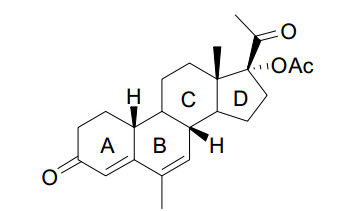
Figure 1.
Schematic representation of Nomegestrol acetate (NOMAC) molecule
Polymorphic Characterization and Bioavailability of Nomegestrol Acetate
Xiao-Feng CHEN , Peng LI , Hui-Ping WANG , Juan XU , Li-Feng NING

The importance of pharmaceutical polymorphism has been increasingly recognized in the scientific community, industry and by regulatory agencies over the past decades[1]. Polymorphism is the ability of one compound to exist in more than one crystal structures in the solid state[2, 3]. Polymorphs have different crystalline structures, physical and chemical attributes, such as compressibility, melting point, crystal habit, color, density, dissolution rate and solubility. These differences may affect the pharmaceutical processing, the stability of drug product and the bioavailability, so the therapeutic efficacy of the drug[4-6]. Crystalline solids that involve the inclusion or incorporation of solvent molecules in the crystal lattice are known as solvates. There are marketed drug products that contain solvates such as darunavir ethanolate (Prezista), indinavir sulfate ethanolate (Crixivan), and warfarin sodium isopropanol solvate (Coumadin)[7, 8].
The most common polymorph screening methods include crystallization, melted substance, vapor and solutions by cooling or evaporating the solvent, addition of an antisolvent to the solution[9-11], slurrying, spray drying, sublimation, grinding and thermal desolvation of solvates[12]. The outcome of a crystallization can be affected by factors such as the solvent (viscosity and polarity), the rate of supersaturation (cooling, adding or evaporation rate), or initial solution concentration[13]. With the development of polymorphic research, many new methods have emerged, such as high throughput screening (HTS)[14], microchannel crystallization[15], macromolecules as additives[16], etc. The polymorph screening methods have greatly promoted the development of polymorphic research.
Polymorphism can be characterized using various analytical techniques such as powder X-ray diffraction (PXRD), single-crystal X-ray diffraction (SXRD), differential scanning calorimetry (DSC), thermo gravimetric analysis (TGA), dynamic vapor sorption (DVS), optical and electron microscopy, infrared (IR), near-IR (NIR), Raman and solid-state nuclear magnetic resonance (ssNMR) spectroscopy[17-21].
Nomegestrol acetate (NOMAC, Fig. 1) is a highly selective progestogen derived from 19-norprogesterone and is structurally related to the naturally occurring progesterone. It has been classified as a "pure" progestogen because it has strong affinity for the human progesterone receptor, strong antigona-dotropic activity and moderate antiandrogenic activity, and is devoid of any estrogen, androgen, glucocorticoid or mineralocorticoid[22-25]. According to the Biopharmaceutics Classification System (BCS), NOMAC belongs to BCS class Ⅱ compounds with very low solubility (0.3~6 μg/mL in water) across physiological pH range. Therefore, increasing the solubility of NOMAC via polymorphism and consequently improving its bioavailability is of interest for the development of new dosage forms of NOMAC.
In this study, two crystalline forms of NOMAC, a solvent-free form (Form Ⅰ) and a dioxane solvate (Form Ⅱ. dioxane is a class 2 solvent in the guidelines of FDA and its use should be limited in pharmaceutical products), were prepared using a variety of screened crystallization methods. The crystal structures of both forms were investigated thoroughly using a variety of relevant techniques, and the biopharmaceutical profiles were further investigated.
NOMAC, the pharmaceutical ingredient (API), was purchased from NEWCHEM Pharmaceuticals Co., Ltd (Verona, Italy, batch number: 20160513). Its chemical purity was more than 99.0% mass fractions, which was determined using high-performance-liquid chromatography (HPLC). All the solvents used for recrystallization were of analytical reagent grade (Fisher Chemical).
Forms Ⅰ and Ⅱ of NOMAC were prepared in our laboratory, while the HPLC grade methanol, acetonitrile and water were obtained from Thermo Fisher Scientific (Waltham, MA, USA). Terfenadine (internal standard IS, 98% purity) was purchased from LIANHUAN Pharmaceuticals Co., Ltd (Jiangsu, China, batch number: 20170603).
Animals
Female Sprague-Dawley rats (weighting 230~250 g) were purchased from Sino-British Experiment Animal (Shanghai, China). The animals were treated in accordance with protocols approved by the Laboratory Animal Ethics Committee at the National Institute of Planned Parenthood Research. All animals were housed five per cage under a 12-h light-dark cycle with free access to food and tap water. Each animal was weighed once weekly and fed a standard rat chow.
The following different crystallization methods were used to comprehensively screen for polymorphism screening, cooling, crystallization, evaporative crystallization, antisolvent addition and slurring, and two crystalline forms were finally obtained.
Preparation of Form Ⅰ: To a 10 mL glass vial, 50 mg NOMAC and 4 mL 2-butanone were added. The mixture was slowly heated to 70 ℃ (1 ℃·min–1), and stirred at this temperature until the solid was dissolved. Then the reaction solution was filtered, and the filtrate solution was slowly cooled to 25 ℃ (1 ℃·min–1) in a cold bath. After that the solution was stirred for approximately 2 h at this temperature, filtered and finally vacuum-dried at room temperature.
Preparation of Form Ⅱ: To a 10 mL glass vial, 50 mg of NOMAC and 3 mL 1,4-dioxane were added. The mixture was slowly heated to 70 ℃ (1 ℃·min–1) and stirred at this temperature until the solid was dissolved, then the reaction solution was filtered. The filtrate solution was cooled down to 25 ℃ (1 ℃·min–1) in a cold bath and then the solution was stirred for approximately 2 h at this temperature, filtered and then vacuum-dried at room temperature.
Single-crystal XR diffraction data were collected by using a Rigaku AFC-10/Saturn 724-CCD diffractometer equipped with graphite-monochromatized MoKa radiation (0.71073 Å) system up to a 2 h limit of 50.0 ℃ at room temperature (25 ℃). Indexing and scaling of the data were performed using DENZO and SCALEPACK. The structure was solved by direct methods and expanded using difference Fourier techniques with Shelxs-97 and refined on F2 by successive full-matrix least-squares techniques for the non-hydrogen atoms. Anisotropic displacement parameters were employed for non-H atoms and H atoms were treated isotropically with Uiso = 1.2 (for those attached to aromatic carbon and N atoms) or 1.5 times (for those bonded to methyl carbons) the Ueq of the parent atoms. All H atoms were located at the expected positions and refined using a riding model. CIF files were obtained from Shelxs-97. The simulated powder patterns were calculated from the single-crystal data using Mercury 3.1[26, 27].
The XRD spectra were recorded using a BRUKER D8 Advance diffractometer (Bruker AXS GmbH, Karlsruhe, Germany) system with CuKaradiation (λ = 1.5406 Å) over an interval of 5~90 °/2θ. The measurement conditions were as follows: target: Cu; filter: Ni; voltage: 40 kV; current: 40 mA; time constant: 0.1 s; angular step: 0.016°, and detector: NaI (Tl) scintillation detector.
TGA-DSC was conducted using a TGA/DSC 3+ equipment (Mettler-Toledo, Switzerland) under a flow of nitrogen (20 mL·min–1) at a scan rate of 10 ℃·min–1 from 25 to 300 ℃. The pan was made of aluminium oxide. The balance was calibrated by the standard weight. And the temperature was calibrated by the melt points of five different metals.
A Spectrum RX Ⅰ FTIR spectrometer (Perkin Elmer, UK) was employed in the KBr diffusereflectance mode (sample concentration 2 mg in 20 mg of KBr) for collecting the IR spectra of samples. Dry KBr (50 mg) was finely ground in mortar, and the sample (1~2 mg) was subsequently added and gently mixed in order to avoid the trituration of crystals. A manual press was used to form the pellet. The spectra were measured over the range of 4000~450 cm–1. Data were analyzed using the Spectrum software.
We studied the dissolution curves of two crystal forms and NOMAC materal (API) using a Chinese pharmacopoeia method. Solid samples were sieved using a Gilson mesh sieve (No. 80) to obtain powders of uniform particle size, then were added in pH 1.0 and 0.2% sodium dodecyl sulfate (SDS) buffer at 37 ℃, respectively.
The experiment was performed using the dissolution apparatus (FADT-800RC, TiandaTianFa. Ltd., China) at the rotation speed set at 100 rpm, and samples (1 mL) were collected at 5, 10, 15, 20, 30, 45, 60, 80, 100, 120 and 180 min. Then the solutin was filtered through a 0.45 μm membrane filter (KeYiLong Ltd, China) and the solution concentrations were measured using an HPLC system (Agilent-1100 Agilent Ltd., USA) with the UV/Vis detector set at 275 nm. A Discovery C18 HPLC column (4.6 × 250 mm, 5 μm) was used with the column oven kept at 25 ℃. The mobile phase consisted of an acetonitrile-phosphate buffer (30 mM, pH 6.4, 20:80, v/v). The flow rate of the mobile phase was 1 ml/min, and the injection volume was 10 μL. For data acquisition and processing, the LC solution software was used.
Dawley rats (200~250 g) were randomly divided into two groups and fasted overnight prior to administration. Following single oral dosing of either NOMAC (0.5 mg, group 1) or Form Ⅰ (0.5 mg, group 2), blood samples (200 µL) were collected pre-dosing and at 0.25, 0.5, 1.0, 2.0, 4.0, 6.0, 8.0, 24.0, 36.0 and 48.0 h post-dosing. Plasma was harvested via centrifugation and stored at −20 ℃ before analysis.
To an aliquot of 50.0 µL rat plasma in a 10 mL glass tube, 20.0 µL of the internal standard (1.00 ng·ml-1 terfenadine), 300 µL of NaOH aqueous (0.1 M), and 3.0 mL of diethyl ether were added. The obtained mixture was vortexed for 5 min, and then centrifuged at 4500 rpm for 10 min. The supernatant was transferred to another glass tube and evaporated to dryness at 40 ℃ under a gentle stream of air. The residue was reconstituted in 100 µL of methanol-water (55/45, v/v). A 40 µL aliquot of the reconstituted solution was introduced into the LC-MS/MS system. Pharmacokinetic parameters for polymorphs were calculated using the DAS 2.0.1 (Mathematical Pharmacology Professional Committee of China, Shanghai, China) software using a non-compartmental analysis. Data were expressed as the means ± SD.
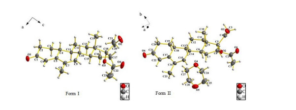
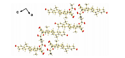
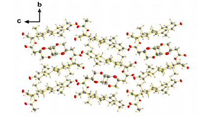
In order to obtain more details of the polymorphic structure information at the atomic level, high quality single crystals of polymorphic Forms Ⅰ and Ⅱ were submitted to single-crystal X-ray diffraction analysis. The molecular structure of the title compound, with atom labelling scheme drawn at 50% probability displacement ellipsoid, is depicted in Fig. 2, and the corresponding packing diagram is shown in Figs. 3 and 4. For better comparison, a summary of the conditions for the data collection and structure refinement parameters is given as follows. Form Ⅰ crystallizes in the monoclinic system, space group P21 with Z = 2, a = 7.0416(14), b = 7.6838(15), c = 18.498(4) Å, γ = 90°, V = 995.3(5) Å3, Dc = 1.231 g·cm–3, formula C23H30O4, μ = 0.083 mm–1, the final R = 0.0578 and wR = 0.0.0518 with I > 2σ(I). Another polymorphic Form Ⅱ belongs to the orthorhombic system, space group P212121 with Z = 4, a = 7.8457(16), b = 14.039(3), c = 22.1944(4) Å, γ = 90°, V = 244.58(8) Å3, Dc = 1.246 g·cm3, formula C27H38O6, μ = 0.087 mm-1, the final R = 0.0537 and wR = 0.1152 with I > 2σ(I). The crystals of Forms Ⅰ and Ⅱ are both blocky but crystallize in different space groups. Form Ⅰ exists in monoclinic chiral space group P21, and each asymmetric unit contains six NOMAC molecules. Form Ⅱ belongs to the orthorhombic chiral P212121 space group with eight NOMAC and eight dioxane molecules in the asymmetric unit. The conformational differences from the rings and the rotating of the single bond of the side-chain substituents are the main causes of the bimolecular phenomena. Form Ⅰ adopts a head-to-head packing configuration and forms a one-dimensional infinite chain running along the b axis with the intermolecular hydrogen bonding interaction. So far only one crystal structure of NOMAC has been reported in the Cambridge Crystallographic Data Centre (CCDC 949571), but it has different cell parameters with Form Ⅰ. For example, they not only have different internal spatial structures, but also pack in different modes, too, indicating their different crystal structures. Form Ⅱ adopts a head-to-hail packing configuration, with NOMAC and solvent molecules (1:1) forming a one-dimensional infinite chain along the b axis. Form Ⅱ belongs to solvate, because it is composed of dioxane. Form Ⅱ molecules connect the dioxane molecule through no-classical hydrogen bonds like C(27A)–H(27A)–O(3). Although ring C shows chair conformation, the other rings adopt envelope conformation in the two polymorphs, respectively. But the twist degrees of the rings are not the same. The dihedral angles between C(13)–C(14)–C(16) and C(10)–C(12)–C(13) of Forms Ⅰ and Ⅱ are 128.20° and 126.89°, respectively. The key parameters that describe doubtlessly the different conformers are the torsional angles of the molecular backbone. The most relevant torsion angles responsible for this polymorphic conformation of NOMAC are compared in Table 1. In Form Ⅰ, atoms C(20) and O(3) locate at each side of the plane formed by atom C(14)~C(18), and the torsion angle of C(20)–C(18)–O(3)–C(22) is 58.47°. In contrast to Form Ⅱ, the torsion angle of C(4)–C(5)–O(2)–C(2) is 52.23°. The number of molecules in the asymmetric unit of the two polymorphs is also different (Fig. 3). From the overlay diagrams, it is distinctive that the conformational differences arise because of the conformers of rings and the rotation of sidechain substituents (-OCOCH3). The differences of these angles confirm the existence of two conformational polymorphs.
 DownLoad:
CSV
DownLoad:
CSV
| Form | Angle | (°) |
| Ⅰ | O(1)–C(1)–C(2)–C(3) | –149.6(3) |
| O(3)–C(18)–C(20)–O(2) | –148.6(3) | |
| C(1)–C(2)–C(3)–C(4) | –56.0(3) | |
| C(12)–O1(3)–C(18)–C(17) | –68.0(3) | |
| C(17)–C(18)–C(20)–O(2) | –23.1(5) | |
| Ⅱ | O(2)–C(5)–C(6)–C(7) | –93.4(2) |
| O(3)–C(4)–C(5)–C(9) | 97.1(4) | |
| C(18)–C(13)–C(14)–C(15) | 55.4(3) | |
| C(20)–C(21)–C(22)–C(17) | 2.2(5) | |
| C(23)–C(16)–C(17)–C(18) | –178.3(3) |
Two-dimensional packing diagrams of Forms Ⅰ and Ⅱ are shown in Figs. 3 and 4. The packing occurs only by non-classical hydrogen bonds. Form Ⅰ packs in a fold line while Form Ⅱ in a wave. Detailed parameters of main hydrogen-bonding interactions with symmetry codes are listed in Table 2. The inclusion of solvents plays a vital role in forming different crystalline forms. The dioxane molecules introduced resulted in the torsional angle difference in two forms, which induced a variation in the arrangement of NOMAC molecules in the unit cell. It is the dioxane molecule that induces the molecules to bend and pack differently in the forms Ⅰ and Ⅱ.
DownLoad:
CSV
| D–H···A | d(D–H) (Å) | d(H···A) (Å) | d(D···A) (Å) | < (D–H–A) (°) | |
| Form Ⅰ | C(13)–H(13)A···O(3)a | 0.99 | 2.40 | 2.794(3) | 103 |
| C(15)–H(15)···O(3)b | 1.00 | 2.56 | 2.916(4) | 100 | |
| C(17)–H(17)A···O(2)c | 0.99 | 2.38 | 2.833(5) | 107 | |
| C(17)–H(17)B···O(4)d | 0.99 | 2.54 | 2.990(5) | 107 | |
| C(21)–H(21)C···O(4)e | 0.98 | 2.49 | 3.282(5) | 137 | |
| Form Ⅱ | C(1)–H(1)B···O(5)f | 0.98 | 2.60 | 3.427(6) | 142 |
| C(3)–H(3)B···O(2)g | 0.98 | 2.47 | 2.935(5) | 109 | |
| C(6)–H(6)A···O(3)h | 0.99 | 2.41 | 2.848(5) | 106 | |
| C(8)–H(8)···O(2)i | 1.00 | 2.51 | 2.870(4) | 101 | |
| C(11)–H(11)A···O(2)j | 0.99 | 2.46 | 2.845(4) | 103 | |
| C(20)– H(20)B···O(1)k | 0.99 | 2.45 | 3.336(4) | 148 | |
| C(25)–H(25)B···O(4)l | 0.99 | 2.51 | 3.427(6) | 154 | |
| C(26)–H(26)A···O(6)m | 0.99 | 2.49 | 3.456(5) | 164 | |
| C(27)–H(27)A···O(3)n | 0.99 | 2.41 | 3.338(4) | 157 | |
| Symmetry codes in Forms Ⅰ and Ⅱ : a2–x, 1/2+y, 1–z; b2–x, –1/2+y, 1–z; c1–x, –1/2+y, 1–z; d–1+x, y, z; e–x, 1/2+y, 2–z; f1/2–x, 1–y, –1/2+z; g1–x, –1/2+y, 1/2–z; h3/2–x, 1–y, –1/2+z; i1/2–x, 1–y, –1/2+z; j1–x, 1/2+y, 1/2–z; k3/2–x, 1–y, 1/2+z; l1/2+x, 3/2–y, 1–z; m1–x, 1/2+y, 3/2–z; n–1+x, y, z | |||||
In Form Ⅰ, the shortest contact for non-classical hydrogen bond is C(17)–H(17A)⋅⋅⋅O(2) (2.38 Å) (i = 1–x, –1/2+y, 1–z). Additionally, the oxygen of carbonyl group acts as an acceptor in another non- classical hydrogen bond of C(15)–H(15)···O(3). There are no hydrogen-bonding interactions on the oxygen of hydroxyl group. In Form Ⅱ, an asymmetric unit consists of eight NOMAC and eight dioxane molecules. Hydrogen-bonding interactions in Form Ⅱ give rise to two-dimensional networks by fostering the non-classical hydrogen bond chains, such as C(1)–H(1B)⋅⋅⋅O(5) (i = 1/2–x, 1–y, –1/2+z), C(25)–H(25B)···O(4) (i = 1/2+x, 3/2–y, 1–z) and C(26)–H(26A)···O(6) (i = 1–x, 1/2+y, 3/2–z).
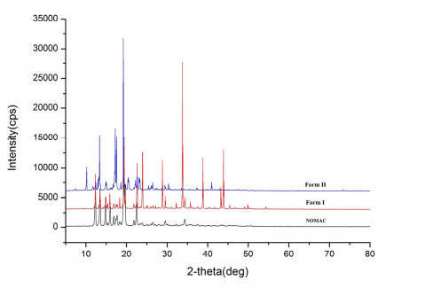
PXRD is always used as method for the identification of polymorphs. The PXRD results of the two novel polymorphs and the API are illustrated in Fig. 5. Form Ⅰ showed the characteristic peaks at 2θ = 12.4, 12.5, 13.6, 14.9, 19.5, 24.2, 28.8, 33.9, 38.9 and 43.9°, whereas Form Ⅱ exhibited them at 2θ = 10.2, 13.3, 15.1, 17.3, 19.2, and 20.5°, indicating the presence of two distinctive polymorphs. The PXRD patterns measured from the powder samples were also in good agreement with those calculated from the single-crystal structures.
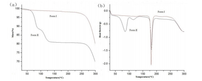
We observed no noticeable weight loss before the compound decomposition occurred, which indicated that Form Ⅰ was a solvent-free crystalline form. The melting point of Form Ⅰ was observed as a pronounced endothermic peak with the extrapolated onset temperature of 181.42 ℃ and an associated heat of absorption of –49.76 J·g–1. In Form Ⅱ, the weight loss occurred before its decomposition, showing that Form Ⅱ contained solvent in the crystalline form. The dehydration (1 mol of dioxane per mol of NOMAC) of the sample showed to be a solvate by the diffraction techniques (Tf = 61 and 116 ℃; ΔmTG = 19.32%, Δmcalc–1 = 19.2%) (Fig. 6), probably because the no-classical weak hydrogen bond force is weak between NOMAC and the dioxane molecule. Form Ⅱ melted with a pronounced endothermic peak, with the extrapolated onset temperature of 182.27 ℃ and the associated heat of absorption of –45.20 J·g–1.
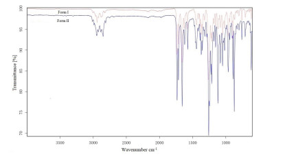
Both polymorphs of NOMAC can be easily identified and assigned by their IR spectra, as shown in Fig. 7. Prominent differences in the IR spectra can be found in the region above 800 cm–1 which reflects the X-H out-of-plane bending vibrations. Form Ⅰ is solvent-free modification and its molecule of crystalline formed hydrogen bonds with carbon or oxygen atoms in NOMAC molecule. There was a strong and sharp band at 1659 cm–1 contributed to the C=O stretching asymmetrical deformation vibration. Form Ⅱ is a dioxane solvate form, in which a strong and sharp band at 1117 cm–1 was contributed to the C-O-C stretching asymmetrical deformation vibration. The pinnacle at 1714 cm–1 is contributed to C=O stretching asymmetrical deformation vibration. The results revealed the two solid forms are different from each other by the analyses of FTIR spectra.
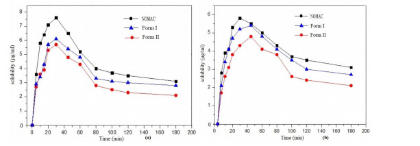
The dissolution profiles of Forms Ⅰ and Ⅱ were analyzed using the dissolution experiments performed in different buffers at 37 ℃. The samples collected at the pre-set time were filtered prior to HPLC analysis and the residual solids after the dissolution experiments were identified using PXRD. The PXRD patterns measured from the solid residues were in good agreement with those of the original forms, indicating that the polymorphs maintained their form during the dissolution experiment. The purity of NOMAC was determined using the area normalization method. As shown in Fig. 8, the concentrations of the two forms in buffers increase rapidly at first, then it approached equilibrium slowly with increasing the time. Due to the strong hydrogen-bond interaction between dioxane and drug molecule, the equilibrium dissolution rate of Form Ⅱ was found to be lower than Form Ⅰ.
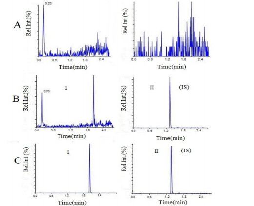
NOMAC was quantified using UHPLC-MS/MS. Typical chromatograms of blank plasma, the lowest limit of quantification (LLOQ) sample, and the test sample is shown in Fig. 9. Under optimized chromatographic conditions, the retention time of the analyte and IS was 2.23 and 1.49 min, respectively, and the total run time was maintained at 3.0 min. The test results demonstrated that there was no significant endogenous interference in the determination of the compound.
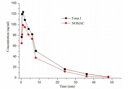
Because the toxic solvent involved in Form Ⅱ, only Form Ⅰ and the NOMAC material (API) were utilized to carry out in the pharmacokinetic experiments. The mean plasma concentration versus time plot for Forms Ⅰ and API after oral administration is shown in Fig. 10. It was found that Form Ⅰ has higher absorption than NOMAC material (API). After peak, plasma concentrations of the two forms began to decline, but remained at a high level, which formed a plateau. Plasma concentration of the two forms can be measured at a higher level till 8 h, a trace level till 36 h, and almost undetectable after 48 h. Overall, the Cmax and AUC of Form Ⅰ were approximately 0.2 times those of the NOMAC material (API).
Two crystal forms of NOMAC were identified and prepared for the first time during the polymorph screening. And their structures were determined by single-crystal X-ray diffraction. The results of the powder dissolution experiments showed that Form Ⅰ dissolved apparently faster and had a little higher equilibrium solubility than the NOMAC material (API). Therefore, Form Ⅰ is the promising candidate for improving the solubility and dissolution rate of NOMAC. Furthermore, we validated a UHPLC-MS quantitative method to rapidly analyze NOMAC in the rat plasma within 3 mins. And the studies revealed that Form Ⅰ improved the AUC and Cmax in an animal model compared with the NOMAC material (API).
Haleblian, J.; McCrone, W. Pharmaceutical applications of polymorphism. J. Pharm. Sci. 1969, 58, 911–929. doi: 10.1002/jps.2600580802
Byrn, S. R.; Pfeiffer, R. R.; Stephenson, G.; Grant, D. J.; Gleason, W. B. Solid-state pharmaceutical chemistry. Chem. Mater. 1994, 6, 1148–1153. doi: 10.1021/cm00044a013
Vippagunta, S. R.; Brittain, H. G.; Grant, D. W. Crystalline solids. Adv. Drug Deliv. Rev. 2001, 48, 3–26. doi: 10.1016/S0169-409X(01)00097-7
Mcgregor, L.; Rychkov, D. A.; Coster, P. L.; Day, S.; Drebushchak, V. A.; Achkasov, A. F.; Nichol, G. S.; Pulham, C. R.; Boldyreva, E. V. A new polymorph of metacetamol. Cryst. Eng. Comm. 2015, 17, 6183–6192. doi: 10.1039/C5CE00910C
Sanphui, P.; Goud, N. R.; Khandavilli, U. B.; Bhanoth, S.; Nanqia, A. New polymorphs of curcumin. Chem. Commun. 2011, 47, 5013–5015. doi: 10.1039/c1cc10204d
Tong, H. Y.; Chow, A. S.; Chan, H. M.; Chow, A. H.; Wan, Y. K.; Williams, L. D.; Shek, F. L.; Chan, C. K. Process-induced phase transformation of berberine chloride hydrates. J. Pharm. Sci. 2010, 99, 1942–1954. doi: 10.1002/jps.21983
Lee, A. Y.; Erdemir, D.; Myerson, A. S. Crystal polymorphism in chemical process development. Annu. Rev. Chem. Biomol. 2011, 2, 259–280. doi: 10.1146/annurev-chembioeng-061010-114224
Byrn, S.; Pfeiffer, R.; Ganey, M.; Hoiberg, C.; Poochikian, G. Pharmaceutical solids: a strategic approach to regulatory considerations. Pharm. Res. 1995, 12, 945–954. doi: 10.1023/A:1016241927429
Morissette, S. L.; Almarsson, Ö.; Peterson, M. L.; Remenar, J. F.; Read, M. J.; Lemmo, A. V.; Ellis, S.; Cima, M. J.; Gardner, C. R. High-throughput crystallization: polymorphs, salts, co-crystals and solvates of pharmaceutical solids. Adv. Drug. Deliv. Rev. 2004, 56, 275–300. doi: 10.1016/j.addr.2003.10.020
Aaltonen, J.; Alleso, M.; Mirza, S.; Koradia, V.; Gordon, K. C.; Rantanen, J. Solid form screening–a review. Eur. J. Pharm. Biopharm. 2009, 71, 23–37. doi: 10.1016/j.ejpb.2008.07.014
Newman, A. Specialized solid form screening techniques. Org. Process Res. Dev. 2013, 17, 457–471. doi: 10.1021/op300241f
Chieng, N.; Rades, T.; Aaltonen, J. An overview of recent studies on the analysis of pharmaceutical polymorphs. J. Pharm. Biomed. Anal. 2011, 55, 618–644. doi: 10.1016/j.jpba.2010.12.020
Getsoian, A.; Lodaya, R. M.; Blackburn, A. C. One-solvent polymorph screen of carbamazepine. Int. J. Pharm. 2008, 348, 3–9. doi: 10.1016/j.ijpharm.2007.06.053
Carter, P. W.; Ward, M. D. Directing polymorph selectivity during nucleation of anthranilic acid on molecular substrates. J. Am. Chem. Soc. 1994, 116, 769–781. doi: 10.1021/ja00081a048
Scottl, C.; Leonard, J. C.; Jeanette, T. D. A metastable polymorph of metformin hydrochloride: isolation and characterization using capillary crystallization and thermal microscopy techniques. Crys Growth Des. 2004, 4, 441–449.. doi: 10.1021/cg034243p
Lang, M.; Grzesiak, L.; Matzger, A. J. The use of polymer hetero nuclei for crystalline polymorph selection. J. Am. Chem. Soc. 2002, 50, 14834–14835.
Aaltonen, J.; Allesr, M.; Mirza, S.; Koradia, V.; Gordon, K.; Rantanen, C. J. Solid form screening a review. Eur. J. Pharm. Biopharm. 2009, 71, 23–37. doi: 10.1016/j.ejpb.2008.07.014
Bugay, D. E. Characterization of the solid-state: spectroscopic techniques. Adv. Drug Deliv. Rev. 2001, 48, 1–2. doi: 10.1016/S0169-409X(01)00096-5
Stephenson, G. A.; Forbes, R. A.; Reutzel, S. M. Characterization of the solid state: quantitative issues. Adv. Drug Deliv. Rev. 2001, 48, 67–90. doi: 10.1016/S0169-409X(01)00099-0
Newman, A. W.; Byrn, S. R. Solid-state analysis of the active pharmaceutical ingredient in drug products. Drug Dis. Today 2003, 8, 898–905. doi: 10.1016/S1359-6446(03)02832-0
Shan, B.; Kakumanu, V. K.; Bansal, A. K. Analytical techniques for quantification of amorphous/crystalline phases in pharmaceutical solids. J Pharm. Sci. 2006, 95, 1641–1665. doi: 10.1002/jps.20644
Sitruk-Ware, R. New progestogens for contraceptive use. Hum. Reprod. Update 2006, 12, 169–178. doi: 10.1093/humupd/dmi046
Mueck, A. O.; Sitru-ware, R. Nomegestol acetate, a novel progestogen for oral contraception. Steroids 2011, 76, 531–539. doi: 10.1016/j.steroids.2011.02.002
Abdel-Aleem, H.; Abol-Oyoun, E. M.; Shaaban, M. M. The use of Nomegestrol acetate subdermal contraceptive implant, uniplant, during lactation. Contraception 1996, 54, 281–286. doi: 10.1016/S0010-7824(96)00180-1
Situk-ware, R. Pharmacological profile of progestins. Maturitas. 2004, 47, 277–283. doi: 10.1016/j.maturitas.2004.01.001
Dolomanov, O. V.; Bourhis, L. J.; Gildea, R. J.; Howard, J. K.; Puschmann, H. J. A complete structure solution, refinement and analysis program. Appl. Cryst. 2009, 42, 339–341. doi: 10.1107/S0021889808042726
Sheldric, G. M. A short history of SHELX. Acta Cryst. 2008, 64, 112–122. doi: 10.1107/S0108767307043930

Figure 2 Molecular view of the compound, showing 50% probability displacement ellipsoids and atom labeling scheme

Figure 5 Experimental powder X-ray diffraction (PXRD) patterns of Nomegestrol acetate NOMAC

Figure 6 Thermal analysis of Forms Ⅰ and Ⅱ (a). Thermogravimetric (TGA) curves of Forms Ⅰ and Ⅱ (b)

Figure 8 Dissolution profile for NOMAC, Forms Ⅰ and Ⅱ in pH 1.0 and 0.2% sodium dodecyl sulfate (SDS) buffer

Figure 9 Multiple reaction (MRM) chromatograms Ⅰ (nomegestrol acetate [NOMAC] and internal standard [IS]: (A) Drug-free plasma, (B) Plasma spiked with 2.5 ng/mL of pharmacokinetic parameters (NOMAC) and 5 μg/mL IS, and (C) Plasma samples 1 h after oral administration to a rat with 5 mg/kg for NOMAC Form Ⅰ

Figure 10 Mean plasma concentration time profile of Form Ⅰ and NOMAC in rat after oral administration
Table 1. Relevant Torsion Angles (°) of Form Ⅱ in Comparison with Those in Form Ⅰ
| Form | Angle | (°) |
| Ⅰ | O(1)–C(1)–C(2)–C(3) | –149.6(3) |
| O(3)–C(18)–C(20)–O(2) | –148.6(3) | |
| C(1)–C(2)–C(3)–C(4) | –56.0(3) | |
| C(12)–O1(3)–C(18)–C(17) | –68.0(3) | |
| C(17)–C(18)–C(20)–O(2) | –23.1(5) | |
| Ⅱ | O(2)–C(5)–C(6)–C(7) | –93.4(2) |
| O(3)–C(4)–C(5)–C(9) | 97.1(4) | |
| C(18)–C(13)–C(14)–C(15) | 55.4(3) | |
| C(20)–C(21)–C(22)–C(17) | 2.2(5) | |
| C(23)–C(16)–C(17)–C(18) | –178.3(3) |
 下载: 导出CSV
Table 2. Hydrogen Bonds of Nomegestrol Acetate (NOMAC) Forms (Å), where D = Donor and A = Acceptor
| D–H···A | d(D–H) (Å) | d(H···A) (Å) | d(D···A) (Å) | < (D–H–A) (°) | |
| Form Ⅰ | C(13)–H(13)A···O(3)a | 0.99 | 2.40 | 2.794(3) | 103 |
| C(15)–H(15)···O(3)b | 1.00 | 2.56 | 2.916(4) | 100 | |
| C(17)–H(17)A···O(2)c | 0.99 | 2.38 | 2.833(5) | 107 | |
| C(17)–H(17)B···O(4)d | 0.99 | 2.54 | 2.990(5) | 107 | |
| C(21)–H(21)C···O(4)e | 0.98 | 2.49 | 3.282(5) | 137 | |
| Form Ⅱ | C(1)–H(1)B···O(5)f | 0.98 | 2.60 | 3.427(6) | 142 |
| C(3)–H(3)B···O(2)g | 0.98 | 2.47 | 2.935(5) | 109 | |
| C(6)–H(6)A···O(3)h | 0.99 | 2.41 | 2.848(5) | 106 | |
| C(8)–H(8)···O(2)i | 1.00 | 2.51 | 2.870(4) | 101 | |
| C(11)–H(11)A···O(2)j | 0.99 | 2.46 | 2.845(4) | 103 | |
| C(20)– H(20)B···O(1)k | 0.99 | 2.45 | 3.336(4) | 148 | |
| C(25)–H(25)B···O(4)l | 0.99 | 2.51 | 3.427(6) | 154 | |
| C(26)–H(26)A···O(6)m | 0.99 | 2.49 | 3.456(5) | 164 | |
| C(27)–H(27)A···O(3)n | 0.99 | 2.41 | 3.338(4) | 157 | |
| Symmetry codes in Forms Ⅰ and Ⅱ : a2–x, 1/2+y, 1–z; b2–x, –1/2+y, 1–z; c1–x, –1/2+y, 1–z; d–1+x, y, z; e–x, 1/2+y, 2–z; f1/2–x, 1–y, –1/2+z; g1–x, –1/2+y, 1/2–z; h3/2–x, 1–y, –1/2+z; i1/2–x, 1–y, –1/2+z; j1–x, 1/2+y, 1/2–z; k3/2–x, 1–y, 1/2+z; l1/2+x, 3/2–y, 1–z; m1–x, 1/2+y, 3/2–z; n–1+x, y, z | |||||
下载: 导出CSV

 扫一扫看文章

扫一扫关注我们


