Citation:
Xu Zhiqiang, Chen Jianhua, Hu Lin-Li, Tan Ying, Liu Sheng-Hua, Yin Jun. Recent advances in formaldehyde-responsive fluorescent probes[J]. Chinese Chemical Letters,
2017, 28(10): 1935-1942.
doi:
10.1016/j.cclet.2017.07.018
Recent advances in formaldehyde-responsive fluorescent probes
College of Chemistry Environmental Engineering, Yangtze University, Jingzhou 434023, China
b.
The State Key Laboratory Breeding Base-Shenzhen Key Laboratory of Chemical Biology, Graduate School at Shenzhen, Tsinghua University, Shenzhen 518055, China
c.
Key Laboratory of Pesticide and Chemical Biology, Ministry of Education, College of Chemistry, Central China Normal University, Wuhan 430079, China
Received Date:
25 June 2017 Accepted Date:
14 July 2017 Revised Date:
10 July 2017 Available Online:
22 October 2017
Abstract:
Formaldehyde, as one of the simplest reactive carbonyl species (RCS), is regarded as a potential carcinogen and a sick house syndrome gas. Recent studies have shown that abnormally high levels of formaldehyde may result in cognitive decline and spatial memory deficits, asthmatic symptoms, Alzheimer's disease, and cancer. Due to the harmfulness of high levels of formaldehyde in nature and humans, it is of great significance to further elucidate the roles and functions of formaldehyde by a non-invasive detection approach. Fluorescence imaging has become a powerful and popular tool in monitoring bio-species owing to their high sensitivity and selectivity, excellent spatiotemporal resolution and non-invasion nature. Therefore, fluorescent probes are widely applied to track and detect formaldehyde in vitro and in vivo which have attracted more and more interest recently. This review focuses on various strategies to design the fluorescent probes for detecting formaldehyde based on different recognition groups.
Formaldehyde is the simplest aldehyde and widely used in the chemical industry for producing detergent, plastics, wood processing, corrosion remover, drugs and so on [1, 2]. Formaldehyde presents in food such as vegetables, fruits, meat and seafood. In addition, formaldehyde is usually used as a food preservative agent, whereas the formaldehyde residues in food may bring about severe threat for human health [3]. Environmental formaldehyde is prevailingly generated from natural and anthropogenic industrial activities and combustion of biofuels. Environmental formaldehyde has emerged as one of the ubiquitous chemical pollutants in indoor environments, which resulted from formaldehyde-containing building materials. Formaldehyde has become a heavy threat to human health. Exposure to high level of exogenous formaldehyde causes lachrymation, sneezing, coughing, nausea and even death. In 2004, formaldehyde was reclassified as a human carcinogen by the International Agency for Research on Cancer (IARC) [4, 5]. In the other hand, formaldehyde is employed as a metabolic intermediate in many living organisms and serves as a tissue fixative and embalming agent. Meanwhile, the occurrence of endogenous formaldehyde in a normal physiological system is produced by histone demethylation, methylation of DNA and semicarbazide sensitive amine oxidase. In a normal physiological brain, the concentration of formaldehyde ranges from 0.2 [28_TD$DIFF]mmol to 0.4 mmol. At this level, formaldehyde is crucial to the memory formation via DNA demethylation cycles and cognitive ability. On the contrary, aberrant accumulation of formaldehyde in patients may induce a variety of diseases, including cancers, neurodegenerative diseases, diabetes, chronic liver and heart disorders [6, 7].
Current methods have been developed practicably for the detection of formaldehyde including colorimetric assay, gas chromatography, mass spectrometry and high-performance liquid chromatography [8-11]. However, they usually suffer from some limitations such as low sensitivity, complicated operation and damage of the intact specimens. Moreover, these methods are not suitable for the detection of formaldehyde in microenvironment. Compared with the above technologies, fluorescent method has overwhelming advantages such as non-destructiveness, good biocompatibility, high sensitivity and convenience [12]. Smallmolecule fluorescent probes for the detection of targets underwent many development stages, including the stage from single emission to ratiometric fluorescence, the stage from visible-light emission to near-infrared emission, and the stage from single photon excitation to two-photon excitation [13-19]. Therefore, fluorescent probes based on organic small molecules for the detection of formaldehyde have attracted an increasing interest in recent years [20]. Especially over past five years, many formaldehyde-responsive fluorescent probes have been reported and utilized to visualize the level of formaldehyde in living cells. In view of the fast development in the field of formaldehyderesponsive fluorescent probes, we summary the recent progress in this work and inquire into the tendency of development in future.
2.
Formaldehyde probes based on different responding sites
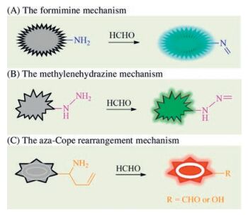
Reactive fluorescent probes based on various chemical reactions have developed to be a main design strategy [21]. Over the past decades, more and more reactive fluorescent probes have been reported and applied in bioscience [22]. Similar strategy based on chemical reaction was also utilized to design the formaldehyde-responsive fluorescent probes. According to the reported formaldehyde-responsive fluorescent probes, it was found that the carbonyl group of formaldehyde and amino group is usually employed as the reaction mechanism to construct the formaldehyde-responsive fluorescent probes. As presented in Fig. 1, the reaction mechanism of formaldehyde with amino group mainly included three types: (1) The condensation of free amino group (— NH2) with formaldehyde to produce the formimine product leading to the changes of fluorescence signal (Fig. 1A); (2) The condensation of hydrazine (— NH-NH2) with formaldehyde to generate the methylenehydrazine product resulting in the changes of fluorescence signal (Fig. 1B); (3) The aza-Cope rearrangement reaction of homoallylamino group with formaldehyde to form the products (such as aldehyde or phenol) inducing the changes of fluorescence signal (Fig. 1C). Accordingly, we summarized the recent advances in formaldehyde-responsive fluorescent probes in terms of different reaction mechanisms.
图 1
图 1
Three types of reaction-based fluorescent probe for the detection of formaldehyde.
Figure 1.
Three types of reaction-based fluorescent probe for the detection of formaldehyde.
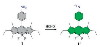
Condensation reaction, known as the reaction between aldehydes and amines moiety, was used to design fluorescent probes for the detection of formaldehyde. The free NH2-labeling fluorescent probes involved in the electron transfer or charge transfer owing to the existence of lone pair electrons on amino group. The formimination of amino group led to the restraint of electron transfer or charge transfer which resulting in the change of fluorescence (Fig. 1A). For instance, Thangadurai group [23] employed an aniline group as the determining group to link a BODIPY dye to form the fluorescent probe 1 in 2012, as shown in Scheme 1. Probe 1 displayed weak fluorescence because the electron transfer from the aniline moiety to the BODIPY fluorophore quenched the excited state of the fluorophore. However, the addition of formaldehyde in degassed dry CH3OH at pH 8 induced a strong green emission at 535 nm upon excitation at 520 nm, ascribing to the sensing mechanism that the formation of an imine product (1') (Scheme 1) suppressed the electron transfer pathway.
Based on the same sensing mechanism, Li and Yu [24] developed a benzoimidazolylpyridine-based fluorescent probe 2 with dual-emission-enhancement property by using the amino group as the recognition moiety. The photo-induced electron transfer (PET) from the amino group to benzoimidazolylpyridine component led to the probe 2 was non-fluorescent. However, upon interaction with formaldehyde, the formation of an imine product (2') (Scheme 2) resulted in the enhancement of the fluorescence intensity centered at 415 nm and 505 nm in mixture solution of water/ethanol (99:1, v/v) with excitation at 365 nm and 400 nm, respectively. The probe 2 could applied to detect formaldehyde quantitatively in the range of concentration 0-2.7 × 10⁻² mol/L with a detection limit of 6 μmol/L. Moreover, the probe 2 was successfully involved in the preliminary application for the detection of residual formaldehyde in food.
The condensation reaction of formaldehyde with arylamine was considered to be an efficient design strategy. Unfortunately, these probes usually suffer from a lower sensitivity to formaldehyde due to the instability of the condensation products under physiological conditions, which limited their further application in biological systems. Compared with the condensation reaction of amine with formaldehyde, formaldehyde-triggered cyclization of formimine product can form more stable imidazolidine or imidazole moieties even under physiological conditions.
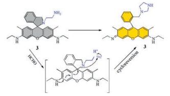
In 2011, Han et al. [25] designed an aldehyde-responsive fluorescent probe by installing ethylenediamine group on rhodamine B fluorophore, in which the aldehydes could induce the formation of imidazolidine along with the cycloreversion of rhodamine B. Subsequently, Lin et al. [26] developed a more sensitive fluorescent probe 3 based on rhodamine 6G derivative for detecting formaldehyde. The ethylenediamine moiety of probe 3 was treated with formaldehyde to firstly form the imino intermediate, and followed by a cyclization of intramolecular deoxylactam and the cycloreversion of rhodamine 6G to generate the sensing product imidazolidine 3', as shown in Scheme 3. Probe 3 exhibited ultra-fast reaction rate with formaldehyde and the fluorescence intensity at 560 nm dramatically enhanced upon the addition of formaldehyde. Imaging of exogenous formaldehyde in living HeLa cells indicated the probe could visualize the level of formaldehyde in biological system. Moreover, the paper-device based on probe 3 was capable of quantitatively detecting formaldehyde in dried shiitake mushrooms and rapidly detect indoor formaldehyde gas by the naked eye.
Scheme 3
图 Scheme 3
The proposed detection mechanism of probe 3 based on rhodamine 6G.
Scheme 3.
The proposed detection mechanism of probe 3 based on rhodamine 6G.
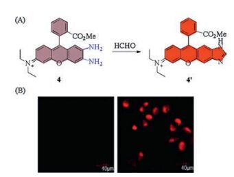
Recently, o-phenylenediamine was also employed as a determining group for the detection of formaldehyde via a similar reaction mechanism by Zeng group [27]. As shown in Fig. 2, the diamine-modified rhodamine 4 had a weak fluorescence emission at 642 nm in EtOH-tris-HCl buffer (3:7, v/v, pH 7.4). The addition of formaldehyde to the solution of probe 4 induced a slightly blueshifted strong fluorescence enhancement at 620 nm due to the formation of imidazole-fused rhodamine 4' (Fig. 2A). When the L929 cells incubated with the probe 4 and formaldehyde, compared with the control group only treated with probe 4, the cell demonstrated a bright red fluorescence. Accordingly, probe 4 was a suitable indicator for labelling the cellular formaldehyde (Fig. 2B).
图 2
图 2
(A) Fluorescent probe 4 based on ortho-diaminorhodamine. (B) Confocal microscope images of probe 4 in L929 cells. Reproduced with permission [27]. Copyright 2017, Elsevier.
Figure 2.
(A) Fluorescent probe 4 based on ortho-diaminorhodamine. (B) Confocal microscope images of probe 4 in L929 cells. Reproduced with permission [27]. Copyright 2017, Elsevier.
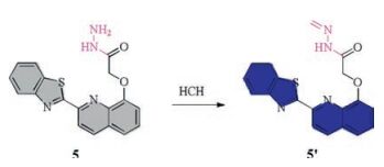
Similar, the hydrazine unit also contains an uncoated amino group, which can also react with formaldehyde to generate the methylenehydrazine, as described in Fig. 1B. The acylation of hydrazine can form hydrazide, and the free amino group on hydrazide reacts with formaldehyde. For example, Yu et al. [28] reported a benzothiazole-modified hydroxyquinoline fluorescent probe 5 in 2015, in which, employed a hydrazide unit as the determining group for formaldehyde. Probe 5 exhibited very weak fluorescence in aqueous solution. Upon the treatment of formaldehyde, a 5.5-fold fluorescence enhancement with a maximum at 467 nm displayed, ascribing to the prohibition of PET process on the molecule of formhydrazide 5' (Scheme 4). And formaldehyde could be detected quantitatively in the concentration range from 0 to 1.3 ×10⁻⁴ mol/L with a detection limit of 0.9 μmol/L. However, formic acid and propylaldehyde also promoted distinct enhancement of fluorescence intensity at 467 nm as well as formaldehyde.
Naphthalimide as a classic fluorophore has also been widely applied in formaldehyde-responsive fluorescent probe. On one hand, one of amino groups of hydrazine moiety can react with naphthalene anhydride to form the carboximide while the other amino group can respond the formaldehyde to induce the fluorescence changes. For instance, Lin et al. [29] developed a hydrazide-based naphthalimides 6a. In MeCN solution containing 5% HOAc at room temperature, the optical properties indicated that the formation of formhydrazide 6a' (Scheme 5) led to the rapid enhancement of fluorescence intensity at 512 nm within 4 min. In this reaction, the synergistic effects that intramolecular charge transfer (ICT) process proceeded from nitrogen atom on piperazine unit to formhydrazide unit and the protonation of the tertiary amino group on piperazine moiety blocked the PET process. The fluorescence intensity at 512 nm was linearly proportional to formaldehyde concentration in the range of 0-1.6 mmol with a detection limit of 0.25 ppm. It demonstrated that the probe 6a could be applied to detect formaldehyde qualitatively.
Meanwhile, a series of similar fluorescent probes 6b-d were developed using a same sensing mechanism by Sun group [30]. In MeCN solution containing 10% HOAc, no obvious fluorescence change was observed when formaldehyde was added to the solution of probe 6b. However, the diamino-dressed probe 6c exhibited a relatively small fluorescence enhancement at 515 nm under excitation at 400 nm and probe 6d presented a remarkable response towards formaldehyde with a low detection limit of around 0.1 μmol/L.
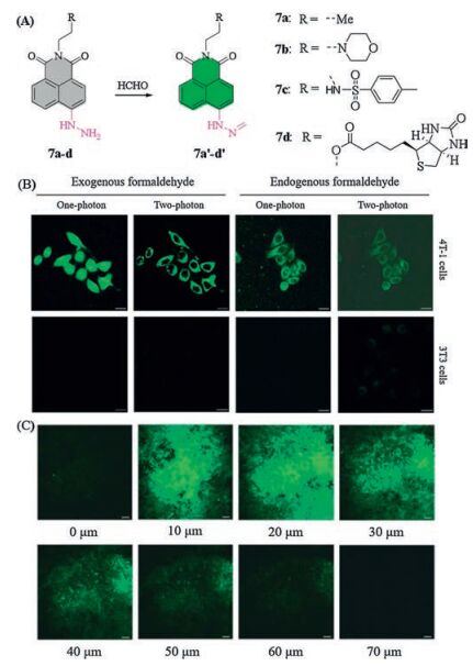
On the other hand, the process of formaldehyde determination involved in the formimination of hydrazide under the acidic condition, which severely restricted their application in physiological level. Accordingly, some groups paid for efforts to introduce the hydrazine unit into the 4-site of 1, 8-naphthalimide backbone. For example, Lin et al. [31] introduced hydrazine unit to the 4-site of 1, 8-naphthalimide scaffold and prepared a two-photon fluorescent probe 7a for the detection of formaldehyde, as shown in Fig. 3. Probe 7a was non-fluorescent in PBS buffer solution (pH 7.4) containing 1% DMSO owing to a PET pathway from hydrazine to the naphthalimide backbone. Along with the addition of formaldehyde, probe 7a responded quickly in physiological pH condition and accompanied by a gradually increasing fluorescence signal at 543 nm because the formation of the product formimine 7a' (Fig. 3A) suppressed the process of PET. The titration of fluorescence confirmed that this probe possessed a low detection limit of 0.71 mmol/L. The bioimaging in HeLa cells showed that probe 7a was capable of detecting exogenous and endogenous formaldehyde in the one-photon and two-photon modes. Further bioimaging analysis in lung tissue slides indicated that probe 7a was capable of tracking endogenous formaldehyde of tissues by two-photon mode. More, they found that sodium bisulfite could be used as an efficacious inhibitor of formaldehyde in biological system.
图 3
图 3
(A) Naphthalimide-based probes 7a-d. (B) Confocal microscope images of exogenous and endogenous formaldehyde with probe 7d in 4T-1 and 3T3 cells by one-photon and two-photon modes. Reproduced with permission [34]. Copyright 2016, Royal Society of Chemistry.
Figure 3.
(A) Naphthalimide-based probes 7a-d. (B) Confocal microscope images of exogenous and endogenous formaldehyde with probe 7d in 4T-1 and 3T3 cells by one-photon and two-photon modes. Reproduced with permission [34]. Copyright 2016, Royal Society of Chemistry.
The generation of formaldehyde is associated with many organelles such as lysosome, endoplasmic reticulum and so on. Lin group [32] employed a 4-(2-ethyl)morpholine unit as lysosomelocating groupto replace the propyl group of probe 7a and developed a lysosome-targeted fluorescent probe 7b. The in vitro results indicated that probe 7b in PBS buffer (pH 7.4) containing 1% DMSO had a highly sensitive response towards formaldehyde along with a 350-fold enhancement of fluorescence intensity at around 543 nm. The bioimaging in Hela cells showed that it was able to visualize the level of cellular formaldehyde. Most importantly, owing to the lysosome-targeting feature of morpholine moiety, probe 7b was firstly used to mark the endogenous formaldehyde in the lysosomes of living cells. Compared with the red channel of commercial Lyso-Tracker Deep Red, it was found that the intensity scatter plot of probe 7b in green channel had a good correlation with a high overlap coefficient of 0.9. Recently, they employed a p-methylbenzenesulfonamide as a targeting group for endoplasmic reticulum that was dressed on the carboximide component, to prepare a formaldehyde-responsive fluorescent probe 7c [33]. The finding of determination showed that probe 7c possessed high sensitivity and selectivity, low cytotoxicity, low detection limit (upon to 5.24 μmol/L) towards formaldehyde. The application in bioimaging suggested that this probe was able to detect the exogenous and endogenous formaldehyde in HeLa cells. Furthermore, the endoplasmic reticulum-targeted feature of probe 7c enabled its use in the visualization of endogenous formaldehyde in the endoplasmic reticulum. In comparison to the commercial endoplasmic reticulum-Tracker Red, the green fluorescence signal of probe 7c overlapped well with the red fluorescence. The intensity scatter plot of the green channel and red channel displayed good correlation with a high overlap coefficient of 0.95. Accordingly, these probes can serve as the efficient targeting probes in biological researches.
Naphthalimide as a two-photon fluorophore has been widely applied in tissue imaging. Recently, Lin and Kim et al. [34] developed a biotin-guided naphthalimide-based formaldehyde probe 7d. Upon treatment with formaldehyde, Probe 7d displayed a 140-fold fluorescence enhancement at 541 nm under physiological conditions due to the inhibition of a PET process from hydrazine unit to naphthalimide moiety. And there was a good linear relationship between the fluorescence intensity and the formaldehyde concentration up to 10 μmol/L. Fluorescence imaging in cancer cells demonstrated that probe 7d was capable of visualizing both exogenous and endogenous levels of formaldehyde in biotin receptor positive 4T-1 cells over biotin negative 3T3 cells by means of one-photon and two-photon excitation, as presented in Fig. 3B. It was worth mentioning that this probe could act as an efficient indicator to track the endogenous formaldehyde in tumor tissue slices upon to 70mm depth by a two-photon model (Fig. 3C). It is expected that this probe will have potential application in disease diagnosis in future.
2.3
The aza-cope rearrangement mechanism
As described above, the reaction product of homoallylamine with formaldehyde can take place a [3, 3]-sigmatropic rearrangement to form the corresponding aldehyde product or undergo a self-immolative β-elimination to release a phenolic compound. Based on such reaction mechanism, some groups employed this strategy to develop a series of formaldehyde-responsive fluorescent probes in recent years.
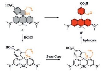
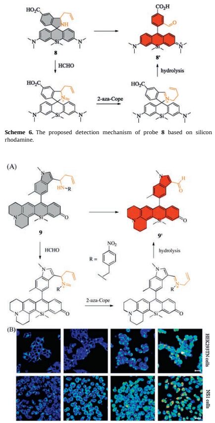
In 2015, Chang group [35] developed a silicon rhodamine-based fluorescent probe 8 which employed homoallylamino unit as a determining group of formaldehyde. In the presence of formaldehyde, probe 8 with weak fluorescence could be transferred to its iminium and followed by the aza-Cope rearrangement to yield the final product 8' (Scheme 6) with a stronger red emission. In the process of determiantion, probe 8 with good solubility exhibited a ~8-fold fluorescence enhancement at 662 nm upon treatment with formaldehyde at pH 7.4 within 1 h. The biological application revealed that it was able to detect exogenous formaldehyde in HEK293T cells and visualize endogenous formaldehyde in MCF7 human breast cancer cells. Almost at the same time, Chan group [36] relied on the same reaction mechanism of aza-Cope rearrangement to prepare an indole-dressed silicon rhodamine 9, which was almost non-fluorescent due to a sensing mechanism of d-PeT that the 4-nitrobenzyl dark quencher reduced background fluorescence. However, a significant turn-on fluorescence response at 649 nm was observed upon the addition of formaldehyde in PBS buffer (pH 7.4), due to the formation of fluorescent product 9' (Fig. 4A). Further analyzing other species showed that it possessed highly selectivity for formaldehyde over potentially competing biological analytes under physiologically relevant levels of formaldehyde. The bioimaging HEK293TN cells and NS1 cells displayed that the addition of formaldehyde could induce a robust dose-dependent signal enhancement of up to 3-fold and 2.3-fold as well as a time-dependent turn-on response at each concentration (Fig. 4B), respectively. These probes provided a better model for the studies of formaldehyde associated with normal and pathological processes.
Scheme 6
图 Scheme 6
The proposed detection mechanism of probe 8 based on silicon rhodamine.
Scheme 6.
The proposed detection mechanism of probe 8 based on silicon rhodamine.
图 4
(A) The proposed detection mechanism of probe 9 based on silicon rhodamine. (B) Confocal microscope images of probe 9 in HEK293TN and NS1 cells. Reproduced with permission [36]. Copyright 2015, American Chemical Society.
Figure 4.
(A) The proposed detection mechanism of probe 9 based on silicon rhodamine. (B) Confocal microscope images of probe 9 in HEK293TN and NS1 cells. Reproduced with permission [36]. Copyright 2015, American Chemical Society.
Quantification of the target analytes using intensity-based fluorescent probes that provide only a single emission feature may afford less reliable information owing to the interference from many analyte-independent factors, such as concentration, temperature, pH, instrumental parameters. In contrast, the ratiometric fluorescent probes rely on analyte-induced changes in the intensity of two or more emission bands, which can alleviate interference from environment concentration and excitation. This strategy has been widely applied in fluorescent probes [37, 38]. Based on the reaction mechanism of 2-aza-Cope rearrangement, several ratiometric formaldehyde-responsive fluorescent probes have been reported.
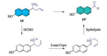
In 2016, Lin et al. [39] firstly reported a ratiometric fluorescent probe 10, which was used to detect formaldehyde in living cells. In PBS buffer containing 1% acetone, probe 10 displayed an emission band centered at 359 nm. Upon the addition of formaldehyde, an obvious red-shift (around 92 nm) to 451 nm and the increasing fluorescence intensity occurred, and concurrently companied by a gradually decrease of fluorescence intensity at 359 nm due to the switching off the intramolecular charge transfer (ICT) process (Scheme 7). The ratio of fluorescent intensity (I451nm/I359nm) showed a good linear relationship with the concentration of formaldehyde ranging from 200 μmol/L to 3000 μmol/L. And probe 10 could visualize the level of formaldehyde in HeLa cells. Almost at the same time, Zeng and co-workers [40] developed a completely same probe as probe 10, which also exhibited similar behavior for the detection of formaldehyde in vitro and in living cells.
Scheme 7
图 Scheme 7
The proposed mechanism of ratiometric probe 10 based on 6-hydroxy naphthalene.
Scheme 7.
The proposed mechanism of ratiometric probe 10 based on 6-hydroxy naphthalene.
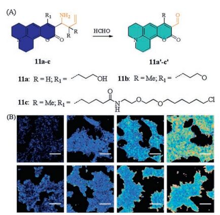
Recently, Chang et al. [41] developed a type of formaldehyderesponsive ratiometric fluorescent probes 11a-c using the same mechanism of 2-aza-Cope rearrangement. As described in Fig. 5, the aminocoumarin-based probes 11a-c employed homoallylamine as the determining group. Upon the addition of formaldehyde, the excitation wavelength displayed an obvious red-shift from 420 nm to 470 nm as a result of formaldehyde-induced products 11a'-c' (Fig. 5A). Among, probe 11a displayed a 1.6-fold excitation ratio change after its incubation with formaldehyde for 2 h, and low reaction rate with a bimolecular rate constant of 0.017 L mol-1 s-1 limited its application in biological systems. In contrast, probe 11b (10 μmol/L) exhibited a 3.2-fold excitation ratio change and probe 11c showed a 6.0-fold excitation ratio change after its incubation with formaldehyde for 2 h, indicating the introduction of a geminal dimethyl substituent could increase the reactivity of formaldehyde-dependent probes. Subsequently, probes 11b and 11c were applied to mark the level of formaldehyde in living cells. The results of fluorescence imaging indicated that probes 11b and 11c could be used for ratiometric bioimaging to monitor the level of formaldehyde fluxes in living cells, as exhibited in Fig. 5B. Moreover, probe 11c was successfully utilized to visualize differences in the resting formaldehyde levels between wild-type cells and models without ADH5.
图 5
图 5
(A) Coumarin-based ratiometric probes 11a-c. (B) Ratiometric confocal microscope images of probes 11b and 11c in HEK293TN cells. Reproduced with permission [41]. Copyright 2017, Royal Society of Chemistry.
Figure 5.
(A) Coumarin-based ratiometric probes 11a-c. (B) Ratiometric confocal microscope images of probes 11b and 11c in HEK293TN cells. Reproduced with permission [41]. Copyright 2017, Royal Society of Chemistry.
Subsequently, N, N-dimethylquinolin-6-amine as a fluorophore was developed into a formaldehyde-responsive ratiometric fluorescent probe 12 by Qian and Zhu group [42]. An appended homoallylamine served as the FA-responsive trigger was used as the determining group based on 2-aza-Cope rearrangement reaction. Probe 12 showed a broad emission peak at 495 nm. Upon the addition of formaldehyde, the intramolecular charge transfer (ICT) pathway was turned off and resulted in a remarkable red-shift of fluorescence emission peak at 570 nm. Meanwhile, the fluorescence intensity at 570 nm gradually enhanced along with a significant decreasing at 495 nm ascribed to the formation of formaldehyde-induced products 12' (Scheme 8). Fluorescence imaging indicated that probe 12 could penetrate cell wall and cell membrane for detecting exogenous formaldehyde in live plant tissues. Furthermore, it was capable of tracking the biologically relevant endogenous formaldehyde in live plant tissues by a ratiometric imaging mode.
Scheme 8
图 Scheme 8
Ratiometric probe 12 based on N, N-dimethylquinolin-6-amine.
Scheme 8.
Ratiometric probe 12 based on N, N-dimethylquinolin-6-amine.
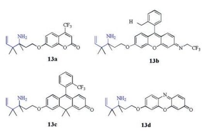
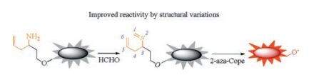
Usually, the fluorescent probes based on the same detecting groups cannot display the same determining properties when the detecting groups are installed on the different dye scaffolds [43]. In order to expand generality of design strategy, Chang group [43] further optimized the structure-activity relationships in the process of aza-Cope rearrangement and found that the homoallyiamine functionality could undergo a subsequent self-immolative β-elimination to yield a phenol on a fluorophore. As presented in Fig. 6, probes react with formaldehyde through a 2-aza-Cope rearrangement to generate the imine. Subsequently, the twocarbon linker is eliminated viaβ-elimination to release phenolic fluorophores along with a turn-on fluorescence signal. This strategy can effectively guarantee that the formaldehyde-triggered products are fluorescent. Utilizing this approach, they developed a series of formaldehyde-responsive selective and sensitive fluorescent probes 13a-d (Scheme 9) based on the fluorescent dyes with different emission color from blue to green to orange to red. Subsequent their bioimaging confirmed that these probes could be utilized as the indicators for visualizing the exogenous and endogenous formaldehyde in living cells. Specifically, probe 13d having a resorufin scaffold displayed the best signal-to-noise response, the most red-shifted and formaldehyde sensitive advantage in comparison to the others. More, it enabled identification of alcohol dehydrogenase 5 as a key enzyme that regulated formaldehyde metabolism in living cells. This strategy will be helpful for the design of fluorescence turn-on probes and further expand the application of determining groups in all kinds of fluorophores.
Scheme 9
图 Scheme 9
Structures of formaldehyde-responsive probes 13a-d.
Scheme 9.
Structures of formaldehyde-responsive probes 13a-d.
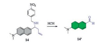
Recently, some formaldehyde-responsive two-photon fluorescent probes were sequential reported and applied in tissues bioimaging due to their deeper tissue penetration. For example, Yuan and Zhang [44] reported a two-photon fluorescent probe 14 based on a classic two-photon backbone in 2016. Upon the treatment of formaldehyde, probe 14 underwent an aza-Cope rearrangement to yield the fluorescent naphthalenone 14' (Scheme 10) and give rise to a strong green fluorescence at 526 nm. Moreover, probe 14 exhibited a turn-on fluorescence response both in one-photon and two-photon models with a low detection limit of 0.2 μmol/L. probe 14 was successfully applied in the two-photon bioimaging of formaldehyde in living cells and rat liver tissues with tissue-imaging depths of 40-170 μm. More, it was capable of tracking the endogenous formaldehyde in live MCF-7 cells.
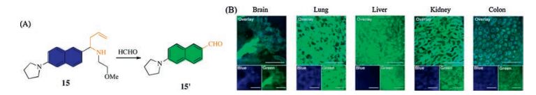
Due to the PET effect of nitrobenzyl in probe 14, subsequently, Ahn group [45] removed the nitrobenzyl group and designed a fluorescent probe 15 having a two-photon scaffold as well as probe 14 in Fig. 7. In PBS buffer (pH 7.4), it exhibited a strong blue emission at 438 nm. Along with the addition of formaldehyde, a marked decrease of the fluorescence intensity at 438 nm was observed, accompaniedbya dramaticfluorescenceenhancement at533 nm as a result of the ICT process (Fig. 7A). The ratio of fluorescence intensity (I533 nm/I438 nm) presented a good linear relationship with the formaldehyde concentration in the range of physiological concentration (0-800 μmol/L). Ratiometric imaging of formaldehyde in mouse tissue slices with a two-photon mode manifested that the different levels of formaldehyde were distributed in differentorgan tissues in Fig. 7B. Additionally, they found that it was possible that theformaldehyde actedas aprotective rolein the small intestine along with the antimicrobials released from the Paneth cells. These results provided definite information for the study of biological function of formaldehde.
图 7
图 7
(A) Two-photon ratiometric fluorescent probe 15. (B) Ratiometric confocal microscope images of formaldehyde with probe 15 in different mouse organ tissues. Reproduced with permission [45]. Copyright 2017, American Chemical Society.
Figure 7.
(A) Two-photon ratiometric fluorescent probe 15. (B) Ratiometric confocal microscope images of formaldehyde with probe 15 in different mouse organ tissues. Reproduced with permission [45]. Copyright 2017, American Chemical Society.
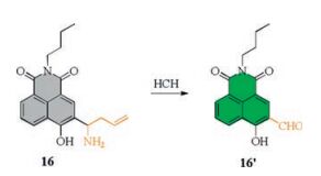
More, another fluorophore naphthalimide with two-photon feature was utilized by Sun and Zhu et al. [46] As shown in Scheme 11, probe 16 employed a 4-hydroxyl 1, 8-naphthalimide as fluorophore and homoallylamino group as the determining group. Probe 16 was a well-known ICT-based platform and possessed a two-photon excitation feature. The bioimaging application suggested that probe 16 could penetrate cells membrane for detecting formaldehyde selectively in living cells. It was worth mentioning that this probe could track the level of formaldehyde in zebrafish via a two-photon mode.
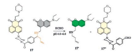
Owing to the fact that lysosome-specific fluorescent probes are easily disturbed by analytes themselves in cytosol and other organelles before probe entered into lysosomes. Recently, Tang group [47] developed an acidic pH-activatable formaldehyderesponsive fluorescent probe 17, which could get rid of the interference effectively. Probe 17 linked a naphthalimide fragment to coumarinfluorophorewith two-photonfeature, introduced4-(2-ethyl)morpholine unit as lysosome-locating group and homoallylamino group as the determining group. Probe 17 showed a weak fluorescence emission at 566 nm in phosphate buffer solution (pH 5.0) owing to the naphthalimide fluorophore. Upon the treatment of formaldehyde, probe 17 underwent an aza-Cope rearrangement to yield the coumarin-containing fluorophore 17' (Scheme 12) and induced the obviously blue-shifted strong fluorescence enhancement at 506 nm, as shown in Scheme 11. The colocalization experiments in HepG2 cells demonstrated that it was able to visualize formaldehyde exclusively in lysosomes and eliminated interference of formaldehyde in neutral cytosol and other organelles. Further bioimaging analysis indicated probe 17 could be utilized to in situ visualize FA in living cells and animals by two-photon mode. Additionally, N-acetylcysteine was found as an efficient scavenger of intracellular formaldehyde in biological system.
Scheme 12
图 Scheme 12
Two-photon fluorescent probe 17 based on coumarin and naphthalimide fragments.
Scheme 12.
Two-photon fluorescent probe 17 based on coumarin and naphthalimide fragments.
In this review, we summarized the recent advances in formaldehyde-responsive fluorescent probes. The reported formaldehyderesponsive fluorescent probes mainly employed three strategies based on different chemical reactions, including the formimine mechanism, the methylenehydrazine mechanism and the aza-Cope rearrangement mechanism, in which, some probes exhibited excellent biological application in living cells, tissues and animals.
Although numerous formaldehyde-responsive fluorescent probes have been reported in recent years, they restrict from some negative factors. For instance, fluorescent probes based on the formimine-and methylenehydrazine-based mechanisms usually suffer from the relatively low selectivity and some analogues of formaldehyde cause serious interference while relatively long reaction kinetics of probes based on the aza-Cope rearrangement mechanism may reduce probe sensitivity. Meanwhile, the emission wavelength of most fluorescent probes locating in the visible region limited their further application in vivo. Future efforts will focus on developing near-infrared fluorescent probes with deep tissue penetration and minimum interference from background autofluorescence. Furthermore, it is expected that biological research closely related to formaldehyde biological function will help us deeply understand its physiological and pathological roles that are related to various diseases in future.
Acknowledgments
The authors acknowledge financial support from National Natural Science Foundation of China (Nos. 21676113, 21402057, 21472059, 81671803); Youth Chen-Guang Project of Wuhan (2016070204010098); the 111 Project B17019 and the MinistryProvince Jointly Constructed Base for State Key Lab-Shenzhen Key Laboratory of Chemical Biology, Shenzhen. This work is also supported by self-determined research funds of CCNU from the colleges' basic research and operation of MOE (No. CCNU16A02004).
[1]
Y.M. Zhang, Y.T. Lin, J.L. Chen, et al., Sens. Actuators B 190(2014) 171-176. doi: 10.1016/j.snb.2013.08.046
[2]
S.E. Sayed, L. Pascual, M. Licchelli, et al., ACS Appl. Mater. Interfaces 8(2016) 14318-14322. doi: 10.1021/acsami.6b03224
Figure 3
(A) Naphthalimide-based probes 7a-d. (B) Confocal microscope images of exogenous and endogenous formaldehyde with probe 7d in 4T-1 and 3T3 cells by one-photon and two-photon modes. Reproduced with permission [34]. Copyright 2016, Royal Society of Chemistry.
Figure 4
(A) The proposed detection mechanism of probe 9 based on silicon rhodamine. (B) Confocal microscope images of probe 9 in HEK293TN and NS1 cells. Reproduced with permission [36]. Copyright 2015, American Chemical Society.
Figure 5
(A) Coumarin-based ratiometric probes 11a-c. (B) Ratiometric confocal microscope images of probes 11b and 11c in HEK293TN cells. Reproduced with permission [41]. Copyright 2017, Royal Society of Chemistry.
Figure 7
(A) Two-photon ratiometric fluorescent probe 15. (B) Ratiometric confocal microscope images of formaldehyde with probe 15 in different mouse organ tissues. Reproduced with permission [45]. Copyright 2017, American Chemical Society.

 下载:
下载:


 下载:
下载:


