
Scheme 1.
Schematic diagram of the treatment of various diseases by adjusting macrophage polarization in pathologic organism microenvironment based on MOF materials.
Metal–organic framework and its composites modulate macrophage polarization in the treatment of inflammatory diseases
Feifei Wang , Hang Yao , Xinyue Wu , Yijian Tang , Yang Bai , Hui Chong , Huan Pang

Inflammation is an important physiological process in the body's defense against pathogenic microbial infection or tissue damage repair [1], it releases inflammatory substances (cytokines, free radicals, hormones and other small molecules), thus helping protect the body from these pathological abnormalities. Redness, swelling, heat, pain and organ dysfunction are the main symptoms [2]. The inflammatory process can be divided into two stages, which are acute inflammation and chronic inflammation [3]. The successful acute inflammatory response will destroy invading pathogens through neutrophils to achieve the effect of reducing inflammation and repairing damaged tissues [4]. During acute inflammation, if harmful stimuli are not removed in time, the inflammatory process will continue and upgrade the "weapons" against invaders and a lot of reactive oxygen species (ROS) will be produced. Chronic inflammation causes tissue necrosis by invading macrophages and lymphocytes, which leads to a variety of chronic diseases, such as arthritis, cancer, cardiovascular disease, diabetes and neurological diseases [5].
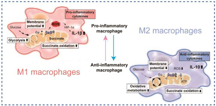
Macrophages played critical role in maintaining the balance of physiological immune environment [6]. It could activate the immune response to inflammatory in the whole process of immune reaction and functioned irreplaceably in the treatment of inflammatory diseases [7]. Silenced macrophages (phenotype of M0) can be stimulated in a variety of ways to produce both classically activated macrophages (M1) and alternatively activated macrophages (M2), respectively [8]. M1 macrophages may release cytokines and chemokines that promote inflammation, which could exacerbate the development of inflammatory diseases [9]. Alternatively, M2 macrophages release cytokines that fight inflammation and repair promoting factors, that could facilitate the recovery of inflammatory diseases [10]. With certain stimulation, interconversion of M1 and M2 subtypes could be achieved [11]. Therefore, inducement of M1 to M2 subtype transformation highly favored the cure of inflammatory diseases [12].
Based on the above therapeutic strategies, many anti-inflammatory drugs have been developed to reduce the inflammatory response, including non-steroidal anti-inflammatory drugs (NSAIDs) [13] and glucocorticoids [14]. However, high doses of these drugs may cause adverse reactions to the body, such as liver damage, pulmonary fibrosis and gastrointestinal discomfort [15]. Finding safe and effective anti-inflammatory drugs remains a top priority in eliminating inflammatory diseases. With the development of nanotechnology, MOFs have attracted more and more attention. MOFs exist in many forms, including polyhedral MOFs and MOF nanosheets, whose shape and size affect performance [16]. MOFs are highly effective porous materials, which are widely used in biomedicine fields, such as wound healing, colitis, osteoarthritis (OA), antibacterial and tumor treatment, due to their adjustable chemical composition, high porosity and large specific surface area [17]. At the same time, some MOFs also have the effect of nano-enzymes, which play a significant role in removing ROS [18].
Therefore, this paper summarized the causes of inflammatory diseases, the relationship between macrophages and inflammatory diseases, the synthesis methods of bioactive MOF or coordination polymers, the relationship between MOFs and macrophage polarization, and the application of MOFs in the treatment of colitis, OA and wound healing (Scheme 1). Finally, we provide ideas for the development of nanomedicine and biomedical materials.
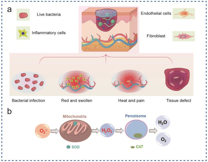
Inflammation is a defense mechanism of tissues against external pathogens or other harmful stimuli. In general, moderate inflammation can play an effective role in protecting against tissue damage and pathogen invasion and is a congenital immune mechanism (Fig. 1). For example, inflammatory congestion can help surface tissues get more oxygen, nutrients, and guarding substances [19]. However, excessive or lack of inflammatory response will cause harm to the body [20]. Some elements of the inflammatory response that are beneficial to the body may be turned into harmful ones under certain conditions. The causes of the inflammation can be divided into exogenous and endogenous [21]. Many substances can trigger the signal of the inflammatory response, which are called inducers [22].
From a mechanistic point of view, multiple factors trigger inflammation, including the release of inflammatory cell mediators (interleukin-6 (IL-6), tumor necrosis factor (TNF), etc.), which act on other cells to cause a series of changes [23]. Exogenous induction can also be divided into two types: microbial and non-microbial, that is, bacteria, and the influence of foreign drugs [24]. Infection of cells by microorganisms can activate an inflammatory response [25]. Infection recognition is mainly mediated by innate pattern recognition receptors (PRRs), including Toll-like receptors, retinoic acid-inducible gene I (RIG-I), NOD like receptors (NLRs), and C-type lectin receptor (CLR). These PRRs cause the transcriptional expression of inflammatory mediators and initiate intracellular signaling pathways to coordinate the clearance of pathogens and infected cells [26].
Non-microbial exogenous inflammatory induction factors mainly include allergens, some irritants and foreign bodies, and some toxic compounds [29]. Some of these foreign substances mimic the toxicity of microbial pathogens, and some foreign substances are difficult to be digested and phagocytosed by macrophages, as a result, the body's immune system will be damaged to a certain extent, causing inflammation [30]. Therefore, some specific drugs are needed to control the inflammatory response, such as curcumin, etc. [31].
The endogenous inducements of inflammation are mainly signals generated by compressed, damaged tissues or other dysfunctional tissues [32]. Studies have shown that ROS can regulate macrophage phenotype (Fig. 2). In RAW264.7 cells, iron overload can induce M1 polarization [33], by increasing ROS generation and inducing P53 acetylation. Formentini et al. showed that partial inhibition of mitochondrial ATP synthase in the gut of transgenic mice induces an anti-inflammatory response through mitochondrial ROS-mediated nuclear factor κB (NF-κB) (mtROS) activation and recruitment of pro-inflammatory M2 macrophages, indicating that mtROS has a new anti-inflammatory effect. properties have an impact [34]. Defective tricarboxylic acid (TCA) cycle is one of the hallmarks of inflammatory macrophage metabolism. In inflammatory mouse macrophages, two points of the TCA cycle, mainly supplied by glycolysis and glutamine metabolites, were disrupted, resulting in the accumulation of TCA intermediates [35]. These breakpoints occur in isocitrate dehydrogenase (IDH) and lead to citrate accumulation. Succinate dehydrogenase (SDH) causes succinate accumulation [36]. Other functions of metabolism and immune signaling then use these accumulated metabolites. First, cumulative citrate was used to support the classical activated macrophage phenotype that drives fatty acid synthesis and nitrogen monoxide (NO) production [37]. Second, accumulated succinate stabilizes hypoxia inducible factor-1α (HIF-1α), resulting in continuous production of IL-1β and ROS. IL-1β works together with TNF-α, IL-6, IL-12, etc., and is one of the inflammatory factors produced by macrophages induced by interferon γ (IFN-γ) [38]. HIF-1α is a key transcription factor regulating macrophage glycolysis and inflammation. Loss of HIF-1α results in reduced macrophage motility, bactericidal activity, and ability to aggregate [39]. These studies suggest that ROS function as a "phenotype switch" that can induce different phenotypes (M1/M2) through the mediation of different signaling pathways. For the mechanism of macrophage polarization, please refer to Fig. S1 (Supporting information).
MOF is an important crystalline porous organic-inorganic hybrid material, composed of inorganic nodes (metal clusters/ions) and coordination bonds, with a periodic lattice structure [40,41]. Compared with traditional porous materials, MOF materials have the characteristics of larger specific surface area and higher porosity (up to 90% free volume), close-fitting pores, fully exposed active sites, and adjustable structure [42]. MOF based materials are mainly composed of three categories, which generally include pure MOF, MOF composites (such as metal nanoparticles [MNPs]@MOFs), and MOF derived materials (metal oxides/nitrogen modified compounds/sulfur modified compounds/phosphates). These strategies both avoid the modification of the original MOF and recommend the merits of advanced functional materials in the composite [43].
In general, the traditional synthesis methods of primitive MOFs mainly include a common solvent method, solvothermal method, microwave synthesis, and sound chemical method.
The common solution method uses a simple reaction vessel (such as a beaker, petri dish), adding the corresponding metal salt and organic ligand into a specific solvent to dissolve, and then standing or stirring at room temperature or slightly higher temperature (< 100 ℃) [44]. As the reaction proceeds, the solvent evaporates, or the temperature decreases, resulting in the crystallization of the target MOF material. Generally, the static method is often suitable for growing large single crystals [45], while the stirring method is suitable for rapidly obtaining a large number of pure-phase microcrystals [46]. If necessary, the pH of the reaction system should be adjusted or other reagents containing coordination ions, such as hydrofluoric acid and ammonia, should be added as reaction buffers, to control the formation of crystal nuclei and their speed, and finally obtain MOF crystal samples with different sizes [47].
The solvothermal method, which involves the reaction in an autoclave (closed vessel) under controlled temperature and pressure, is the most commonly used method to obtain target MOFs (Fig. 3) [48]. This method generally employs high boiling point solvents such as DMF, DEF, acetonitrile, acetone, ethanol or methanol [49]. Alternatively, problems related to differences in solubility of reactants can also be addressed with solvent mixtures [50]. However, this method has certain limitations, namely long reaction time, high temperature, and expensive solvents [51]. Therefore, to mask the solvent cost, a greener and cheaper hydrothermal method was adopted.
Microwave synthesis is a rapid MOF synthesis involving microwave heating (MW) [52]. This process results in a highly crystalline and porous structure, small particle size and well-controlled shape. Cr-MIL-100 (MIL: Lavoisier materials) is the first MOF to use this approach. This approach depends on several factors. Power, exposure time, temperature, solvent concentration, substrate composition (Fig. 3) [53].
MOF composites consist of one or more materials with different MOF compositions by combining different MOFs with carbon [54], polymers [55], enzymes [56], graphene, metallic NPS, etc. The integration of materials improves the performance of MOFs [57].
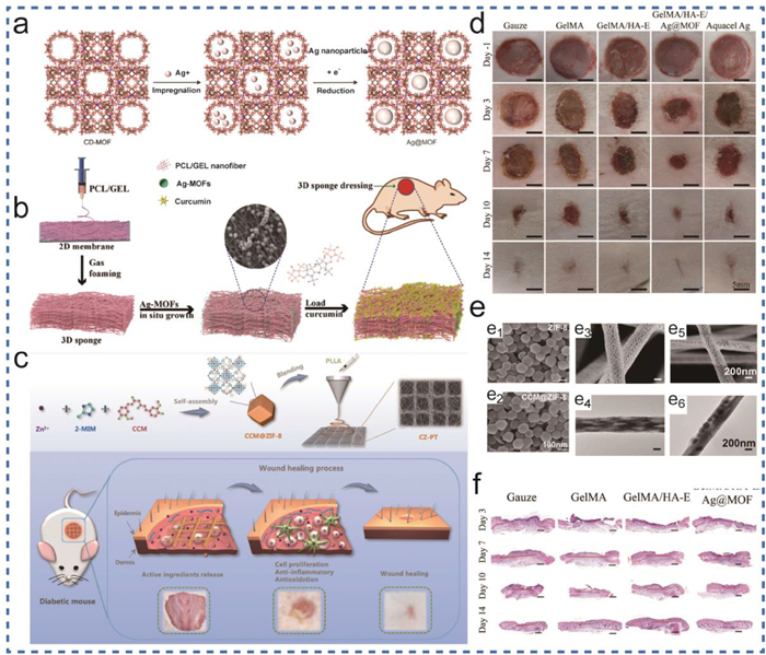
An important fabrication method is materials grown in situ on MOFs. In this method, the structural framework of MOF affects the growth direction and growth rate of nanoparticles, and prevents the problem of easy agglomeration of nanoparticles, thus improving the stability of nanoparticles [58]. For instance, Chen et al. expanded 2D polycaprolactone/gelatin (PCL/GEL) films into 3D nanofiber sponges by gas foaming method, and in situ grew Ag-MOFs (3D-Ag+ MOF) and loaded curcumin (3D-Ag-cur) on the sponges by impregnation-lyophilization. Ag-MOF has antimicrobial properties. The layered nanostructure of the sponge not only achieves a uniform distribution of Ag, but also prevents the aggregation of Ag-MOF nanoparticles, resulting in strong antibacterial properties of the dressing. Curcumin has excellent antioxidant and anti-inflammatory properties, and its functionalization significantly promotes hemostasis and tissue regeneration, promoting wound healing [59]. Opinions of Zhao et al. PLMOF is formed by in-situ growth of ZIF-8 on luminous nanoparticles (PLNPS) (Fig. 4). PLMOF has good anticancer effects, high loading capacity, and pH responsive drug release behavior, and significant antitumor effect. This study expanded the role of loading materials and provided new ideas for many future drugs that require loading [60]. The template method is often used to regulate the morphology of nanomaterials. We know that conventional organic polymers have abundant directional channels, which can be used to allow MOF to grow in their channels, and MOF nanomaterials with specific morphology can be obtained after removing the template [61]. For example, Cheng et al. prepared a new MOF material with a hierarchical porous structure by template method. They used salicylic acid and melamine as hydrogel precursors, blended with MOF precursors and synthesized hydrogel and MOF composite materials by reverse-phase emulsion polymerization method, and then removed the hydrogel template by simple hot water dissolution. MOFs with both microporous and mesoporous structures were prepared. They skillfully took advantage of the thermal instability of the hydrogel template, and the process was simple. The loading of glucose oxidase (GOx), horseradishadic peroxidase (HRP) into the MOFs material significantly improved the catalytic efficiency of the enzyme, and the MOF material has a broad application prospect in the field of biomedical carriers (Figs. 5a and b) [62].

Coating one MOF or COF on top of another (MOF@MOF/COF) is an advanced method due to the unique structural properties of MOFs, epitaxial growth is the formation of core-shell or layered A good approach to MOF@others is for MOF/COF composites (Figs. 4a and b) [63]. When one frame material is introduced into the synthetic solution of another frame material, the layered MOF-on-MOF heterogeneous structure formed can improve the layered porosity, acticity, and stability of MOF composites [64]. In general, the successful growth of secondary MOFs on prefabricated MOFs is attributed to their low-boundary cell lattices. The zeolite imidazolate framework (ZIF) is a subfamily of MOF topologically isomorphic to zeolites by combining transition metal cations (mainly Zn or Co) with imidazolyl ligands [65]. As mentioned earlier, building MOF-ON-MOF structures relies on good cellular lattice matching between the two MOFs. Given two MOFs with the same structural characteristics, the second MOF grows on the surface of the first MOF, forming a core-shell structure [66].
MOF itself contains both metallic elements and organic components, and can be used as a metal source, carbon source, and dopant source at the same time [67]. At the same time, its own regular porous skeleton structure can be inherited by derived materials to a certain extent to play the role of a templating agent [41]. Using MOFs as a multifunctional self-sacrificing template agent and precursor, a wide variety of derivative nanomaterials can be obtained, including porous carbon materials, metal oxides, metal hydroxides, metal sulfides, metal phosphide, and other metal compounds [68]. Compared with the precursor MOFs, the stability (especially water stability) of these derived materials is significantly improved and inherits some advantages of the original MOFs. High specific surface area, tunable pore structure, easy structure/composition control, and uniform composition distribution [69]. Thermal decomposition is another method that is most used to synthesize MOF derivatives [70]. In the process of pyrolysis of MOF materials, the gold inorganic ions and organic ligands can be directly transformed into uniformly distributed metal oxide nanoparticles under the influence of temperature [71]. On the other hand, the organic ligands in MOF can also be converted into porous carbon structures during pyrolysis [72]. For instance, Cao et al. via a simple one-pot heat treatment of Ce-MOFs yielded Nano-Cell particles with oxidase-like activity. Unusually, this preparation prevents agglomeration of metal oxides at the nanoscale and enhances the catalytic activity of the material (Figs. 5c–e) [73].
It has been found that nano-MOF materials can induce inflammation and immune responses in vivo and in vitro [74]. Nano MOF materials can be internalized by macrophages, induce cell surface changes, and secrete cytokines and chemokines [8]. Understanding the mechanism of interaction between nano-MOF materials and macrophages will help to effectively design a specific therapeutic strategy for nano-MOF materials. Stimulation of macrophages by different nanomaterials can induce the polarization of M0 macrophages into M1 or M2 types. The different polarization of macrophages plays different roles in the outbreak and elimination of inflammation and the growth and inhibition of tumor [75].
M1 macrophages show strong microbial killing and antitumor activity [76]. Nano MOF materials induce the generation of inflammation by inducing macrophages to become M1-polarized and ROS generation (Fig. 6) [77]. The polarized generation of M1 macrophages was found to be related to the chemical composition [78], size [79] and material shape [80] of nanomaterials.
The other direction of polarization of macrophages is to become M2 macrophages. M2-type macrophages are mainly used as anti-inflammatory cells to protect the body from inflammation and ROS, which is conducive to wound healing. It was found that some surface treatments of nanomaterials, such as increasing the roughness of the materials and enhancing the surface hydrophilicity of the materials, can enhance the anti-inflammatory effect.
Inflammation is an organism's defense response to noxious stimuli [83]. Oxidative stress of inflammatory cells can counteract the damage of the external environment to the body, but at the same time, excessive oxidative stress will destroy the endothelial barrier, thus accelerating the migration and infiltration of inflammatory cells and aggravating tissue damage [84]. An important pathophysiological change occurring in various inflammatory diseases is the overproduction of ROS, including H2O2, hydroxyl radical (∙OH), and superoxide anion (OOH) [85]. On the one hand, they destroy cellular components, on the other hand, they also act as signaling molecules that promote infiltration of immune cells and release inflammatory mediators [86]. We summarized the treatment of different inflammatory diseases using MOF materials (Table S1 in Supporting information) and observed and compared the changes in markers of different phenotypes of macrophages.
Inflammatory bowel disease (IBD) is a chronic autoimmune disease with recurrent manifestations of subcutaneous barrier dysfunction and intestinal ulcers [87]. It comes in two forms, ulcerative colitis (UC) and Crohn's disease (CD) [88]. Typical symptoms, such as diarrhea, abdominal, pain, and hemorrhagic stool, seriously affect patients' quality of life [89]. A key contributor to outbreaks of IBD is oxidative stress, which manifests itself in abdominal pain and diarrhea [90]. During the inflammatory attack, neutrophils and macrophages will be recruited to gather in large numbers in the intestinal mucosa at the site of IBD, and then infiltrate and release lots of ROS and pro-inflammatory cytokines [91]. Excessive production of ROS can lead to oxidative damage to DNA, proteins, and lipids, thereby promoting the development and progression of IBD [92]. Current treatment strategies for IBD mainly rely on the remission of chemotherapy agents, such as amino salicylate and glucocorticosteroids, and adjuvant surgery [93]. However, long-term and high-dose chemotherapy drugs can cause serious side effects, such as hyperglycemia and the occurrence of atherosclerosis [94]. The limitations of these therapies, the complexity of the disease, and individual differences make the current treatment of UC far from satisfactory. Therefore, it is necessary to develop new IBD treatment modes that are more effective and safer for the body.
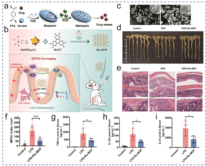
In recent years, nanomaterials with catalytic activity, including carbon nanoparticles [95], noble metal nanoparticles [96], metal oxides [97], and polyphenol nanoparticles [98], have been considered as alternative methods to remove ROS in the treatment of disease damage caused by oxidative stress. This type of material is called nanozyme, which has the catalytic activity of catalase (CAT), superoxide dismutase (SOD), peroxidase (POD) and other enzymes. MOF materials are known for their large surface area and porosity [99]. The framework structure composition of MOF is optimized at the molecular/atomic level to optimize active site exposure and allow efficient and sufficient interactions with reactants that can be ordered in sequence (porous structure and regular channels) [100]. More importantly, MOF can be physically and chemically programmed by suitable metals and ligands to have structures or catalytic powers, demonstrating the special capacity of MOF to be engineered as nanozymes [101]. Liu et al. found that ruthenium (Ru) is a transition metal element with redox activity. The interconversion between Ru polyvalent structures can form REDOX capacity, which can be used to eliminate reactive oxygen ROS (Fig. 7) [102]. They synthesized a Ru foundation organic framework (Ru-MOF) as a nano-antioxidant for the cure of IBD. The porous Ru-MOF not only eliminates but also significantly reduces colonic damage dextran sulfate sodium salt (DSS)-induced acute colitis in vivo by down-regulating pro-inflammatory cytokines by inhibiting TNF-associated inflammatory pathways.
The rupture of the mucous layer of the inflammatory colonic mucosa tends to attract positively charged proteins [103], as for transferrin and eosinophilic cationic proteins, so that positive charges tend to accumulate in the damaged epithelial cells. This provides a molecular target the adhesion of negatively charged nanoparticles through electrostatic interactions (Figs. 7a and c) [104]. Furthermore, the characterization of the low pH (pH < 7) inflammatory microenvironment during inflammatory overload allows the development of nanoparticles for more targeted therapeutics. Yin et al. found through foregoing studies that cerium-based materials with high Ce content showed better SOD activity and could be used as ROS scavengers. Therefore, they chose Ce-MOF as nanocarriers for drug delivery in inflammatory tissues [104]. Ce-MOF consists omixed-valenceence cerium center and a p-phthalic acid (PTA) ligand. After modification with poly(4-styrene sodium sulfonate) (PSS), the resulting negatively charged Ce-MOF@PSS would preferentially adhere to the inflamed gut. When cerium ions participate in the reaction of excessive ROS production at the inflammatory site, they feel the low pH in the inflammatory microenvironment. The change of its value changes the bonding mode between PTA and cerium, further generates the conversion of Ce-MOF from mesoporous to macroporous, controls the release of anti-inflammatory drugs, reduces inflammatory cytokines, regulates the significantly down proportion of M1 macrophages, and repairs the intestinal barrier function.
As described above, nanoparticles of different sizes also have different effects on macrophage polarization. In IBDs, the researchers found the same thing. Nanoparticles with suitable sizes are easier to target in the inflamed colon, and easier to extend the retention time of the material [76]. In this aspect, Prussian blue nanoparticles (PPBs) are generally about 60 nm in size and preferentially aggregate in the epithelial mucosa of the colon with inflammation, increasing the permeability and retention of the material. In addition, PPB could modulate macrophage polarization to the M2 phenotype through its mainly acts on TLR signaling and exerts unique therapeutic effects on DSS induced colitis mice without adverse side effects [76].
OA is a chronic degenerative disease that causes rickets, synovitis, and subchondral bone remodeling [105]. OA causes pain and dysfunction of the knee joint and poses a significant medical and economic burden [106]. In OA, inflammation up-regulates matrix metalloproteinases, promotes the degradation of cartilage tissue, and will lead to bone loss, self-inflammatory pain, and will ultimately lead to reduced daily exercise in patients [107]. The proliferation of macrophages infiltrated the lubricating membrane during the progression of OA, hinting that macrophages play a key role in the occurrence and growing of OA [108]. LPS induces M1 macrophages to elevate inflammation through the secretion of various pro-inflammatory factors, thereby causing the degeneration of chondrocytes [109]. However, M2 macrophages can inhibit inflammation and promote tissue recovery [110]. A key factor in OA is the lack of oxygen and the production of too much ROS at the site of inflammation (Fig. 8) [111]. Decreased mitochondrial aerobic respiration leads to hypoxia in the mitochondrial microenvironment, which tends to produce lots of ROS, increases oxidative damage and HIF-1α expression, and increases mitochondrial DNA mutation, leading to significant changes in mitochondrial structure, dynamics and genomic stability [112]. Former studies have shown that high expression of HIF-1α induces M1-type polarization of macrophages, which induces OA (Fig. 8b) [113]. ROS directly induces the synthesis of matrix metalloproteinases in fibroblast-like synoviocytes, inhibits the generation of extracellular matrix (ECM), and promotes bone resorption [114]. ROS induce M1 macrophage transformation by regulating the expression of HIF-1α, accelerating its synthesis and restraining its degradation. Therefore, simultaneously reducing hypoxia and ROS levels by balancing the various subtypes of macrophages may be an effective strategy for the treatment of OA.

Previous studies have shown that early intervention in cartilage degradation can effectively prevent the progression of OA, and some therapeutic medicines, like NSAIDs, have been broadly put to use for subjective symptom relief [115]. However, these drugs do not fundamentally impede the progression of OA and may be associated with certain side effects, such as damage to the digestive and cardiovascular systems [116,117]. Injections of anti-inflammatory drugs into the joints are a good way to improve OA, but free drug can be removed from the joint space, which reduces drug availability and increases complications [118]. Therefore, exploring more new treatments to lead to good outcomes is a top priority for managing OA.
As a new type of porous material [119], MOF materials have been widely used in drug delivery [120], in vivo imaging [121] and biological detection [122]. MIL-100(Fe) has been widely used in nanodrug delivery systems due to its strong porous morphology, mass, responsiveness to pH. Moreover, it has been shown to have good biocompatibility through cell experiments and animal experiments. A bottleneck of MIL-100(Fe) is its low stability in water, which makes it difficult to use in the treatment of OA [123]. The dispersity and hydrophobicity of MIL-100(Fe) can be improved by modification with hyaluronic acid (HA) [124]. Artificially added HA is widely used due to its good anti-inflammatory effect in OA. These include reducing inflammation, hydrating the body, protecting chondrocytes from reactive oxygen species, alleviating pain, protecting joints, and promoting cartilage regeneration [125]. Xiong et al. reported on an improved pH-responsive system is described using Te MOF@HA nanoparticles to encapsulate anti-inflammatory protocatechuic acid (PCA), modified with the natural cartilage humectant HA. Xiong et al. reported that using the pH sensitivity of MOF@HA, MOF materials can change the structural characterization and release PCA under acidic conditions to achieve the function of slow release of drugs, so that the residence time in the joint cavity can be extended, and thus improve the possibility of curing OA [126]. In addition, PCA is a phenolic compound derived from plants that fights inflammation [127]. This is because PCA significantly reduces enzymes, including inducible nitric oxide synthase (incyclohexyl-2xy-2 (COX2) and chromoprotein), which contain inflammatory factors, thereby reducing the level of M1 macrophages.
Because the onset of OA can attract the outbreak of lots of nanozyme-modified field OA remodeling microenvironment is an effective strategy to protect chondrocytes and delay OA (Figs. 8a and d–f) [128]. A major disadvantage of most studied nanozymes is their poor biodegradation performance, which prevented them from being completely cleared from the body. This defect hinders the further application of nanozymes in clinic. Therefore, it is necessary to develop and improve the biodegradability and biosafety of nanozymes, although this is still a big challenge. Xiong et al. through template mediated biomineralization method, bovine serum protein (BSA) as a template, to build a pH sensitive response, and had good biodegradation performance hollow manganese Prussian blue nanozymes (HMPBzymes) [129]. The polyvalent convertible ability of Mn and Fe in HMPBzymes enabled the material to have good REDOX performance, thus inhibiting ROS and reducing oxidative damage. By regulating mitochondrial respiration and reducing catabolism, it could promote the synthesis and metabolism of ECM in cartilage and induced the polarization of synovium macrophages from M1 type to M2 type. HMPBzymes catalyzed hydrogen peroxide to produce O2, alleviated the harm caused by hypoxia, induced HIF-1α increasing, and then promoted the polarization of macrophages to change from M1 type to M2 type, which promoted the development and application of nanozymes in biomedical materials.
As the largest organ of the human, the skin acts as a barrier against the external environment [130]. Any damage to the skin may allow microorganisms to infiltrate our bodies and cause infection [131]. Wound infections caused by various accidental injuries accompanied by foul smell, severe wound pain, and slow healing often bring physical trauma and psychological adverse effects to patients. Therefore, in the field of clinical research, such as surgery, emergency trauma and even battlefield rescue, more and more attentions are paid to wound healing. Diabetes-induced wound is one of the most serious complications of diabetes, which has high incidence, poor cure rate and high medical costs [132]. Patients with diabetes-induced wound wounds are often accompanied by chronic inflammation, which is easy to cause problems such as insufficient angiogenesis and abnormal collagen metabolism. Ordinary treatment methods are often unable to cure, so it is urgent to find an efficient treatment method for diabetic wound wounds [133]. Diabetic wounds not only produce a large amount of ROS and inflammatory cytokines, but there are also often chronic bacterial infections that lead to an overreaction of inflammation at the wound site and prolonged wound healing time [134]. There are also many bacteria, such as S. aureus and E. coli, that can be found in infected wound sites [135]. The accumulation of bacteria can easily lead to the formation of bacterial membranes in the skin wound, which can inhibit the host's immune response and antibacterial treatment, resulting in bacterial infection. Moreover, E. coli infection has been covered to reduce the levels of these wound proteins, hydroxyproline, collagen type Ⅰ, and collagen type Ⅲ, which may delay wound healing. Although the use of antibiotics can effectively reduce the possibility of bacterial infection, its effectiveness is still decreasing year by year with the emergence of multiple drug-resistant bacteria, which poses a challenge to the development of antibacterial materials for medical use [136]. To address this problem, an effective strategy must be explored that simultaneously improves antibacterial, ROS clearance, and anti-inflammatory capabilities to promote skin wound healing.
Curcumin is a natural polyphenol that has been shown to reduce inflammation and combat oxidative stress. Previous studies have found that curcumin not only clears ROS, but also reduces the production of pro-inflammatory cytokines, thereby regulating the polarization of macrophages [137]. Recent studies have found that curcumin has beneficial results on the treatment of wound healing, endothelial injury, and mucosal injury [138]. However, curcumin's clinical use is limited due to its insolubility in water, low physical and chemical stability, and low bioavailability under physiological conditions [139]. Therefore, Wang et al. used crystal engineering methods (CCM@ZIF-8) (Fig. 9) [140]. The MOF centered on Zn2+ was synthesized with curcumin as ligand, and its structure was similar to ZIF-8. By using ZIF-8 as a drug carrier and loading anti-inflammatory drug CCM, the solubility of CCM in aqueous solution was improved. When the composite material reaches the inflammatory site of the wound, it receives the influence of weak acidic conditions in the inflammatory area, and ZIF-8 material begins to decompose, gradually releasing the anti-inflammatory drug CCM, achieving a sustained release function. CCM induces wound inflammatory macrophages to polarize from M1 phenotype to M2 phenotype, thereby reducing inflammatory response.
However, CCM@ZIF-8 can release Zn2+ during the degradation process, showing that it has good anti-inflammatory effect and can promote cell proliferation. To better apply to diabetic wound healing and improve the slow-release ability of the material, CCM@ZIF-8 was also loaded into a graded micro/nanofiber scaffold made of poly(lactic acid) as raw material. In addition to increasing the roughness of the material, increasing the cell contact area, promoting cell adhesion, and increasing protein adsorption sites, this composite structure can also promote epidermal regeneration, angiogenesis, and collagen deposition, and relieve oxidative stress according to wound inflammation.
The problem of bacterial infection in wound healing has been a constant concern of researchers. Silver nanoparticles (Ag-NPs) are widely used as antibacterial agents for skin wound repair due to their good antibacterial activity and small vulnerability to bacterial resistance [141]. However, Ag+ bursts of silver ions (Ag+) may increase cytotoxicity [142]. In addition, small Ag-NPs lack stability due to the clustering of clusters. Therefore, Xiong et al. constructed the cyclodextrin MOF (CD-MOF), and added silver nanoparticles to MOF materials to maintain the stability of silver nanoparticles and gradually release them, which is an effective way to treat the antibacterial problem of diabetes wounds. Solving antibacterial and anti-inflammatory issues should not be underestimated. We combined epigallocatechin gallate (EGCG) with HA and coated it on CD-MOF. By utilizing the versatility and efficient drug loading function of MOF materials, EGCG begins to encounter inflammatory macrophages when the composite material reaches the wound inflammatory area, thereby inhibiting their production of pro-inflammatory cytokines. On the other hand, it regulates the transformation of macrophages to the M2 phenotype and enhances the production of anti-inflammatory cytokines. Promote angiogenesis and ultimately accelerate wound healing (Fig. 9) [143].
In addition to the elimination of inflammatory factors by loading anti-inflammatory drugs, researchers have found that Artificial enzymes called "nanozymes" offer great potential in the treatment of wound healing [144]. Among them, several nanozymes with transition metal or lanthanide metal as main metal elements showed good anti-inflammatory activity in vivo [145]. Wan et al. developed MOF nanoenzymes with antibacterial and inflammatory regulatory functions by uniformly doping Mn2+/Mn4+ in the ZIF-8 (Mn-ZIF-8) skeleton. They achieved bactericidal and anti-inflammatory functions in MOF structures and reduced the complexity of nanomaterials [146]. In this platform, Zn2+contained in ZIF-8 is released during hydrolysis, and has excellent antibacterial performance against bacteria such as S. aureus and E. coli. In this simple MOF structural design, Mn-ZIF-8 exhibits the coexistence of Mn2+/Mn4+, which endows nanocomposites with anti-inflammatory capabilities that can be regulated through redox environments. Mn containing nanoenzymes can functionally mimic three major antioxidant enzymes, such as SOD, CAT and GPx, to maintain the redox balance in cells, to combat oxidative stress and inhibit inflammation of overreaction. With the in-depth study of anti-inflammatory mechanisms, it has been found that Mn-ZIF-8 nanoenzymes can not only regulate the polarization of immune cells (macrophages) by reducing the expression of pro-inflammatory factors, but also reverse the expression of pro-inflammatory factors. The Mn ZIF-8 nanoenzyme platform can serve as a potential new type of nanomedicine material and play an important role in wound healing/skin infection treatment.
Biosafety refers to the impact of nanomaterials on human health and the environment, which is crucial for converting nanomaterials into clinical applications. We provide a detailed description of this aspect in Supporting information [147–150">150].
In this paper, the synthesis of bioactive MOF or coordination polymer, including common solution method, in-situ growth method and template method, is reviewed. Based on the type of MOF, its adjustable composition, structure, and pore size, as well as the adsorption and release of biomolecules, as well as a series of nanoenzyme activities of the material itself, their effects on different polarization of macrophages were discussed. In summary, we have drawn the following conclusions:
1. Metal ions/clusters and organic ligands in MOF materials can be selected differently, resulting in different arrangement patterns that provide rich catalytic sites for nanoenzyme activity and enhance enzyme like activity.
2. The porous structure of MOF material makes it an appropriate carrier for controlled release materials to promote the entry of small molecular substances and contact with active site, thus facilitating the transport and diffusion of substances, improving drug loading and release capacity, achieving synergistic treatment effect, and improving gas diffusion, such as oxygen generated during ROS clearance.
3. The large specific surface area of MOF materials enhances their biological activity and chemical/colloidal stability, makes surface modification more effective, improves biological distribution, prolongs blood retention, and targets specific tissues.
Because MOF materials can improve the physical properties of drugs, making them more suitable for the cellular environment or the presence of the body, enhancing their clinical application, they can be effectively applied to the treatment of inflammatory diseases. In conclusion, the development of bio-based MOF for regulating macrophage polarization and removing reactive oxygen species is progressing rapidly, and it plays a key role in various biomedical applications. Although this treatment model is significantly different from traditional treatments, its high activity and enormous potential for application make it worth more important research. We hope that these technologies and biosafety issues will gradually be addressed. In addition, we hope that this review will provide broad prospects and broaden the road for the development of nanomedicine and biomedical materials, and will be conducive to the rapid development of nanomedicine and biomaterials in the foreseeable future.
The authors declare that they have no known competing financial interests or personal relationships that could have appeared to influence the work reported in this paper.
F. W and H. Y contributed equally to this work. This work was supported by the National Natural Science Foundation of China (No. U1904215), Natural Science Foundation of Jiangsu Province (Nos. BK20200044, BK20190903 and BK20190905), and the "Young Scientists Lifting Project" of Jiangsu Province, China (No. TJ-2022-072).
Supplementary material associated with this article can be found, in the online version, at doi:
C. Nathan, Immunity 55 (2022) 592–605. doi: 10.1016/j.immuni.2022.03.016
R. Medzhitov, Science 374 (2021) 1070–1075. doi: 10.1126/science.abi5200
H. Zhao, L. Wu, G. Yan, et al., Signal. Transduct. Target Ther. 6 (2021) 1. doi: 10.1038/s41392-020-00451-w
M.A. Cassatella, N.K. Östberg, N. Tamassia, et al., Trends Immunol. 40 (2019) 648–664. doi: 10.1016/j.it.2019.05.003
D. Furman, J. Campisi, E. Verdin, et al., Nat. Med. 25 (2019) 1822–1832. doi: 10.1038/s41591-019-0675-0
M.D. Park, A. Silvin, F.G.P. Envelope, et al., Cell 185 (2022) 4259–4279. doi: 10.1016/j.cell.2022.10.007
R.N. Yi, M. Stakenborg, S.H. Seok, et al., Nat. Rev. Gastroenterol. Hepatol. 16 (2019) 531–543. doi: 10.1038/s41575-019-0172-4
G. Hu, M. Guo, J. Xu, et al., Front Immunol. 10 (2019) 1998. doi: 10.3389/fimmu.2019.01998
Y.R. Na, M. Stakenborg, S.H. Seok, et al., Nat. Rev. Gastroenterol. Hepatol. 16 (2019) 531–543. doi: 10.1038/s41575-019-0172-4
A.C. Moldoveanu, M. Diculescu, C.F. Braticevici, Rom. J. Intern. Med. 53 (2015) 118–127.
F. Zanconato, M. Forcato, G. Battilana, et al., Nat. Cell. Biol. 17 (2015) 1218–1227. doi: 10.1038/ncb3216
T. Sun, C.H.T. Kwong, C. Gao, et al., Theranostics 10 (2020) 10106–10119. doi: 10.7150/thno.48448
A. Zhao, D. Zou, H. Wang, et al., Med. Gas. Res. 9 (2019) 145–152. doi: 10.4103/2045-9912.266990
A. Lee, C. De Mei, M. Fereira, et al., Theranostics 7 (2017) 3653–3666. doi: 10.7150/thno.18183
Wen. Zhang, Y. Chen, Q. Liu, et al., J. Control. Release 345 (2022) 851–879. doi: 10.1016/j.jconrel.2022.04.001
P. Geng, L. Wang, M. Du, et al., Adv. Mater. 34 (2022) 2107836. doi: 10.1002/adma.202107836
F. Yang, M. Du, K. Yin, et al., Small 18 (2022) 2105715. doi: 10.1002/smll.202105715
N. Singh, S.K. Naveenkumar, M. Geethika, Angew. Chem. Int. Ed. 60 (2021) 3121–3130. doi: 10.1002/anie.202011711
S.I. Grivennikov, F.R. Greten, M. Karin, Cell 140 (2010) 883–899. doi: 10.1016/j.cell.2010.01.025
B. Ritter, F.R. Greten, J. Exp. Med. 216 (2019) 1234–1243. doi: 10.1084/jem.20181739
S. Steinmann, M. Schoedsack, F. Heinrich, et al., Sci. Rep. 10 (2020) 1071. doi: 10.1038/s41598-020-57985-w
T.E. Adolph, M. Meyer, J. Schwärzler, et al., Nat. Rev. Gastroenterol. Hepatol. 19 (2022) 753–767. doi: 10.1038/s41575-022-00658-y
L. Chen, H. Deng, H. Cui, et al., Oncotarget 9 (2018) 7204–7218. doi: 10.18632/oncotarget.23208
Z.E. Hum, J. Sanchez, H.D. Lim, et al., Sci. Signal. 10 (2017) eaai8529. doi: 10.1126/scisignal.aai8529
A.Y. Andreyev, Y.E. Kushnareva, N.N. Starkova, et al., Biochemistry 85 (2020) 1650–1667.
Ö. Kayisoglu, N. Schlegel, S. Bartfeld, J. Mol. Med. 99 (2021) 517–530. doi: 10.1007/s00109-021-02043-9
T. Wang, Z. Zhou, J. Liu, et al., J. Nanobiotechnol. 19 (2021) 275. doi: 10.1186/s12951-021-01014-z
Y. Liu, Y. Cheng, H. Zhang, et al., Sci. Adv. 6 (2020) eabb2695. doi: 10.1126/sciadv.abb2695
M. Brusilovsky, M. Rochman, Y. Rochman, et al., Nat. Immunol. 22 (2021) 1316–1326. doi: 10.1038/s41590-021-01011-2
A.S. Chikina, F. Nadalin, I.D. Iliev, et al., Cell 183 (2020) 1–18. doi: 10.1016/j.cell.2020.09.027
M. Zhang, X. Zhang, T. Tian, et al., Bioact. Mater. 8 (2022) 368–380.
Z. Zeng, X. He, C. Li, et al., Biomaterials 271 (2021) 120753. doi: 10.1016/j.biomaterials.2021.120753
Y. Zhou, K. Que, Z. Zhang, et al., Cancer Med 7 (2018) 4012–4022. doi: 10.1002/cam4.1670
L. Formentini, F. Santacatterina, C.N. Arenas, et al., Cell Rep. 19 (2017) 1202–1213. doi: 10.1016/j.celrep.2017.04.036
G.M. Tannahill, A.M. Curtis, J. Adamik, et al., Nature 496 (2013) 238–242. doi: 10.1038/nature11986
X. Chen, P. Yang, Y. Qiao, et al., Sci. Rep. 12 (2022) 18830. doi: 10.1038/s41598-022-23659-y
L.A. O'Neill, R.J. Kishton, J. Rathmel, Nat. Rev. Immunol. 16 (2016) 553–565. doi: 10.1038/nri.2016.70
D.M. Mosser, J.P. Edwards, Nat. Rev. Immunol. 8 (2008) 958–969. doi: 10.1038/nri2448
T. Cramer, Y. Yamanishi, B.E. Clausen, et al., Cell 112 (2003) 645–657. doi: 10.1016/S0092-8674(03)00154-5
E.L. Mills, B. Kelly, A. Logan, et al., Cell 167 (2016) 457–470. doi: 10.1016/j.cell.2016.08.064
Y. Xue, S. Zheng, H. Xue, et al., J. Mater. Chem. A 7 (2019) 7301–7327. doi: 10.1039/C8TA12178H
Q.L. Zhu, Q. Xu, Chem. Soc. Rev. 43 (2014) 5468–5512. doi: 10.1039/C3CS60472A
P. Falcaro, A.J. Hill, K.M. Nairn, et al., Nat. Commun. 2 (2011) 237. doi: 10.1038/ncomms1234
G. Chen, X. Kou, S. Huang, et al., Angew. Chem. Int. Ed. 59 (2019) 2867–2874.
J. Xu, J. Ma, Y. Peng, et al., Chin. Chem. Lett. 34 (2023) 107527. doi: 10.1016/j.cclet.2022.05.041
A. Morozan, F. Jaouen, Environ. Res. 5 (2012) 9269–9290.
S. Zheng, Y. Sun, H. Xue, et al., Natl. Sci. Rev. 7 (2021) nwab197.
J.H. Wang, R.L. Huang, W. Qi, et al., J. Hazard. Mater. 5 (2022) 128404.
J. Bedia, V. Muelas-Ramos, M. Peñas-Garzon, et al., Catalysts 9 (2019) 52. ´ doi: 10.3390/catal9010052
X. Shi, Y. Shan, M. Du, et al., Coord. Chem. Rev. 444 (2021) 214060. doi: 10.1016/j.ccr.2021.214060
X. Huang, S. Zhang, Y. Tang, et al., Coord. Chem. Rev. 449 (2021) 214216. doi: 10.1016/j.ccr.2021.214216
Y. Shan, L. Chen, H. Pang, et al., Small Struct. 2 (2021) 2000078. doi: 10.1002/sstr.202000078
J. Wu, Y. Yu, Y. Cheng, et al., Angew. Chem. Int. Ed. 60 (2021) 1227–1234. doi: 10.1002/anie.202010714
X. Xu, W. Shi, P. Li, et al., Chem. Mater. 29 (2017) 6058–6065. doi: 10.1021/acs.chemmater.7b01947
Y. Huang, M. Zhao, S. Han, et al., Adv. Mater. 29 (2017) 1700102. doi: 10.1002/adma.201700102
X.Z. Lian, Y. Fang, E. Joseph, et al., Chem. Soc. Rev. 46 (2017) 3386–3401. doi: 10.1039/C7CS00058H
J.D. Xiao, L.L. Han, J. Luo, et al., Angew. Chem. Int. Ed. 57 (2018) 1103–1107. doi: 10.1002/anie.201711725
X. Xiao, G. Zhang, Y. Xu, et al., J. Mater. Chem. A 7 (2019) 17266–17271. doi: 10.1039/C9TA05409J
J. Chen, Z. Huang, H. Zhang, et al., Chem. Eng. J. 443 (2022) 136234. doi: 10.1016/j.cej.2022.136234
H. Zhao, G. Shu, J. Zhu, et al., Biomaterials 217 (2019) 119332. doi: 10.1016/j.biomaterials.2019.119332
R. Gao, N. Ye, X. Kou, et al., Chem. Commun. 58 (2022) 12720–12723. doi: 10.1039/D2CC04895G
K. Cheng, F. Svec, Y. Lv, et al., Small 15 (2019) 1902927. doi: 10.1002/smll.201902927
L. Zhang, Z. Liu, Q. Deng, et al., Angew. Chem. Int. Ed. 60 (2021) 3469–3474. doi: 10.1002/anie.202012487
O. Shekhah, M. Eddaoudi, Chem. Commun. 49 (2013) 10079. doi: 10.1039/c3cc45343j
C. Zhang, W.J. Koros, J. Phys. Chem. Lett. 6 (2015) 3841. doi: 10.1021/acs.jpclett.5b01602
R.R. Salunkhe, Y.V. Kaneti, J. Kim, et al., Chem. Res. 49 (2016) 2796. doi: 10.1021/acs.accounts.6b00460
Z. Zhao, J. Ding, R. Zhu, et al., J. Mater. Chem. A 7 (2019) 15519–15540. doi: 10.1039/C9TA03833G
W. Yang, X. Li, Y. Li, et al., Adv. Mater. 31 (2019) 1804740. doi: 10.1002/adma.201804740
Y. Li, Y. Xu, W. Yang, et al., Small 14 (2018) 1704435. doi: 10.1002/smll.201704435
L. Xiao, Z. Wang, J. Guan, Coord. Chem. Rev. 472 (2022) 214777. doi: 10.1016/j.ccr.2022.214777
H. Zhao, F. Wang, L. Cui, et al., Nano-Micro Lett. 13 (2021) 208. doi: 10.1007/s40820-021-00734-z
A. Radwan, H. Jin, D. He, et al., Nano-Micro Lett. 13 (2021) 132. doi: 10.1007/s40820-021-00656-w
F. Cao, Y. Zhang, Y. Sun, et al., Chem. Mater. 30 (2018) 7831–7839. doi: 10.1021/acs.chemmater.8b03348
X. Ge, R. Wong, A. Anisa, et al., Biomaterials 281 (2022) 121322. doi: 10.1016/j.biomaterials.2021.121322
N.R. Anderson, N.G. Minutolo, S. Gill, et al., Cancer Res. 81 (2021) 1201–1208.
G.R. Gunassekaran, S.M.P. Vadevoo, M.C. Baek, et al., Biomaterials 278 (2021) 121137. doi: 10.1016/j.biomaterials.2021.121137
M. Sylvestre, C.A. Crane, S.H. Pun, Adv. Mater. 32 (2020) 1902007. doi: 10.1002/adma.201902007
M. Hashimoto, H. Toshima, T. Yonezawa, et al., J. Biomed. Mater. Res. A 102 (2014) 18381849.
J. Ma, R. Liu, X. Wang, et al., ACS Nano 9 (2015) 10498–10515. doi: 10.1021/acsnano.5b04751
A. Garapaty, J.A. Champion, PLoS One 14 (2019) e0217022. doi: 10.1371/journal.pone.0217022
Z. Fan, H. Liu, Y. Xue, et al., Bioact. Mater. 6 (2021) 312–325.
Y. Wang, L. Shi, W. Wu, et al., Adv. Funct. Mater. 31 (2021) 2010241. doi: 10.1002/adfm.202010241
C.C. Winterbourn, Nat. Chem. Biol. 4 (2008) 278–286. doi: 10.1038/nchembio.85
S.A. MacParland, K.M. Tsoi, B. Ouyang, et al., ACS Nano 11 (2017) 2428–2443. doi: 10.1021/acsnano.6b06245
H. Guo, J.B. Callaway, J.P. Ting, Nat. Med. 21 (2015) 677–687. doi: 10.1038/nm.3893
Y. Lei, K. Wang, L. Deng, et al., Med. Res. Rev. 35 (2015) 306–340. doi: 10.1002/med.21330
A.N. Ananthakrishnan, Nat. Rev. Gastroenterol. Hepatol. 12 (2015) 205. doi: 10.1038/nrgastro.2015.34
G.G. Kaplan, Nat. Rev. Gastroenterol. Hepatol. 12 (2015) 720. doi: 10.1038/nrgastro.2015.150
J.D. Feuerstein, A.C. Moss, F.A. Farraye, Mayo Clin. Proc. 94 (2019) 1357–1373. doi: 10.1016/j.mayocp.2019.01.018
A. Bhattacharyya, R. Chattopadhyay, S. Mitra, et al., Physiol. Rev. 94 (2014) 329–354. doi: 10.1152/physrev.00040.2012
A. Piechota-Polanczyk, J. Fichna, Naunyn Schmiedebergs Arch. Pharmacol. 387 (2014) 605–620. doi: 10.1007/s00210-014-0985-1
H. Zhu, Y.R. Li, Exp. Biol. Med. 237 (2012) 474–480. doi: 10.1258/ebm.2011.011358
S.S. Seyedian, F. Nokhostin, M.D. Malamir, J. Med. Life 12 (2019) 113–122. doi: 10.25122/jml-2018-0075
A. Kornbluth, D.B. Sachar, Practice Parameters Committee, Am. J. Gastroenterol. 105 (2010) 501–523. doi: 10.1038/ajg.2009.727
C. Wang, J. Xie, X. Dong, et al., Small 16 (2020) 1906915. doi: 10.1002/smll.201906915
Z. Miao, S. Jiang, M. Ding, et al., Nano Lett. 20 (2020) 3079–3089. doi: 10.1021/acs.nanolett.9b05035
T. Liu, B. Xiao, F. Xiang, et al., Nat. Commun. 11 (2020) 2788. doi: 10.1038/s41467-020-16544-7
S. Lin, Y. Cheng, H. Zhang, et al., Small 16 (2020) e1902123. doi: 10.1002/smll.201902123
J. Li, X. Wang, G. Zhao, et al., Chem. Soc. Rev. 47 (2018) 2322–2356. doi: 10.1039/C7CS00543A
S.S. Ding, L. He, X.W. Bian, et al., Nano Today 35 (2020) 100920. doi: 10.1016/j.nantod.2020.100920
L. Ma, F. Jiang, X. Fan, et al., Adv. Mater. 32 (2020) 2003065. doi: 10.1002/adma.202003065
J. Liu, L. Shi, Y. Wang, et al., Nano Today 47 (2022) 101627. doi: 10.1016/j.nantod.2022.101627
S. Zhang, R. Langer, G. Traverso, Nano Today 16 (2017) 82–96. doi: 10.1016/j.nantod.2017.08.006
Y. Yin, J. Yang, Y. Pan, et al., Adv. Healthc. Mater. 10 (2021) 2000973. doi: 10.1002/adhm.202000973
H.J. Faust, S.D. Sommerfeld, S. Rathod, et al., Biomaterials 183 (2018) 93–101. doi: 10.1016/j.biomaterials.2018.08.045
H. Zhang, C. Lin, C. Zeng, et al., Ann. Rheum. Dis. 77 (2018) 1524. doi: 10.1136/annrheumdis-2018-213450
E.B. Davidson, A. Van Caam, P. Van Der Kraan, Osteoarthr. Cartil. 25 (2017) 175–180. doi: 10.1016/j.joca.2016.09.024
C. Manferdini, F. Paolella, E. Gabusi, et al., Osteoarthr. Cartil. 25 (2017) 1161–1171. doi: 10.1016/j.joca.2017.01.011
M. Song, T. Liu, C. Shi, et al., ACS Nano 10 (2016) 633. doi: 10.1021/acsnano.5b06779
C. Li, Y. Zhao, J. Huang, Adv. Sci. 6 (2019) 1900610. doi: 10.1002/advs.201900610
F. Zhou, J. Mei, S. Yang, et al., ACS Appl. Mater. Interfaces 12 (2020) 2009. doi: 10.1021/acsami.9b16327
R.B. Vega, J.L. Horton, D.P. Kelly, Circ. Res. 116 (2015) 1820. doi: 10.1161/CIRCRESAHA.116.305420
H.L. Zhang, H. Xiong, W. Ahmed, et al., Chem. Eng. J. 409 (2021) 128147. doi: 10.1016/j.cej.2020.128147
J. Tsay, Z. Yang, F.P. Ross, et al., Blood 116 (2010) 2582–2589.
L. Fraenkel, E. Buta, L. Suter, et al., JAMA Intern. Med. 180 (2020) 1194–1202. doi: 10.1001/jamainternmed.2020.2821
B.R. da Costa, S. Reichenbach, N. Keller, et al., Lancet 390 (2017) e21–e33. doi: 10.1016/S0140-6736(17)31744-0
T. Grosser, E. Ricciotti, G.A. FitzGerald, Trends Pharmacol. Sci. 38 (2017) 733–748. doi: 10.1016/j.tips.2017.05.008
P. Maudens, C.A. Seemayer, C. Thauvin, et al., Small 14 (2018) 1703108. doi: 10.1002/smll.201703108
Y. Zhang, L. Wang, L. Liu, et al., ACS Mater. Interfaces 10 (2018) 41035–41045. doi: 10.1021/acsami.8b13492
M.L. Kathryn, P. Taylor, D.R. Joseph, et al., J. Am. Chem. Soc. 131 (2009) 14261–14263. doi: 10.1021/ja906198y
P. Horcajada, T. Chalati, C. Serre, et al., Nat. Mater. 9 (2010) 172–178. doi: 10.1038/nmat2608
P. Ling, J. Lei, H. Ju, Biosens. Bioelectron. 71 (2015) 373–379. doi: 10.1016/j.bios.2015.04.046
K. Kim, S. Lee, E. Jin, et al., ACS Appl. Mater. Interfaces 11 (2019) 27512–27520. doi: 10.1021/acsami.9b05736
X. Fu, Z. Yang, T. Deng, et al., J. Mater. Chem. B 8 (2020) 1481–1488. doi: 10.1039/C9TB02482D
Y. Zheng, J. Yang, J. Liang, et al., Biomacromol 20 (2019) 4135–4142. doi: 10.1021/acs.biomac.9b00964
F. Xiong, Z. Qin, H. Chen, et al., J. Nanobiotechnol. 18 (2020) 139. doi: 10.1186/s12951-020-00694-3
K. Sahil, B. Souravh, ISRN Pharmacol. 2014 (2014) 952943.
W. Hou, C. Ye, M. Chen, et al., Bioact. Mater. 6 (2021) 2439.
H. Xiong, Y. Zhao, Q. Xu, et al., Small 18 (2022) 2203240. doi: 10.1002/smll.202203240
M. Rahimi, R. Ahmadi, H. Samadi Kafil, et al., Mater. Sci. Eng. C: Mater. Biol. Appl. 101 (2019) 360–369. doi: 10.1016/j.msec.2019.03.092
M.T. Khorasani, A. Joorabloo, A. Moghaddam, et al., Int. J. Biol. Macromol. 114 (2018) 1203–1215. doi: 10.1016/j.ijbiomac.2018.04.010
J. Xiao, Y. Zhu, S. Huddleston, et al., ACS Nano 12 (2018) 1023–1032. doi: 10.1021/acsnano.7b01850
L.N. Kasiewicz, K.A. Whitehead, Biomater. Sci. 5 (2017) 1962–1975. doi: 10.1039/C7BM00264E
D. Gao, T. Chen, S. Chen, et al., Nanomicro. Lett. 13 (2021) 99.
J. Sonamuthu, Y. Cai, H. Liu, et al., Int. J. Biol. Macromol. 153 (2020) 1058–1069. doi: 10.1016/j.ijbiomac.2019.10.236
I. Levin-Reisman, I. Ronin, O. Gefen, et al., Science 355 (2017) 826–830. doi: 10.1126/science.aaj2191
B. Kloesch, T. Becker, E. Dietersdorfer, et al., Int. Immunopharmacol. 15 (2013) 400–405. doi: 10.1016/j.intimp.2013.01.003
Y. Wang, J. Luo, S.Y. Li, ACS Appl. Mater. Interfaces 11 (2019) 3763–3770. doi: 10.1021/acsami.8b20594
S. Chen, Q. Li, D.J. McClements, et al., Food Hydrocoll. 99 (2020) 105334. doi: 10.1016/j.foodhyd.2019.105334
Y. Wang, T. Ying, J. Li, et al., Chem. Eng. J. 402 (2020) 126273. doi: 10.1016/j.cej.2020.126273
H. Chen, R. Cheng, X. Zhao, et al., NPG Asia Mater. 11 (2019) 3. doi: 10.1038/s41427-018-0103-9
Z. Lu, J. Gao, Q. He, et al., Carbohydr. Polym. 156 (2017) 460–469. doi: 10.1016/j.carbpol.2016.09.051
Y. Xiong, Y. Xu, F. Zhou, et al., Bioeng. Transl. Med. 8 (2022) e10373.
S. Wang, H. Zheng, L. Zhou, et al., Nano Lett. 20 (2020) 5149. doi: 10.1021/acs.nanolett.0c01371
X. Sun, J. Sun, Y. Sun, et al., Adv. Funct. Mater. 31 (2021) 2101040. doi: 10.1002/adfm.202101040
Y. Wan, J. Fang, Y. Wang, et al., Adv. Healthc. Mater. 10 (2021) 2101515. doi: 10.1002/adhm.202101515
M. Yu, H. Wu, Y. Meng, et al., Appl. Clay Sci. 246 (2023) 107185. doi: 10.1016/j.clay.2023.107185
X. Chang, L. Chen, B. Liu, et al., Chin. Chem. Lett. 33 (2022) 3303–3314. doi: 10.1016/j.cclet.2022.03.057
J. Jua, Y. Chena, Z. Liu, et al., Chin. Chem. Lett. 34 (2023) 107820. doi: 10.1016/j.cclet.2022.107820
J. Zhong, X. Chen, M. Zhang, et al., Chin. Chem. Lett. 31 (2020) 769–773. doi: 10.1016/j.cclet.2020.01.007

Scheme 1 Schematic diagram of the treatment of various diseases by adjusting macrophage polarization in pathologic organism microenvironment based on MOF materials.


Figure 2 Schematic diagram of correlation between ROS production and mitochondrial respiratory movement. Reproduced with permission [40]. Copyright 2016, Elsevier.

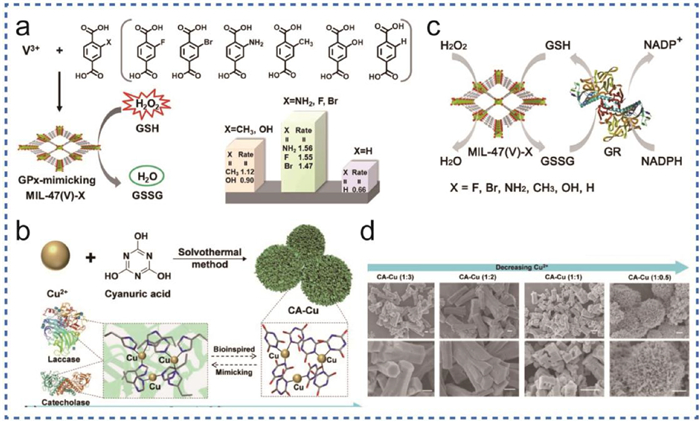
Figure 3 (a) Schematic diagram of synthesis of MIL-47(V)-X MOFs with nanozyme activity by microwave synthesis. (b) Schematic diagram of synthesis of CA-Cu nanozymes with enzyme-like activity. (c) Schematic diagram of MIL-47(V)-X MOF mimicking glutathione reductase. (d) SEM image diagram of CA-Cu nanozyme. (a, c) Reproduced with permission [53]. Copyright 2020, Wiley-VCH. (b, d) Reproduced with permission [48]. Copyright 2022, Elsevier.

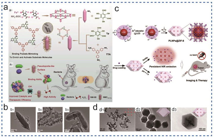
Figure 4 (a) Synthesis of NMCTP-TTA nano-MOF with nanozyme activity and its application to inhibit damage by harmful bacteria in mice. (b) TEM data images of (b1) NMCTTATFB, (b2) NMCTP-TAPB, and (b3) NMCTTAP-TFB. (c) A schematic diagram of the synthesis of PLNPs@ZIF-8 and the study of infrared imaging of drug release. (d) The TEM image of PLNPs@ZIF-8. PLNPs (d1), ZIF-8 (d2) and PLNPs@ZIF-8 (d3). (a, b) Reproduced with permission [63]. Copyright 2020, Wiley-VCH. (c, d) Reproduced with permission [60]. Copyright 2019, Elsevier.

Figure 5 (a) Schematic diagram of the synthesis of stratified micro and mesoporous zeolite imidazole framework (HZIFs) by template method. (b) TEM images of different materials: (b1) mesoporous HZIF-8 (0.1), (b2) microporous ZIF-8 prepared using conventional solvothermal method, (b3) mesoporous HZIF-8 (0.075), (b4) mesoporous HZIF-8 (0.05), (b5) GOx-HRP@HZIF-8 (0.1), (b6) GOx-HRP-on-ZIF-8. (c) Schematic diagram of n-CeO2 NSs synthesized by Ce MOFs through pyrolysis. (d) Schematic illustration of nanoenzyme activity of CeO2 nanoparticles. (e) SEM (e1) and TEM (e2) images of n-CeO2 NSs. (a, b) Reproduced with permission [62]. Copyright 2019, WILEY-VCH. (c, d, e) Reproduced with permission [73]. Copyright 2018, American Chemical Society.

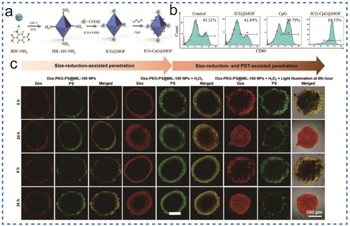
Figure 6 (a) A composite diagram of ICG-CpG@MOF. (b) Flow cytometry was used to detect the expression of CD80 in peritoneal macrophages with different MOF materials. (c) Schematic diagram of the penetration effect of Dox-PEG-PS@MIL-100 NPs of different sizes into cells at different times. (a, b) Reproduced with permission [81]. Copyright 2020, Elsevier. (c) Reproduced with permission [82]. Copyright 2021, Wiley-VCH.

Figure 7 (a) Synthesis and encapsulation of 5-ASA@Ce-MOF@PSS. (b) Synthesis of Ru-MOF and schematic diagram for the treatment of colitis. (c) SEM morphology changes of Ce-MOF before and after H2O2 treatment. (d) Images of complete colons isolated from mice after colitis modeling in different experimental groups. (e) H&E staining was used to detect colonic tissue characterization of mice in different experimental groups. (f) Quantitative study on the detection of MPO positive cells in mouse liver after different treatment groups. (g–i) Serum was used to quantitatively detect the influence of different experimental groups on the levels of inflammatory cytokines TNF-α, IL-1β and IL-6 secreted by macrophages in vivo. (a, c) Reproduced with permission [104]. Copyright 2020, Wiley-VCH. (b, d, e, f, g, h, i) Reproduced with permission [102]. Copyright 2022, Elsevier.

Figure 8 (a) Schematic diagram of the synthesis of Prussian blue nanozyme. (b) Therapeutic mechanism of OA. (c) Schematic diagram of saffron O detecting the efficacy of MOF@HA@PCA in the treatment of OA. (d) Data diagram of TEM experimental result of Prussian blue nanozymes. (e) The effect of Prussian blue nanozyme on iNOS protein expression was detected by immunofluorescence. (f) IVIS imaging of nano MOF in vivo after injection through the articular cavity. (a, d, e) Reproduced with permission [128]. Copyright 2021, Elsevier. (f) Reproduced with permission [111]. Copyright 2019, American Chemical Society. (b) Reproduced with permission [113]. Copyright 2020, Elsevier.

Figure 9 (a) A schematic diagram of the synthesis of Ag-MOF from CD-MOF guided by template method. (b) Schematic diagram of the preparation of biomedical dressings for wound healing by in-situ growth of Ag-MOF combined with PCL/GEL loaded with the anti-inflammatory drug curcumin. (c) Research schematic diagram of the treatment of diabetic wound with biological dressing prepared by self-assembly method of CCM@ZIF-8 MOFs combined with electrostatic spinning. (d) Data graph of wounds treated with different materials at regular intervals to detect wound healing. (e) Electron microscopy data diagram of materials with different preparation processes. SEM data of ZIF-8 (e1) and CCM@ZIF-8 (e2), SEM of NW (e3) and CZ-PT (e5), TEM data of NW (e4) and CZ-PT (e6). (f) H&E detected wound healing on the 3rd to 14th day treated with different materials. (a, d, f) Reproduced with permission [143]. Copyright 2022, Wiley. (b) Reproduced with permission [59]. Copyright 2022, Elsevier. (c, e) Reproduced with permission [140]. Copyright 2020, Elsevier.

 扫一扫看文章

扫一扫关注我们
 DownLoad:
 下载:
下载: