Citation:
Ying WANG, Rui XUE, Sheng-Li HUANG, Guo-Yu YANG. Luminescent heterobimetallic probes for selectively locating DNA in the nucleus and mitochondria of live cells[J]. Chinese Journal of Inorganic Chemistry,
2023, 39(8): 1463-1470.
doi:
10.11862/CJIC.2023.124
Received Date:
26 March 2023 Revised Date:
20 May 2023 Available Online:
10 August 2023
Abstract:
Nucleus probes for real-time imaging of DNA in live cells are very rare. Herein, the combined use of Ru(N^N)3, Ir(C^N)2(N^N), Re(CO)3Cl(N^N), and Pt(C^N)(N^N) produced four heterobimetallic complexes. Two different functional metal sites are arranged at both ends of linear bis-chelating linkers (N^N)-(N^N), leading to asymmetric dumbbell-like molecules M1-M2, namely, Ru-Re, Ru-Pt, Ir-Re, and Ir-Pt. The red emissive probes of Ru-Re and Ru-Pt offer the benefits of large Stokes shift, excellent nucleus membrane penetration, and DNA binding ability with good photostability. Moreover, DNA imaging of Ru-Re and Ru-Pt probes was comparable to the commercial dye of Hoechst 33342 which is specially used for staining DNA in the nucleus of live cells. However, the Ir-based probes of Ir-Re and Ir-Pt directly targeted mitochondria. The emission properties and selective intracellular localizations of different probes are highly dependent on the metal species used in the heterobimetallic complexes.
In the eukaryotic nucleus, DNA is tightly wrapped around histones in the form of chromosomes, each of which contains the instruction for making protein. The visualization of DNA facilitates its structural and functional study inside the nucleus[1]. In this regard, fluorescence microscopy has become a powerful tool in visualizing DNA due to the use of fluorescent dye with accurate DNA binding[2-6]. Currently, commercial fluorescent dyes such as Hoechst 33342 and DAPI are wildly used for staining DNA in the nucleus, however, they always suffer from small Stokes shifts, low photostability, and weak cell penetration.
The photoluminescent metal-containing complexes have large Stokes shifts, good photostability, and membrane permeability, that have been widely applied as biological probes[7-11]. In particular, the heteronuclear complexes can not only provide rich emission tuning but also combine the identity of different metal centers into one complex, promisingly producing probe candidates with exciting emission properties, improved cellular uptake, and selective cellular localization. In contrast to the well-developed d-f heteronuclear system[12-16], only a few d-d heteronuclear complexes were reported[17-21] because of the similar coordination identity of d-metal centers commonly resulting in the formation of homonuclear complexes. The d-metal-based units of Ru(N^N)3[22], Ir(C^N)2(N^N)[23], Re(CO)3Cl(N^N)[24], and Pt(C^N)(N^N)[25] are the four most widely used photosensitive moieties due to their important photoluminescent nature with long lifetimes and high yields, as well as the ligand-associated photoluminescence tuning. They have varied photo-excited states, emission spectrum, and cellular affinity, and the combination of different units mayendow the heteronuclear complexes with novel properties[26].
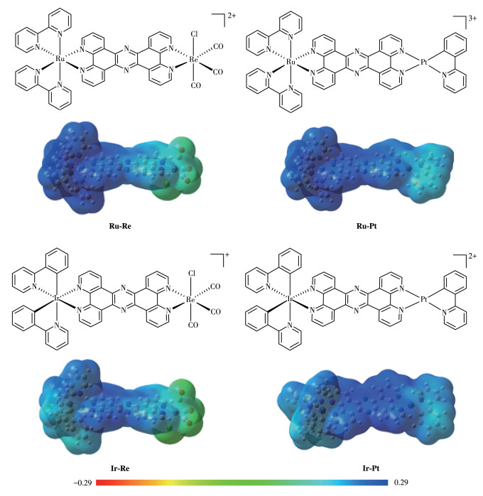
In pursuit of heteronuclear DNA probes, the conjugate tpphz (tpphz=tetrapyrido[3, 2-a: 2′, 3′-c: 3″, 2″-h: 2‴, 3‴-j] phenazine) should be an inborn bridge linker because of its highly efficient electron intercommunication and mild synthetic condition from the condensation of M1-(5, 6-diamino-1, 10-phenanthroline) and M2-(5, 6-dione-1, 10-phenanthroline). Furthermore, the nitrogen-rich and planar structure of tpphz make it easily intercalate DNA and intrigue the light-switching effect[27-29]. Che et al. reported that the Pt(C^N)-tpphz scaffold could effectively interact with the terminal face of the telomeric G-quadruplex[25]. LuminescentRe(CO)3(pyridine)(N^N) complex was explored forduplex DNA imaging[30]. Thomas and co-workers reported mononuclear Ru-tpphz[31] and homonuclear Ru-tpphz-Ru[29, 32] (Fig.S1, Supporting information) can stain nucleus DNA, but with high concentration and long incubation time. The mononuclear Ir-tpphz and homonuclear Ir-Ir were also reported for staining the nucleus and mitochondria, respectively[33]. The reported homonuclear probes need high concentration and long incubation time, and they only image in fixed cells. Promoting the uptake of probes in live cells whilst remaining nucleus targeting is still a significant challenge. Herein, the combined use of Ru(N^N)3, Ir(C^N)2(N^N), Re(CO)3Cl(N^N), and Pt(C^N)(N^N) produced four heterobimetallic complexes M1-tpphz-M2, whereM1=Ru(Ⅱ) or Ir(Ⅲ), and M2=Re(Ⅰ) or Pt(Ⅱ), namely, Ru-Re, Ru-Pt, Ir-Re, and Ir-Pt (Fig. 1). Through this metal hybridization, we hope to achieve (ⅰ) photoluminescence tuning, (ⅱ) improved nucleus DNA bindingaffinity, (ⅲ) selective cellular localization, and (ⅳ) highly efficient bio-imaging in live cells.
Figure 1
Figure 1.
Structures of heterobimetallic probes Ru-Re, Ru-Pt, Ir-Re, and Ir-Pt and corresponding molecular electron density surface (isovalue=0.0004) mapped with electrostatic potential (ESP)
The most positive potential regions are shown in deep blue.
Heterobimetallic complexes (M1-tpphz-M2) were readily obtained from two different synthetic routes. Mononuclear and homonuclear complexes (Ru-tpphz, Ir-tpphz, Ru-Ru, and Ir-Ir) were prepared according to the literature[31, 33]. Re(CO)5Cl was added to Ru-tpphz solution to reflux for 8 h. After refluxing, the solvent was evaporated. The crude product was dissolved into acetonitrile, filtered, and recrystallized with diethyl ether several times. The solids were dried in a vacuum oven to give dark red Ru-Re. The syntheses of Ru-Pt, Ir-Re, and Ir-Pt were similar to Ru-Re. The details of the synthetic steps can be found in the Supporting information (Scheme S1).
2.
Results and discussion
Heterobimetallic structures of M1-tpphz-M2 were characterized by 1H NMR (Fig.S1-S4) and 13C NMR. The HRMS spectra gave further evidence for the existence of M1-tpphz-M2 (Fig.S5-S8). These peaks correspond to an intact entity with different charge states. Specifically, the peaks at m/z=1 253.015 0, 648.067 5, 1 191.114 3, and 1 383.189 4, corresponding to [M-OTf]+ for Ru-Re, [M-2OTf]2+ for Ru-Pt, [M-OTf]+ for Ir-Re, and [M-OTf]+ for Ir-Pt, respectively, were observed. These peaks were isotopically resolved, which were in line with their calculated theoretical distributions.
In UV-Vis spectra, tpphz exhibited sharp absorption bands at 273 nm with a molar absorption coefficient (ε) of 1.37×10⁴ L·mol-1·cm-1. After metal coordination, metal-centered (MC) and MLCT (metal-to-ligand charge transfer) transitions led to the increased ε. The lowest energy bands (π-π*) of Ru-Re, Ru-Pt, Ir-Re, and Ir-Pt had a slightly red shift with ε=1.53×10⁴ L·mol-1·cm-1 (280 nm), 2.44×10⁴ L·mol-1·cm-1 (281 nm), 1.98×10⁴ L·mol-1·cm-1 (277 nm) and 1.9×10⁴ L·mol-1·cm-1 (277 nm), respectively (Fig.S9). Furthermore, MLCT transitions of Ru-Re and Ru-Pt over 400 nm were observed as well, which could effectively avoid the overlap with the co-staining commercial nuclei dye Hoechst 33342. Photoexcitation of Ru-Re, Ru-Pt, and Ir-Re had a broad featureless peak at about 600 nm, whereas Ir-Pt resulted in an emission band with a prominent shoulder at 470 nm and a broad shoulder at 580 nm (Fig.S10). A ca. 200 nm Stokes shift for four heterobimetallic complexes was observed, which provides an inborn advantage for cell imaging probes.
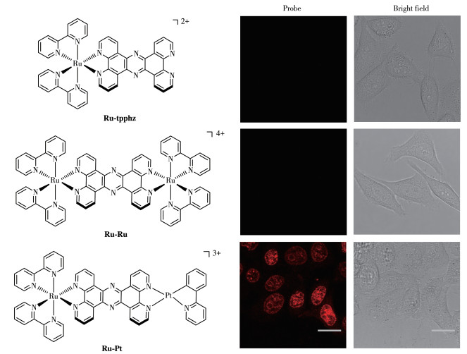
To compare the cellular uptake abilities among mononuclear, homonuclear, and heteronuclear Ru-tpphz analogues, live HeLa cells were treated with Ru-tpphz, Ru-Ru, and Ru-Pt in a 6-well plate at the concentration of 50 μmol·L-1 for 20 min. The confocal scanning laser microscope (CLMS) imaging of Ru-tpphz and Ru-Ru did not capture any signal, whereas Ru-Pt completely stained nucleus DNA with redsignals (Fig. 2). Experimentally, heterobimetallic probeRu-Pt exhibited better nucleus membrane permeation and DNA binding ability than mononuclear Ru-tpphz (200 μmol·L-1, 1h in fixed cells)[31] and homonuclear Ru-Ru (500 μmol·L-1, 1 h in fixed cells)[32]. The comparison in cellular uptake ability among Ir-tpphz analogues was carried out as well. When HeLa cells were treated with 25 μmol·L-1Ir-tpphz, Ir-Ir, and Ir-Pt for 15 min, all probes only were accumulated in the cytoplasm (Fig.S11). Cell uptake results show that (ⅰ) heteronuclear Ru-Pt is more effective in staining nucleus DNA than Ru-tpphz and Ru-Ru; (ⅱ) Ru-tpphz analogues could stain nucleus DNA while Ir-tpphz analogues stay in the cytoplasm.
Figure 2
Figure 2.
Cell labeling with mononuclear Ru-tpphz (50 μmol·L-1), homonuclear Ru-Ru (50 μmol·L-1), and heterobimetallic Ru-Pt (50 μmol·L-1) for 20 min in HeLa cell
The confocal microscopy images were collected at an excitation wavelength of 470 nm; All scale bars are 20 μm.
To study the impact of metal moiety on intracellular localization, density functional theory (DFT) calculation was performed in all heterobimetallic complexes (Fig. 1). Compared with Re(CO)3Cl(N^N) and Pt(C^N)(N^N) moieties, Ru(N^N)3 moiety is more electron deficient, resulting in the imbalanced electron density in Ru-Re and Ru-Pt. The hugely uneven electron distribution of Ru-Re and Ru-Pt should be responsible for their efficient crossing nucleus membrane and specified DNA location. In combination with cell imagingresults, although Ru-tpphz, Ru-Ru, Ru-Re, and Ru-Pt can target nucleus DNA under different conditions, the required concentration and incubation time of heterobimetallic complexes Ru-Re and Ru-Pt were decreased. This facilitates the heterobimetallic nucleus probe to be applied in other bio-imaging studies. In contrast to +2 charged Ru(N^N)3, +1 charged Ir(C^N)2(N^N) unit decreased electron distribution of two terminal metal centers in Ir-Re and Ir-Pt. The relatively even electron distribution of Ir-Re and Ir-Pt increased electrostatic interactions between probes and the surface of mitochondria, leading to their preferable staying in mitochondria. The electron distribution of the probe should be a key factor to determine its selective cellular targeting.
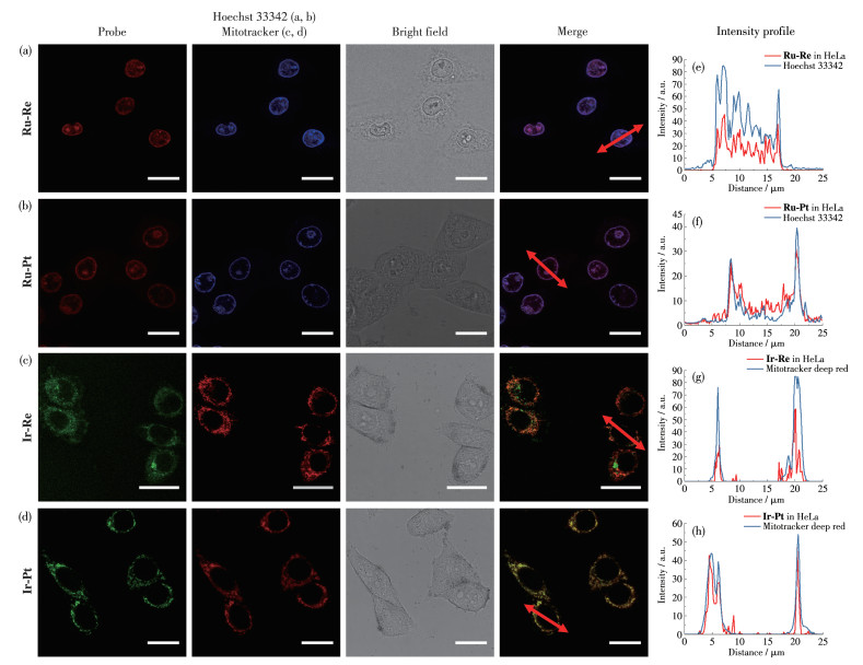
Cellular uptake experiments of Ru-Re, Ru-Pt, Ir-Re, and Ir-Pt were carried out for making a comparison with commercial dye. Intense emissive signals of Ru-Pt and Ru-Re were detected in the nuclei region as a result of successfully intercalating DNA and intriguing light-switching effect (Fig. 3a, 3b, and S12). The red emissive spots from Ru-Pt and Ru-Re were almost completely colocalized with the luminescence of Hoechst 33342 which could bind to the minor groove of double-stranded DNA. The Pearson′s R-values for both the emissions of heterobimetallic probes and Hoechst 33342 were over 0.80. In addition, intensity profile curves of Ru-Re and Ru-Pt strongly agreed with Hoechst 33342 under co-staining conditions (Fig. 3e and 3f). Thus, heterobimetallic complexes Ru-Pt and Ru-Re are alternative nucleus DNA probes of live cells. Interestingly, at 75 μmol·L-1 above the concentration of Ru-Re, most nuclei were stained very bright within 20 min, making it impossible to obtain Hoechst33342 co-staining, because Ru-Re preferably occupies all DNA binding sites.
Figure 3
Figure 3.
(a, b) Cell labeling with Ru-Re (50 μmol·L-1) and Ru-Pt (50 μmol·L-1) for 20 min and further incubating with Hoechst 33342 for 15 min in HeLa cell; (c, d) Cell labeling with Ir-Re (25 μmol·L-1) and Ir-Pt (25 μmol·L-1) for 15 min and further incubating with Mitotracker Deep Red for 15 min in HeLa cell; (e-h) Intensity profile of selected region
The confocal microscopy images were collected at excitation wavelengths of 405 nm (Ir-Re, Ir-Pt, and Hoechst 33342), 470 nm (Ru-Re and Ru-Re), and 500 nm (Mitotracker Deep Red); All scale bars are 20 μm.
Different from Ru-Pt and Ru-Re, as shown in Fig. 3c, 3d, and S13, Ir-Re and Ir-Pt only accumulated in the cytoplasm. Co-staining was conducted with Mitotracker Deep Red which exhibited a deep red emission but an excitation wavelength (> 500 nm) that was well separated from the Ir-Pt and Ir-Re. In live HeLa and MCF-7 cells, strong colocalization between Ir-Pt and Mitotracker was observed at 25 μmol·L-1 concentration with Pearson′s R-value of 0.85 and 0.83, respectively. Co-staining between Ir-Re and Mitotracker yielded Pearson′s R-values of 0.76 in HeLa cells and 0.75 in MCF-7 cells. The obtained results show that probes Ir-Re and Ir-Pt entered the mitochondria of live cells.
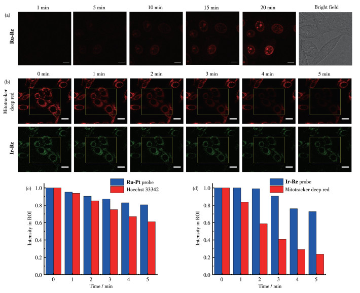
To evaluate the cytotoxicity of all heteronuclear probes, the MTT assay was performed in HeLa and MCF-7. As shown in Fig.S17, four heterobimetallic probes were toxic at 200 μmol·L-1 concentration overnight. In the above cell imaging studies, the staining conditions were under 50 μmol·L-1Ru-Re and Ru-Pt for 20 min of incubation and 25 μmol·L-1Ir-Re and Ir-Pt for 15 min of incubation. This condition did not cause significant cytotoxicity. The kinetic tracking of nucleus staining with Ru-Re and mitochondria staining with Ir-Pt was respectively monitored by continuous imaging of live HeLa cells. In a live imaging chamber under 37 ℃ and a CO2 volume fraction of 5%, weak luminescence was observed after incubation with Ir-Pt in 1 min (Fig.S14). With increasing time, a continuous accumulation in mitochondria was observed. At the 15th minute, the emission from Ir-Pt was bright enough to obtain a high-quality image. In contrast, the nucleus DNA probe Ru-Re generated emission signals not as rapidly as the mitochondria probe Ir-Pt. Due to the weak and dark signal of Ru-Re, we didn′t capture any signal from the membrane to the cytoplasm (Fig. 4a). From the 5th minute, the red emission signal gradually increased at the nucleus area until the 20th minute.
Figure 4
Figure 4.
(a) Time-lapse imaging in situ of HeLa cells incubated with Ru-Re (50 μmol·L-1); (b) Photostability imaging of Mitotracker Deep Red and Ir-Pt at the square window in HeLa cells; (c) Intensity fraction of Hoechst 33342 and Ru-Pt at different irradiation times; (d) Intensity fraction of Mitotracker Deep Red and Ir-Pt at different irradiation times
Excitation wavelengths were 405 nm for Ir-Pt and Hoechst 33342, 470 nm for Ru-Pt and Mitotracker Deep Red; All scale bars are 20 μm.
To investigate the cellular uptake mechanism of nucleus probe Ru-Re, HeLa cells were incubated under four different conditions (Fig.S16). When incubated at 4 ℃ for 20 min, no emissive signal was detected. The endocytic inhibitors chloroquine and NH4Cl were used to explore the contribution of the endocytic pathway for Ru-Re uptake[34], and the cell imagingexhibited the luminescence intensity in the nucleus was not affected in the presence of endocytic inhibitors. Colchicine, which disrupts the polymerization of microtubules and prevents membrane and endosomal trafficking, didn′t show any inhibition of Ru-Re uptake. Thus, all results suggest that the cellular uptake ofnucleus probe Ru-Re passes through an energy-dependent and non-endocytic pathway.
Heterobimetallic probes derived from the mechanism of phosphorescence exhibited excellent photostability compared with commercial organic dye[35-37], in particular, under intense laser conditions. Ir-Re and Ru-Pt were selected to study the photobleaching effect. As shown in Fig. 4b and 4d, the square region was continuously irradiated using double channels: 405 nm 30% laser for Ir-Re and 620 nm 30% laser for Mitotracker Deep Red. During 5 min, the luminescentintensity of Mitotracker Deep Red was reduced to below 25%, whereas Ir-Re kept over 65% luminescent intensity. The same trends were found in comparison nucleus probe Ru-Pt with Hoechst 33342 (Fig. 4c, S15). Thus, heterobimetallic probes are more suitable for high-intensity and continuous imaging than organic dye.
3.
Conclusions
In conclusion, a set of heterobimetallic probes were successfully synthesized and achieved with high-quality imaging of mitochondria and nucleus DNA in live cells. These heterobimetallic probes possess large Stokes shifts and good photostability compared to organic probes, and they are promising tools for the cell imaging study. The controlled study of heterobimetallic probes Ru-Re and Ru-Pt showed excellentnucleus DNA imaging ability of live cells over mononuclear and homonuclear probes. In a co-stain study with commercial dye Mitotracker Deep Red and Hoechst 33342, the four probes exhibited good colocalization images. Especially, Ru-Re and Ru-Pt showed very good overlay with Hoechst 33342 in live cell nuclei, which could enable the probe to locate nucleus DNA in various biological studies. Moreover, Ru-Re and Ru-Pt selectively image DNA in the nucleus, however, Ir-Re and Ir-Pt preferably stain mitochondria. The exploration of heterobimetallic probes provides more ideas for how to design probes with high cellular uptake and selective DNA targeting in live cells.
Supporting information is available at http://www.wjhxxb.cn
[1]
Zeraati M, Langley D B, Schofield P, Moye A L, Rouet R, Hughes W E, Bryan T M, Dinger M E, Christ D. I-motif DNA structures are formed in the nuclei of human cells[J]. Nat. Chem.,
2018, 10:
631-637.
doi: 10.1038/s41557-018-0046-3
[2]
Wu Z K, Fan H H, Satyavolu N S R, Wang W J, Lake R, Jiang J H, Lu Y. Imaging endogenous metal ions in living cells using a DNAzyme-catalytic hairpin assembly probe[J]. Angew. Chem. Int. Ed.,
2017, 56:
8721-8725.
doi: 10.1002/anie.201703540
[3]
Peng X J, Wu T, Fan J L, Wang J Y, Zhang S, Song F L, Sun S G. An effective minor groove binder as a red fluorescent marker for live-cell DNA imaging and quantification[J]. Angew. Chem. Int. Ed.,
2011, 50:
4266-4269.
[4]
Rieder U, Luedtke N W. Alkene-tetrazine ligation for imaging cellular DNA[J]. Angew. Chem. Int. Ed.,
2014, 53:
9322-9326.
[5]
Zhang L, Chu M G, Ji C L, Wei J W, Yang Y B, Huang Z N, Tan W H, Tan J, Yuan Q. In situ visualization of epidermal growth factor receptor nuclear translocation with circular bivalent aptamer[J]. Anal. Chem.,
2022, 94(50):
17413-17421.
doi: 10.1021/acs.analchem.2c02762
[6]
Zeng W, Lin M H, Zhu L Y, Lin M J. A triphenylphosphonium functionalized AIE conjugated macrocyclic tetramaleimide for mitochondrial-targeting bioimaging[J]. Chin. J. Chem.,
2022, 40(1):
39-45.
doi: 10.1002/cjoc.202100580
[7]
Huang H, Yu B, Zhang P Y, Huang J J, Chen Y, Gasser G, Ji L N, Chao H. Highly charged ruthenium(Ⅱ) polypyridyl complexes as lysosome-localized photosensitizers for two-photon photodynamic therapy[J]. Angew. Chem. Int. Ed.,
2015, 54:
14255-14258.
[8]
Liu J P, Zhang C, Rees T W, Ke L B, Ji L N, Chao H. Harnessing ruthenium(Ⅱ) as photodynamic agents: Encouraging advances in cancer therapy[J]. Coord. Chem. Rev.,
2018, 363:
17-28.
doi: 10.1016/j.ccr.2018.03.002
[9]
Wang J, Xue J, Yan Z, Zhang S C, Qiao J, Zhang X R. Photoluminescence lifetime imaging of synthesized proteins in living cells using an iridium-alkyne probe[J]. Angew. Chem. Int. Ed.,
2017, 56:
14928-14932.
doi: 10.1002/anie.201708566
[10]
欧阳艾, 罗雨珩, 陆农, 胡仁涛, 张平玉, 张黔玲. 基于粘度响应的线粒体靶向铱(Ⅲ)配合物用于肿瘤的光动力治疗[J]. 无机化学学报,
2021,37,(3): 401-411.
OUYANG A, LUO Y H, LU N, HU R T, ZHANG P Y, ZHANG Q L. Viscosity responsive and mitochondria targeted iridium(Ⅲ) complexes for photodynamic therapy of tumors[J]. Chinese J. Inorg. Chem.,
2021, 37(3):
401-411.
[11]
陈曦, 李思媛, 王元, 吴伟娜, 陈忠. 一种席夫碱Al3+荧光探针的制备及细胞成像应用[J]. 无机化学学报,
2022,38,(10): 1993-1998.
doi: 10.11862/CJIC.2022.190CHEN X, LI S Y, WANG Y, WU W N, CHEN Z. A Schiff base fluorescent probe for Al3+: Synthesis and application in living cells imaging[J]. Chinese J. Inorg. Chem.,
2022, 38(10):
1993-1998.
doi: 10.11862/CJIC.2022.190
[12]
Huang S L, Jia A Q, Jin G X. Pd(diimine)Cl2 embedded heterometallic compounds with porous structures as efficient heterogeneous catalysts[J]. Chem. Commun.,
2013, 49:
2403-2405.
doi: 10.1039/c3cc38714c
[13]
Plecnik C E, Liu S, Shore S G. Lanthanide-transition-metal complexes: From ion pairs to extended arrays[J]. Acc. Chem. Res.,
2003, 36:
499-508.
doi: 10.1021/ar010050o
[14]
Szeto K C, Kongshaug K O, Jakobsen S, Tilset M, Lillerud K P. Design, synthesis and characterization of a Pt-Gd metal-organic framework containing potentially catalytically active sites[J]. Dalton Trans.,
2008, (15):
2054-2060.
doi: 10.1039/b719766g
[15]
Tan Y X, Yang X, Li B B, Yuan D Q. Rational design of a flu-type heterometallic cluster-based Zr-MOF[J]. Chem. Commun.,
2016, 52:
13671-13674.
doi: 10.1039/C6CC08191F
[16]
Kobayashi A, Suzuki Y, Ohba T, Noro S I, Chang H C, Kato M. Ln-Co-based rock-salt-type porous coordination polymers: Vapor response controlled by changing the lanthanide ion[J]. Inorg. Chem.,
2011, 50:
2061-2063.
doi: 10.1021/ic102361d
[17]
Huang S L, Jin G X. Selective CO2 capture by a 3d-4d coordination polymer material with 1D channel[J]. CrystEngComm,
2011, 13:
6013-6016.
doi: 10.1039/c1ce05474k
[18]
Huang S L, Lin Y J, Hor T S A, Jin G X. Cp*Rh-based heterometallic metallarectangles: size-dependent borromean link structures and catalytic acyl transfer[J]. J. Am. Chem. Soc.,
2013, 135:
8125-8128.
doi: 10.1021/ja402630g
[19]
Huang S L, Liu N, Ling Y, Luo H K. IrⅢ-based octahedral metalloligands derived primitive cubic frameworks for enhanced CO2/N2 separation[J]. Chem.-Asian J.,
2017, 12:
3110-3113.
doi: 10.1002/asia.201701339
[20]
Huang S L, Hor T S A, Jin G X. Metallacyclic assembly of interlocked superstructures[J]. Coord. Chem. Rev.,
2017, 333:
1-26.
doi: 10.1016/j.ccr.2016.11.009
[21]
Huang S L, Lin Y J, Li Z H, Jin G X. Self-assembly of molecular borromean rings from bimetallic coordination rectangles[J]. Angew. Chem. Int. Ed.,
2014, 53:
11400-11404.
[22]
Burke C S, Byrne A, Keyes T E. Highly selective mitochondrial targeting by a ruthenium(Ⅱ) peptide conjugate: Imaging and photoinduced damage of mitochondrial DNA[J]. Angew. Chem. Int. Ed.,
2018, 57:
12420-12424.
doi: 10.1002/anie.201806002
[23]
Lai P, Brysacz C H, Alam M K, Ayoub N A, Gray T G, Bao J M, Teets T S. Highly efficient red-emitting bis-cyclometalated iridium complexes[J]. J. Am. Chem. Soc.,
2018, 140:
10198-10207.
doi: 10.1021/jacs.8b04841
[24]
Hedstrom S, Chaudhuri S, Porte N T L, Rudshteyn B, Martinez J F, Wasielewski M R, Batista V S. Thousandfold enhancement of photoreduction lifetime in Re(bpy)(CO)3 via spin-dependent electron transfer from a perylenediimide radical anion donor[J]. J. Am. Chem. Soc.,
2017, 139:
16466-16469.
doi: 10.1021/jacs.7b09438
[25]
Ma D, Che C, Yan S. Platinum(Ⅱ) complexes with dipyridophenazine ligands as human telomerase inhibitors and luminescent probes for G-quadruplex DNA[J]. J. Am. Chem. Soc.,
2009, 131:
1835-1846.
doi: 10.1021/ja806045x
[26]
Chen Y, Guan R, Zhang C, Huang J J, Ji L N, Chao H. Two-photon luminescent metal complexes for bioimaging and cancer phototherapy[J]. Coord. Chem. Rev.,
2016, 310:
16-40.
doi: 10.1016/j.ccr.2015.09.010
[27]
Baggaley E, Gill M R, Green N H, Turton D, Sazanovich I V, Botchway S W, Smythe C, Haycock J W, Weinstein J A, Thomas J A. Dinuclear ruthenium(Ⅱ) complexes as two-photon, time-resolved emission microscopy probes for cellular DNA[J]. Angew. Chem. Int. Ed.,
2014, 53:
3435-3439.
[28]
Holmberg R C, Thorp H H. Digital simulation of catalytic cyclic voltammograms for oxidation of DNA by a heterobimetallic dimer: Effects of DNA binding and mass transport[J]. Anal. Chem.,
2003, 75:
1851-1860.
doi: 10.1021/ac0204653
[29]
Wragg A, Gill M R, Turton D, Adams H, Roseveare T M, Smythe C, Su X D, Thomas J A. Tuning the cellular uptake properties of luminescent heterobimetallic iridium(Ⅲ)-ruthenium(Ⅱ) DNA imaging probes[J]. Chem.-Eur. J.,
2014, 20:
14004-14011.
doi: 10.1002/chem.201403693
[30]
Yam V W W, Lo K K W, Cheung K K, Kong R Y C. Deoxyribonucleic acid binding and photocleavage studies of rhenium(Ⅰ) dipyridophenazine complexes[J]. J. Chem. Soc.-Dalton Trans.,
1997, (12):
2067-2072.
doi: 10.1039/a700828g
[31]
Gill M R, Derrat H, Smythe C G W, Battaglia G, Thomas J A. Ruthenium(Ⅱ) metallo-intercalators: DNA imaging and cytotoxicity[J]. ChemBioChem,
2011, 12:
877-880.
doi: 10.1002/cbic.201000782
[32]
Gill M R, Garcia-Lara J, Foster S J, Smythe C, Battaglia G, Thomas J A. A ruthenium(Ⅱ) polypyridyl complex for direct imaging of DNA structure in living cells[J]. Nat. Chem.,
2009, 1:
662-667.
doi: 10.1038/nchem.406
[33]
Liu S, Liang H, Zhang K Y, Zhao Q, Zhou X B, Xu W J, Huang W. A multifunctional phosphorescent iridium(Ⅲ) complex for specific nucleus staining and hypoxia monitoring[J]. Chem. Commun.,
2015, 51:
7943-7946.
doi: 10.1039/C5CC01978H
[34]
Ziegler H K, Unanue E R. Decrease in macrophage antigen catabolism caused by ammonia and chloroquine is associated with inhibition of antigen presentation to T cells[J]. Proc. Natl. Acad. Sci. U. S. A.,
1982, 79:
175-178.
doi: 10.1073/pnas.79.1.175
[35]
Nicol A, Qin W, Kwok R T K, Burkhartsmeyer J M, Zhu Z F, Su H F, Luo W W, Lam J W Y, Qian J, Wong K S, Tang B Z. Functionalized AIE nanoparticles with efficient deep-red emission, mitochondrial specificity, cancer cell selectivity and multiphoton susceptibility[J]. Chem. Sci.,
2017, 8:
4634-4643.
doi: 10.1039/C7SC00908A
[36]
Xu W, Zeng Z B, Jiang J H, Chang Y T, Yuan L. Discerning the chemistry in individual organelles with small-molecule fluorescent probes[J]. Angew. Chem. Int. Ed.,
2016, 55:
13658-13699.
doi: 10.1002/anie.201510721
[37]
Yuan Y Y, Xu S D, Zhang C J, Liu B. Light-responsive AIE nanoparticles with cytosolic drug release to overcome drug resistance in cancer cells[J]. Polym. Chem.,
2016, 7:
3530-3539.
doi: 10.1039/C6PY00449K
Figure 1
Structures of heterobimetallic probes Ru-Re, Ru-Pt, Ir-Re, and Ir-Pt and corresponding molecular electron density surface (isovalue=0.0004) mapped with electrostatic potential (ESP)
The most positive potential regions are shown in deep blue.
Figure 2
Cell labeling with mononuclear Ru-tpphz (50 μmol·L-1), homonuclear Ru-Ru (50 μmol·L-1), and heterobimetallic Ru-Pt (50 μmol·L-1) for 20 min in HeLa cell
The confocal microscopy images were collected at an excitation wavelength of 470 nm; All scale bars are 20 μm.
Figure 3
(a, b) Cell labeling with Ru-Re (50 μmol·L-1) and Ru-Pt (50 μmol·L-1) for 20 min and further incubating with Hoechst 33342 for 15 min in HeLa cell; (c, d) Cell labeling with Ir-Re (25 μmol·L-1) and Ir-Pt (25 μmol·L-1) for 15 min and further incubating with Mitotracker Deep Red for 15 min in HeLa cell; (e-h) Intensity profile of selected region
The confocal microscopy images were collected at excitation wavelengths of 405 nm (Ir-Re, Ir-Pt, and Hoechst 33342), 470 nm (Ru-Re and Ru-Re), and 500 nm (Mitotracker Deep Red); All scale bars are 20 μm.
Figure 4
(a) Time-lapse imaging in situ of HeLa cells incubated with Ru-Re (50 μmol·L-1); (b) Photostability imaging of Mitotracker Deep Red and Ir-Pt at the square window in HeLa cells; (c) Intensity fraction of Hoechst 33342 and Ru-Pt at different irradiation times; (d) Intensity fraction of Mitotracker Deep Red and Ir-Pt at different irradiation times
Excitation wavelengths were 405 nm for Ir-Pt and Hoechst 33342, 470 nm for Ru-Pt and Mitotracker Deep Red; All scale bars are 20 μm.



 下载:
下载:

 下载:
下载:



