Table 1.
Selected Bond Lengths (Å) and Bond Angles (°)
Citation:
Wei-Sheng LIN, Jian-Gen HUANG, Yun-Xiang WEN, Hui LUO, Wen-Tong CHEN. Photophysical Performance and Energy Transfer Mechanism of a 1-D Chain-like Complex[J]. Chinese Journal of Structural Chemistry,
2020, 39(4): 747-755.
doi:
10.14102/j.cnki.0254-5861.2011-2486

Photophysical Performance and Energy Transfer Mechanism of a 1-D Chain-like Complex
English
Photophysical Performance and Energy Transfer Mechanism of a 1-D Chain-like Complex
Abstract:
By means of solvothermal reactions, a novel lanthanide-mercury compound, {[Ho(IA)3(H3O)2]2n[2n(HgCl4)][n(HgCl5)]}·3nH3O·nH2O (1, HIA = isonicotinc acid) was prepared and structurally characterized by single-crystal X-ray diffraction. Complex 1 crystallizes in the C2/c space group of monoclinic system with a = 24.2147(5), b = 20.8106(4), c = 15.3060(3) Å, β = 128.326(2)°, V = 6050.8(2) Å3, C36H45Cl13Hg3Ho2N6O20, Mr = 2274.26, Z = 4, Dc = 2.497 g/cm3, μ(MoKα) = 10.817 mm–1 and F(000) = 4232. It exhibits a one-dimensional (1-D) chain-like structure. Solid-state photoluminescence measurement shows that it displays brown light emission bands. These emission bands originate from the characteristic emissions of the 4f electrons intrashell transitions of 5S2 → 5I8 and 5F5 → 5I8 of the holmium(Ⅲ) ions in 1. The photoluminescence emission energy transfer mechanism is elucidated by the energy level diagrams of the holmium(Ⅲ) ions and isonicotinic acid ligand. Complex 1 has CIE chromaticity coordinates of (0.4361, 0.4992). Solid-state UV/Vis diffuse reflectance spectra reveal that it possesses wide optical band gap of 4.94 eV.
-
Key words:
- chromaticity coordinate
- / energy transfer
- / isonicotinc acid
- / lanthanide
- / Photoluminescence
-
1. INTRODUCTION
Lanthanide compounds have a wide range of applications, such as fluorescent probes, electrolumine-scent devices, magnetic materials, sensors, catalysts, cell imaging, and so on[1-5]. Therefore, lanthanide compounds have attracted more and more attention in recent decades. So far, a large number of lantha-nide compounds have been studied on their structures and physical and chemical properties[6-9]. The physical and chemical properties of lanthanide compounds are closely related to their 4f electrons.
For example, the reason that lanthanide compounds usually display photoluminescence is related to the transition of their 4f electrons between orbitals. If the 4f electrons of a lanthanide compound can have an effective transition, it is possible for it to show pho-toluminescence.
However, because the absorption coefficient of lanthanide ions is usually very low and cannot cause effective interorbital transition of 4f electrons, lanthanide compounds are usually unable to exhibit ideal photoluminescence. Therefore, in order to improve the absorption coefficient of lanthanide ions and make the transition between orbitals of 4f electrons possible, many researchers adopted a so-called "antenna effect" strategy[10, 11], that is, the use of organic ligands with conjugate structure, such as heterocyclic molecules, aromatic carboxylic acids, etc., as coordination ligands with lanthanide ions to synthesize novel compounds. The "antenna effect" is the use of these conjugated structures of organic molecules to absorb the energy of the excited light and transfer the energy to lanthanide ions, causing the latter to show photoluminescence. Compared with the photoluminescence of lanthanide com-pounds, the research on semiconductor properties of lanthanide compounds is very little, and it still needs to be studied[12].
As far as we know, isonicotinic acid (HIA) is an important organic ligand, which can be used as a useful building unit, because it has two carboxyl oxygen atoms at one end and one nitrogen atom at the other end. These chelating atoms give HIA the ability to coordinate with multiple metal centers, thus constructing extended structures with different coordination modes. It is believed that lanthanide compounds with HIA ligands should have interesting extended structures and novel physical and chemical properties. Based on the above considerations, we have been studying the crystal engineering of lanthanide HIA compounds in recent years[13]. In this paper, we report the solvothermal synthesis, single-crystal X-ray structure, photophysical performance, as well as energy transfer mechanism of a novel lanthanide-mercury compound, {[Ho(IA)3(H3O)2]2n-[2n(HgCl4)][n(HgCl5)]}·3nH3O·nH2O (1, HIA = isonicotinc acid). It is characterized by a 1-D chain-like structure and a three-dimensional (3-D) supramolecular network.
2. EXPERIMENTAL
2.1 General procedures
In this study, all reagents and chemicals for the synthesis of the compound was analytically pure, commercially purchased, and were not further purified before use. The infrared spectra were measured on a PE Spectrum-One FT-IR spectro-photometer with KBr pellets. The solid-state photo-luminescence was measured by F97XP photolumi-nescence spectrometer using powder samples. The solid-state UV-visible diffuse reflectance spectra were measured on the computer-controlled TU1901 UV/Vis spectrometer equipped with an integrating sphere in the wavelength range of 190~900 nm.
2.2 Synthesis of 1
Compound 1 was prepared by mixing HgCl2 (2 mmol, 542 mg), HoCl3·6H2O (1 mmol, 379 mg), isonicotinic acid (3 mmol, 369 mg), 0.1 mL HCl and 10 mL distilled water in a 25 mL stainless-steel Teflon-lined reactor. After heating at 433 K for 10 days, colorless bulk crystals were obtained when the temperature was reduced to room temperature. The crystals were filtered, cleaned with distilled water and obtained in the yield of 37% (based on holmium). IR peaks (cm−1): 3444(s), 3204(w), 3140(w), 3071(m), 2882(w), 2801(w), 1704(m), 1595(vs), 1502(w), 1416(vs), 1234(m), 848(m), 759(s), 680(m), 549(w) and 414(s).
2.3 X-ray structural determination
The single-crystal X-ray diffraction data of the title compound were measured on a SuperNova CCD X-ray diffractometer by using carefully selected suitable single crystals. The X-ray source is a gra-phite-monochromatic Mo-Kα ray with a wavelength of 0.71073 Å and the data were corrected by using CrystalClear software for data reduction and empiri-cal absorption. The single-crystal structure was resolved by direct methods using SHELXS software[14] and refined with a full-matrix least-squares refinement on F2. All non-hydrogen atoms were determined by difference Fourier peaks and refined using anisotropic refinement. All hydrogen atoms are identified by theoretical models and added to their parent atoms, and refined using assigned isotropic thermal parameters. Reflections measured are 16056; the final R = 0.0258 for 384 parameters and 4713 observed reflections with I > 2σ(I) and wR = 0.0617 (w = 1/[σ2(Fo2) + (0.0290P)2 + 42.1789P], where P = (Fo2 + 2Fc2)/3); S = 1.039, (Δρ)max = 1.235, (Δρ)min = –1.369 e/Å3 and (Δ/σ)max = 0.002. The selected bond distances and bond angles are shown in Table 1.
Table 1

 DownLoad:
CSV
DownLoad:
CSV
Bond Dist. Bond Dist. Hg(1)–Cl(1) 2.470(4) Ho(1)–O(1) 2.324(3) Hg(1)–Cl(2) 2.443(4) Ho(1)–O(3) 2.334(3) Hg(1)–Cl(2)#1 2.443(4) Ho(1)–O(4)#2 2.305(3) Hg(1)–Cl(3) 2.288(3) Ho(1)–O(2)#3 2.318(3) Hg(1)–Cl(3)#1 2.624(3) Ho(1)–O(5) 2.341(3) Hg(2)–Cl(4) 2.4799(16) Ho(1)–O(6)#3 2.350(3) Hg(2)–Cl(5) 2.4487(15) Ho(1)–O(1W) 2.460(3) Hg(2)–Cl(6) 2.4621(15) Ho(1)–O(2W) 2.434(3) Hg(2)–Cl(7) 2.5649(14) Angle (°) Angle (°) Cl(1)–Hg(1)–Cl(2) 90.75(9) O(4)#2–Ho(1)–O(2)#3 79.11(12) Cl(1)–Hg(1)–Cl(3) 123.06(8) O(4)#2–Ho(1)–O(1) 145.49(12) Cl(1)–Hg(1)–Cl(3)#1 110.38(7) O(2)#3–Ho(1)–O(1) 76.82(13) Cl(2)–Hg(1)–Cl(3) 109.50(12) O(2)#3–Ho(1)–O(3) 139.63(13) Cl(2)–Hg(1)–Cl(3)#1 110.93(12) O(1)–Ho(1)–O(3) 81.87(12) Cl(3)–Hg(1)–Cl(3)#1 110.41(13) O(2)#3–Ho(1)–O(5) 74.27(13) Cl(4)–Hg(2)–Cl(5) 113.85(5) O(1)–Ho(1)–O(5) 114.42(12) Cl(4)–Hg(2)–Cl(6) 110.03(5) O(3)–Ho(1)–O(5) 146.10(13) Cl(4)–Hg(2)–Cl(7) 105.60(5) O(4)#2–Ho(1)–O(6)#3 138.71(12) Cl(5)–Hg(2)–Cl(6) 117.57(6) O(6)#3–Ho(1)–O(2W) 69.60(12) Cl(5)–Hg(2)–Cl(7) 103.80(5) O(2)#3–Ho(1)–O(1W) 70.15(12) Cl(6)–Hg(2)–Cl(7) 104.63(5) O(2W)–Ho(1)–O(1W) 124.83(12) Symmetry codes: #1: –x+1, y, –z+5/2; #2: –x+1/2, –y+1/2, –z+1; #3: –x, y, –z+1/2 3. RESULTS AND DISCUSSION
The FT-IR bands of the title compound are dominantly located in the frequency range of 540~1710 cm–1. The strong absorption band residing at 3444 cm–1 is originated from the νO–H stretching vibration of coordination water molecules. The absorption band locating at 3071 cm–1 can be ascribed to the νC–H stretching vibration of pyridyl rings of the isonicotinic acid ligand. The very strong absorption bands residing at 1595 and 1416 cm–1 result from the νC–O stretching vibration of the coordinating carboxylic groups, indicating that all carboxylic groups are bound to the metal centers. The strong absorption band at 759 cm–1 is originated from the νC–H bending modes of the pyridyl rings.
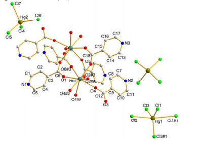
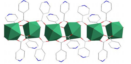
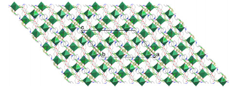
The title compound was synthesized from the reaction of HgCl2, HoCl3·6H2O, isonicotinic acid, hydrochloric acid and distilled water through a hydrothermal reaction. Single-crystal X-ray diffrac-tion results unveiled that compound 1 crystallizes in the monoclinic system with space group C2/c and the Z value being four. The asymmetric unit of com-pound 1 contains one Ho(Ⅲ) ion, one and a half Hg(Ⅱ) ions, six and a half Cl ions, three isonicotinic acid ligands, and two coordinating water and two lattice water molecules, as presented in Fig. 1. There are two kinds of crystallographically independent mercury ions in different coordination environments. The Hg(1) ion displays a five-coordinated triangular bipyramidal configuration, while Hg(2) exhibits four-coordinated tetrahedral coordination geometry. The bond lengths of Hg–Cl are in the span of 2.288(3)~2.624(3) Å with a mean length of 2.4693(16) Å, which is in the normal range and comparable with those documented[15-17]. The bond angles of Cl–Hg–Cl vary from 90.75(9)° to 123.06(8)°. The Ho(Ⅲ) ion shows a slightly distor-ted square anti-prism coordination configuration. It is surrounded by eight oxygen atoms, of which six are from six isonicotinic acid molecules and two from two coordinating water molecules. The Ho3+–OIA bond lengths in compound 1 fall in the range of 2.305(3)~2.350(3) Å with a mean length of 2.328(3) Å, while the bond lengths of Ho3+–Owater are much longer (2.434(3) and 2.460(3) Å). As a result, the Ho3+ ion displays much stronger affinity to the isonicotinic acid molecules than to water molecules. All of the Ho3+–O bond lengths locate in the normal span and are comparable with that reported[18-26]. The bond angles of O–Ho–O change from 69.60(12)° to 146.10(13)°. Every two neighboring Ho3+ ions are interconnected by four or two isonicotinic acid ligands to form a one-dimensional (1D) infinite -Ho–(IA)4–Ho–(IA)2–Ho–(IA)4–Ho-chain running along the a axis, as given in Fig. 2. The distances of neighboring Ho3+···Ho3+ are 4.5096(3) and 5.0333(3) Å. In 1, as shown in Table 2, there are some N–H···Cl, N–H···O, O–H···O and O–H···Cl hydrogen bonding interactions, which interlink such 1D infinite chains together to generate a 3D supramole-cular network (Fig. 3). Moreover, there are some π···π interactions existing in the title compound. The centroid to centroid distance is 3.827(4) Å for rings Cg(1) and Cg(1)#1, while 3.602(4) Å for rings Cg(2) and Cg(2)#4. The perpendicular separation between Cg(1) and Cg(1)#1 is 3.530 Å; while that between Cg(2) and Cg(2)#4 is 3.343 Å. Cg(1) slides from Cg(1)#1 by about 1.48 Å, while Cg(2) from Cg(2)#4 by about 1.34 Å. The dihedral angles are 0º and 0.03º between Cg(1) and Cg(1)#1 and between Cg(2) and Cg(2)#4, indicating that they are almost parallel. Both these π···π interactions and hydrogen bonding interactions consolidate the crystal packing structure.
Figure 1
 Figure 1. Structure of 1 with lattice water and hydrogen atoms being omitted for clarity. Symmetry codes: #1: –x+1, y, –z+5/2; #2 –x+1/2, –y+1/2, –z+1; #3 –x, y, –z+1/2
Figure 2
 Figure 2. 1D infinite -Ho–(IA)4–Ho–(IA)2–Ho–(IA)4–Ho-chain in 1
Table 2
Table 2. Hydrogen Bonding Interactions and π···π Interactions (Cg(1) and Cg(2) Stands for the Centre of Gravity of Rings N1(C(1)~C(5)) and N3(C(13)~C(17)), respectively)DownLoad:
CSV
D–H···A D–H, (Å) H···A, (Å) D···A, (Å) D–H···A, (º) N(1)–H(1B)···Cl(7)#1 0.86 2.34 3.142(9) 156 N(2)–H(2B)···Cl(4)#2 0.86 2.54 3.270(7) 143 N(3)–H(3A)···O(4W)#3 0.86 1.99 2.834(13) 165 O(1W)–H(1WA)···O(3W) 0.82 1.97 2.740(8) 157 O(1W)–H(1WB)···Cl(7) 0.85(6) 2.39(6) 3.214(4) 164(5) O(4W)–H(4WA)···Cl(5) 0.82(9) 2.58(10) 3.323(8) 151(8) O(3W)–H(3WA)···Cl(5) 0.82(10) 2.49(9) 3.224(8) 150(9) π···π interactions Dist. (Å) Cg(1)···Cg(1)#1 3.827(4) Cg(2)···Cg(2)#4 3.602(4) Symmetry codes: #1: –x, 1–y, 1–z; #2: 1/2–x, –1/2+y, 3/2–z; #3: 1/2–x, –1/2+y, 1/2–z; #4: –x, –y, –z Figure 3
 Figure 3. Packing diagram of 1 with dashed lines representing hydrogen bonding interactions (Å, °): N(1)–H(1B)···Cl(7) (–x, 1–y, 1–z) 3.142(9), 156; N(2)–H(2B)···Cl(4) (1/2–x, –1/2+y, 3/2–z) 3.270(7), 143; N(3)–H(3A)···O(4W) (1/2–x, –1/2+y, 1/2–z) 2.834(13), 165; O(1W)–H(1WA)···O(3W) 2.740(8), 157; O(1W)–H(1WB)···Cl(7) 3.214(4), 164(5); O(4W)–H(4WA)···Cl(5) 3.323(8), 151(8); O(3W)–H(3WA)···Cl(5) 3.224(8), 150(9). The green are HoO8 polyhedra
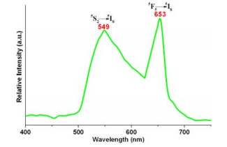
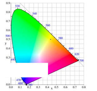
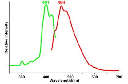
So far, a large number of photoluminescent ter-bium and holmium compounds have been repor-ted[27-29]. In addition, as far as we know, compounds containing mercury tend to emit photoluminescence. In view of the above, we used solid sample of the title compound to test the photoluminescence perfor-mance. As shown in Fig. 4, compound 1 can emit two photoluminescence peaks between 500 and 700 nm when excited by a light of 318 nm, located at 549 nm (green region) and 653 nm (red region), respectively, and the latter is stronger. The emission bands at 549 and 653 nm can be ascribed to the characteristic emissions of the 4f electrons intrashell transitions of the Ho3+ ions, i.e. both emission bands must come from the 5S2 → 5I8 and 5F5 → 5I8 (Ho3+)[30, 31]. As for compound 1, its CIE chromaticity coordinate is (0.4361, 0.4992), as presented in Fig. 5. As a result, the title compound is a potential light emitting material.
Figure 4
 Figure 4. Solid-state photoluminescence spectrum of compound 1 measured at room temperature with λex = 318 nm
Figure 5
 Figure 5. CIE chromaticity figure and chromaticity coordinates of the photoluminescence emission spectrum of 1
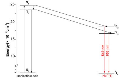
In order to explore the nature of photolumine-scence of the title compound in more depth, we carried out a phosphorescence spectrum test at 77 K for isonicotinic acid and the result is shown in Fig. 6. From the phosphorescence spectrum of the isonico-tinic acid, we can estimate that its onset is about 434 nm and the lowest triplet level of the isonicotinic acid is around 23041 cm–1. As a result, the energy level difference between the lowest triplet state of the isonicotinic acid molecule and the resonant energy level of the Ho3+ ion (5S2, 18300 cm–1) is 4741 cm–1 for the title compound (Fig. 7). According to the intramolecular energy level transition mechanism proposed by Dexter and Sato et al.[32, 33], the intramolecular energy level transition efficiency of compounds mainly depends on two factors. On one hand, energy is forward transferred from the lowest triplet energy level of ligand molecule to the resonance corresponding energy level of the central lanthanide ion through Dexter's resonance exchange; on the other hand, energy is reversely transferred from the central lanthanide ion to ligand molecule by a thermal deactivation process. These two processes are obviously opposite and are closely related to energy level matching or energy level difference between the lowest triplet energy level of the ligand molecules and the resonant energy level of the central lanthanide ions. If the energy level difference is too small, the reverse transfer process of energy is easy to occur, while if it is too large, the forward transfer of energy is difficult to occur. In order to determine whether the energy level difference is too large or too small, a suitable energy gap value, about 3000 ± 500 cm–1, is proposed by the mechanism of intramolecular energy level transfer[34]. Any large deviation from the value of this energy gap may lead to poor photoluminescence performance of a compound.
Figure 6
 Figure 6. Phosphorescence spectra of the isonicotinic acid measured at 77 K
Figure 7
 Figure 7. Schematic and partial energy level diagram of the main energy absorption transfer and phosphorescence processes in 1 and the isonicotinic acid ligand
Based on the above discussion, the energy level difference of holmium ion in 1 is 4741 cm–1, which is obviously greater than the optimal band gap value. The photoluminescence properties of compound 1 should not be well-shaped. This is also in good agreement with the photoluminescence spectra of compound 1. Because of the photoluminescence emission curve of compound 1, there are two emission peaks which are not good shape and not well separated, as shown in Fig. 4. Therefore, we can conclude that isonicotinic acid is not an appropriate ligand to excite holmium ions in compound 1, that is, not an appropriate "antenna molecule".
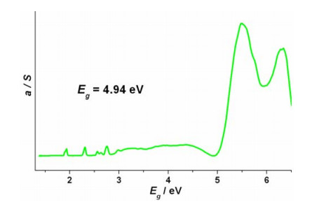
In general, mercury compounds can show semiconductor properties. In order to further study the photophysical properties of the title compound, the solid UV-vis diffuse reflectance spectra were measured with powder samples at room temperature. The data of solid UV-vis diffuse reflectance spectra are analyzed by the Kubelka-Munk formula α/S = (1 – R)2/2R which is commonly used in this field. In this formula, α is the absorption coefficient, S the scattering coefficient, and R the reflectance. The optical band gap of the compound is determined by the linear epitaxy method with the maximum absorption edge on the α/S vs energy diagram. In this way, compound 1 has a band gap of 4.94 eV, as presented in Fig. 8. On the curve, there are some small peaks between 1.5 and 2.8 eV due to the holmium ions. Judging from the bandgap value, it is possible that the title compound can be used as a wide band gap semiconductor material. It can also be seen from the graph that the maximum absorption edge of the curve is not sharp, which means that there is an indirect transition in the compound[35].
Figure 8
 Figure 8. Solid-state UV/Vis diffuse reflectance curve of 1 Photophysical performance and energy transfer mechanism of a 1-D chain-like complex
A novel lanthanide-mercury compound was reported. It displays a 1D chain-like structure. Solid-state photoluminescence measurements show that it can emit brown light. The emission bands can be ascribed to the characteristic emissions of 4f elec-trons intrashell transitions of 5S2 → 5I8 and 5F5 → 5I8 of the holmium(Ⅲ) ions. The photo-luminescence emission energy transfer mechanism is elucidated by the energy level diagrams of the holmium(Ⅲ) ions and isonicotinic acid ligand. It possesses CIE chro-maticity coordinates of (0.4361, 0.4992); so, it is a potential candidate for light emitting materials. Solid-state UV/Vis diffuse reflectance spectra reveal that it has a wide optical band gap 4.94 eV.
-
-
[1]
Qiu, L. Y.; Yu, C. F.; Wang, X. L.; Xie, Y. B.; Kirillov, A. M.; Huang, W.; Li, J. P.; Gao, P.; Wu, T.; Gu, X. W.; Nie, Q.; Wu, D. Y. Tuning the solid-state white light emission of postsynthetic lanthanide-encapsulated double-layer MOFs for three-color luminescent thermometry applications. Inorg. Chem. 2019, 58, 4524–4533. doi: 10.1021/acs.inorgchem.9b00084
-
[2]
Zhao, L. H.; Chen, H. M.; Yang, A. H.; Wu, D. F.; Gou, J.; Cui, J. Z.; Gao, H. L. Synthesis, characterization and properties of lanthanide complexes with different ancillary ligands. Inorg. Chim. Acta 2019, 490, 240–245. doi: 10.1016/j.ica.2019.03.030
-
[3]
Yao, X.; An, G. H.; Li, Y. X.; Yan, P. F.; Li, W. Z.; Li, G. M. Effect of nuclearity and symmetry on the single-molecule magnets behavior of seven-coordinated β-diketonate Dy(Ⅲ) complexes. J. Solid State Chem. 2019, 274, 295–302. doi: 10.1016/j.jssc.2019.03.044
-
[4]
Yu, L. H.; Li, G. W.; Liu, Y. S.; Jiang, F. L.; Hong, M. C. Lanthanide-doped KGd2F7 nanocrystals: controlled synthesis, optical properties, and spectroscopic identification of the optimum core/shell architecture for highly enhanced upconverting luminescence. Cryst. Growth Des. 2019, 19, 2340–2349. doi: 10.1021/acs.cgd.9b00040
-
[5]
D'Vries, R. F.; Gomez, G. E.; Mondragon, L. P.; Onna, D.; Barja, B. C.; Soler-Illia, G. J. A. A.; Ellena, J. 1D lanthanide coordination polymers based on lanthanides and 4'-hydroxy-4-biphenylcarboxylic acid: synthesis, structures and luminescence properties. J. Solid State Chem. 2019, 274, 322–328. doi: 10.1016/j.jssc.2019.02.043
-
[6]
Xu, L.; Pu, N.; Li, Y. Z.; Wei, P. P.; Sun, T. X.; Xiao, C. L.; Chen, J.; Xu, C. Selective separation and complexation of trivalent actinide and lanthanide by a tetradentate soft-hard donor ligand: solvent extraction, spectroscopy, and DFT calculations. Inorg. Chem. 2019, 58, 4420–4430. doi: 10.1021/acs.inorgchem.8b03592
-
[7]
Ren, K.; Wu, S. H.; Guo, X. F.; Wang, H. Lanthanide organic framework as a reversible luminescent sensor for sulfamethazine antibiotics. Inorg. Chem. 2019, 58, 4223–4229. doi: 10.1021/acs.inorgchem.8b03284
-
[8]
Wang, K.; Zhang, J. Q.; Lu, J.; Jing, P.; Li, L. C. Slow magnetic relaxation in Cu–Ln heterometallic Schiff base complexes containing Ln(hfac)-4 as counterions magnetic relaxation in Cu–Ln heterometallic Schiff base complexes containing Ln(hfac)-4 as counterions. Inorg. Chim. Acta 2019, 490, 51–56. doi: 10.1016/j.ica.2019.02.030
-
[9]
Martin-Caballero, J.; Artetxe, B.; Reinoso, S.; San Felices, L.; Vitoria, P.; Larranaga, A.; Vilas, J. L.; Gutierrez-Zorrilla, J. M. Thermostructural behavior in a series of lanthanide-containing polyoxotungstate hybrids with copper(Ⅱ) complexes of the tetraazamacrocycle cyclam: a single-crystal-to-single-crystal transformation study. Inorg. Chem. 2019, 58, 4365–4375. doi: 10.1021/acs.inorgchem.8b03471
-
[10]
Clementino, R. F. P.; de Souza Santos, A. B.; Marques, O. J. B. J.; Ratkovski, D. R.; Gatto, C. C.; Malvestiti, I.; de Araujo Machado, F. L.; Falcao, E. H. L. Structural description, luminescent and magnetic properties of novel 2-D coordination polymers containing thiazolo[5, 4-d]thiazole rings and trivalent lanthanide ions. J. Solid State Chem. 2018, 268, 94–101. doi: 10.1016/j.jssc.2018.07.033
-
[11]
Cerfontaine, S.; Marcelis, L.; Laramee-Milette, B.; Hanan, G. S.; Loiseau, F.; De Winter, J.; Gerbaux, P.; Elias, B. Converging energy transfer in polynuclear Ru(Ⅱ) multiterpyridine complexes: significant enhancement of luminescent properties. Inorg. Chem. 2018, 57, 2639–2653. doi: 10.1021/acs.inorgchem.7b03040
-
[12]
Wang, W.; Peng, D. F.; Zhang, H. L.; Yang, X. H.; Pan, C. F. Mechanically induced strong red emission in samarium ions doped piezoelectric semiconductor CaZnOS for dynamic pressure sensing and imaging. Opt. Commun. 2017, 395, 24–28. doi: 10.1016/j.optcom.2016.03.046
-
[13]
Kuang, H. M.; Huang, J. G.; Lin, L. Z.; Zhong, Q. X.; Chen, W. T. Structure and luminescence of two new mercury-lanthanide complexes. Inorg. Chim. Acta 2019, 489, 48–53. doi: 10.1016/j.ica.2019.02.006
-
[14]
Sheldrick, G. M. SHELXS, Program for X-ray Crystal Structure Solution. University of Göttingen, Germany 1997.
-
[15]
Yi, X. G.; Zhang, Z. X.; Chen, W. T.; Lin, L. Z.; Chen, H. L. Photoluminescence and semiconductor properties of two novel lanthanide-mercury compounds with one-dimensional chain-like structures. J. Solid State Chem. 2018, 266, 16–22. doi: 10.1016/j.jssc.2018.07.004
-
[16]
Lin, L. Z.; Zhong, Q. X.; Hong, J. T.; Chen, H. L.; Chen, W. T. Syntheses, structures, photoluminescence and semiconductor properties of two novel mercury-lanthanide complexes with a three-dimensional open framework. Inorg. Chim. Acta 2018, 479, 30–35. doi: 10.1016/j.ica.2018.04.039
-
[17]
Luo, Q. Y.; Luo, H.; Kuang, H. M.; Chen, W. T.; Wen, Y. X. A novel samarium material: synthesis, structure, photophysical properties and photoluminescence energy transfer mechanism. J. Solid State Chem. 2019, 270, 200–204. doi: 10.1016/j.jssc.2018.09.040
-
[18]
Gupta, S. K.; Langley, S. K.; Sharma, K.; Murray, K. S.; Murugavel, R. Pentanuclear lanthanide mono-organophosphates: synthesis, structure, and magnetism. Inorg. Chem. 2017, 56, 3946–3960. doi: 10.1021/acs.inorgchem.6b03014
-
[19]
Zhang, L. Y.; Lu, L. P.; Zhu, M. L.; Feng, S. S. Self-assembly of lanthanide(Ⅲ) coordination polymers from a bifunctional 2-(pyridin-2-yl)-1H-imidazole-4, 5-dicarboxylate ligand with the assistance of oxalate: syntheses, structures, luminescence, and magnetic properties. CrystEngComm. 2017, 19, 1953–1964. doi: 10.1039/C7CE00149E
-
[20]
Coban, M. B.; Amjad, A.; Aygun, M.; Kara, H. Sensitization of HoIII and SmIII luminescence by efficient energy transfer from antenna ligands: magnetic, visible and NIR photoluminescence properties of GdIII, HoIII and SmIII coordination polymers. Inorg. Chim. Acta 2017, 455, 25–33. doi: 10.1016/j.ica.2016.10.010
-
[21]
Wu, J.; Li, X. L.; Zhao, L.; Guo, M.; Tang, J. Enhancement of magnetocaloric effect through fixation of carbon dioxide: molecular assembly from Ln4 to Ln4 cluster pairs. Inorg. Chem. 2017, 56, 4104–4111. doi: 10.1021/acs.inorgchem.7b00094
-
[22]
An, L.; Zhou, J.; Zou, H. H.; Xiao, H.; Zhao, R.; Ding, Q. Syntheses, structures and properties of a series of new lanthanide chalcoarsenates(Ⅲ) containing crown-shaped [As3Q6]3- (Q = S, Se) clusters. J. Alloy. Compd. 2017, 702, 594–600. doi: 10.1016/j.jallcom.2017.01.284
-
[23]
Schmidt, S. F. M.; Koo, C.; Mereacre, V.; Park, J.; Heermann, D. W.; Kataev, V.; Anson, C. E.; Prodius, D.; Novitchi, G.; Klingeler, R.; Powell, A. K. A three-pronged attack to investigate the electronic structure of a family of ferromagnetic Fe4Ln2 cyclic coordination clusters: a combined magnetic susceptibility, high-field/high-frequency electron paramagnetic resonance, and 57Fe Mössbauer study. Inorg. Chem. 2017, 56, 4796–4806. doi: 10.1021/acs.inorgchem.6b02682
-
[24]
Zhang, J. W.; Jiang, Y.; Xie, Y. R.; Chu, J.; Liu, B. Q. Syntheses, structures, photoluminescence, and magnetism of a series of discrete heavy lanthanide complexes based on a tricarboxylic acid. Inorg. Chim. Acta 2016, 453, 257–262. doi: 10.1016/j.ica.2016.08.020
-
[25]
Ridenour, J. A.; Carter, K. P.; Butcher, R. J.; Cahill, C. L. RE-p-halobenzoic acid-terpyridine complexes, part Ⅱ: structural diversity, supramolecular assembly, and luminescence properties in a series of p-bromobenzoic acid rare-earth hybrid materials. CrystEngComm. 2017, 19, 1172–1189. doi: 10.1039/C6CE02355J
-
[26]
Botezat, O.; van Leusen, J.; Ch Kravtsov, V.; Kogerler, P.; Baca, S. G. Ultralarge 3d/4f coordination wheels: from carboxylate/amino alcohol-supported {Fe4Ln2} to {Fe18Ln6} rings. Inorg. Chem. 2017, 56, 1814–1822. doi: 10.1021/acs.inorgchem.6b02100
-
[27]
Rojas-Hernandez, R. E.; Santos, L. F.; Almeida, R. M. Tb3+/Yb3+ doped aluminosilicate phosphors for near infrared emission and efficient down-conversion. J. Lumin. 2018, 197, 180–186. doi: 10.1016/j.jlumin.2018.01.020
-
[28]
Gao, Y.; Sun, X. R.; Feng, Z. S.; Zhu, L. Y.; Zhang, J.; Gao, W. L.; Zhou, X. J.; Cong, R. H.; Yang, T. Tb3+ and Eu3+ co-doped Ba6Bi9B79O138: color-tunable phosphors by utilizing the host-sensitization effect of Bi3+ and enhancement of red emission upon heating. New J. Chem. 2017, 41, 2037–2045. doi: 10.1039/C6NJ03603A
-
[29]
Shi, H. W.; Zhu, Y. P.; Zhao, Y. M.; Liu, C.; Ren, X. Z.; Hao, J. G.; Li, W. Field-induced large strain and strong green photoluminescence in (Ho, Sb)-modified (Bi0. 5Na0. 5)0. 945Ba0. 065TiO3 multifunctional ferroelectric ceramics. J. Alloy. Compd. 2018, 767, 666–674. doi: 10.1016/j.jallcom.2018.07.135
-
[30]
Krishnan, R.; Thirumalai, J. Up/down conversion luminescence properties of (Na0. 5Gd0. 5)MoO4: Ln3+ (Ln = Eu, Tb, Dy, Yb/Er, Yb/Tm, and Yb/Ho) microstructures: synthesis, morphology, structural and magnetic investigation. New J. Chem. 2014, 38, 3480–3491. doi: 10.1039/C4NJ00165F
-
[31]
Barrera, E. W.; Pujol, M. C.; Carvajal, J. J.; Mateos, X.; Sole, R.; Massons, J.; Speghini, A.; Bettinelli, M.; Cascales, C.; Aguilo, M.; Diaz, F. White light upconversion in Yb-sensitized (Tm, Ho)-doped KLu(WO4)2 nanocrystals: the effect of Eu incorporation. Phys. Chem. Chem. Phys. 2014, 16, 1679–1686. doi: 10.1039/C3CP53847H
-
[32]
Dexter, D. L. A theory of sensitized luminescence in solids. J. Chem. Phys. 1953, 21, 836–850. doi: 10.1063/1.1699044
-
[33]
Sato, S.; Wada, M. Relations between intramolecular energy transfer efficiencies and triplet state energies in rare earth β-diketone chelates. Bull. Chem. Soc. Jpn. 1970, 43, 1955–1962. doi: 10.1246/bcsj.43.1955
-
[34]
Xu, B.; Yan, B. Photophysical properties of novel lanthanide (Tb3+, Dy3+, Eu3+) complexes with long chain para-carboxyphenol ester p-L-benzoate (L = dodecanoyloxy, myristoyloxy, palmitoyloxy and stearoyloxy). Spectrochim. Acta A 2007, 66, 236–242. doi: 10.1016/j.saa.2006.02.047
-
[35]
Huang, F. Q.; Mitchell, K.; Ibers, J. A. New layered materials: syntheses, structures, and optical and magnetic properties of CsGdZnSe3, CsZrCuSe3, CsUCuSe3, and BaGdCuSe3. Inorg. Chem. 2001, 40, 5123–5126. doi: 10.1021/ic0104353
-
[1]
-
Figure 1 Structure of 1 with lattice water and hydrogen atoms being omitted for clarity. Symmetry codes: #1: –x+1, y, –z+5/2; #2 –x+1/2, –y+1/2, –z+1; #3 –x, y, –z+1/2
Figure 3 Packing diagram of 1 with dashed lines representing hydrogen bonding interactions (Å, °): N(1)–H(1B)···Cl(7) (–x, 1–y, 1–z) 3.142(9), 156; N(2)–H(2B)···Cl(4) (1/2–x, –1/2+y, 3/2–z) 3.270(7), 143; N(3)–H(3A)···O(4W) (1/2–x, –1/2+y, 1/2–z) 2.834(13), 165; O(1W)–H(1WA)···O(3W) 2.740(8), 157; O(1W)–H(1WB)···Cl(7) 3.214(4), 164(5); O(4W)–H(4WA)···Cl(5) 3.323(8), 151(8); O(3W)–H(3WA)···Cl(5) 3.224(8), 150(9). The green are HoO8 polyhedra
Figure 4 Solid-state photoluminescence spectrum of compound 1 measured at room temperature with λex = 318 nm
Figure 5 CIE chromaticity figure and chromaticity coordinates of the photoluminescence emission spectrum of 1
Figure 7 Schematic and partial energy level diagram of the main energy absorption transfer and phosphorescence processes in 1 and the isonicotinic acid ligand
Figure 8 Solid-state UV/Vis diffuse reflectance curve of 1 Photophysical performance and energy transfer mechanism of a 1-D chain-like complex
Table 1. Selected Bond Lengths (Å) and Bond Angles (°)
Bond Dist. Bond Dist. Hg(1)–Cl(1) 2.470(4) Ho(1)–O(1) 2.324(3) Hg(1)–Cl(2) 2.443(4) Ho(1)–O(3) 2.334(3) Hg(1)–Cl(2)#1 2.443(4) Ho(1)–O(4)#2 2.305(3) Hg(1)–Cl(3) 2.288(3) Ho(1)–O(2)#3 2.318(3) Hg(1)–Cl(3)#1 2.624(3) Ho(1)–O(5) 2.341(3) Hg(2)–Cl(4) 2.4799(16) Ho(1)–O(6)#3 2.350(3) Hg(2)–Cl(5) 2.4487(15) Ho(1)–O(1W) 2.460(3) Hg(2)–Cl(6) 2.4621(15) Ho(1)–O(2W) 2.434(3) Hg(2)–Cl(7) 2.5649(14) Angle (°) Angle (°) Cl(1)–Hg(1)–Cl(2) 90.75(9) O(4)#2–Ho(1)–O(2)#3 79.11(12) Cl(1)–Hg(1)–Cl(3) 123.06(8) O(4)#2–Ho(1)–O(1) 145.49(12) Cl(1)–Hg(1)–Cl(3)#1 110.38(7) O(2)#3–Ho(1)–O(1) 76.82(13) Cl(2)–Hg(1)–Cl(3) 109.50(12) O(2)#3–Ho(1)–O(3) 139.63(13) Cl(2)–Hg(1)–Cl(3)#1 110.93(12) O(1)–Ho(1)–O(3) 81.87(12) Cl(3)–Hg(1)–Cl(3)#1 110.41(13) O(2)#3–Ho(1)–O(5) 74.27(13) Cl(4)–Hg(2)–Cl(5) 113.85(5) O(1)–Ho(1)–O(5) 114.42(12) Cl(4)–Hg(2)–Cl(6) 110.03(5) O(3)–Ho(1)–O(5) 146.10(13) Cl(4)–Hg(2)–Cl(7) 105.60(5) O(4)#2–Ho(1)–O(6)#3 138.71(12) Cl(5)–Hg(2)–Cl(6) 117.57(6) O(6)#3–Ho(1)–O(2W) 69.60(12) Cl(5)–Hg(2)–Cl(7) 103.80(5) O(2)#3–Ho(1)–O(1W) 70.15(12) Cl(6)–Hg(2)–Cl(7) 104.63(5) O(2W)–Ho(1)–O(1W) 124.83(12) Symmetry codes: #1: –x+1, y, –z+5/2; #2: –x+1/2, –y+1/2, –z+1; #3: –x, y, –z+1/2  下载: 导出CSV
Table 2. Hydrogen Bonding Interactions and π···π Interactions (Cg(1) and Cg(2) Stands for the Centre of Gravity of Rings N1(C(1)~C(5)) and N3(C(13)~C(17)), respectively)
D–H···A D–H, (Å) H···A, (Å) D···A, (Å) D–H···A, (º) N(1)–H(1B)···Cl(7)#1 0.86 2.34 3.142(9) 156 N(2)–H(2B)···Cl(4)#2 0.86 2.54 3.270(7) 143 N(3)–H(3A)···O(4W)#3 0.86 1.99 2.834(13) 165 O(1W)–H(1WA)···O(3W) 0.82 1.97 2.740(8) 157 O(1W)–H(1WB)···Cl(7) 0.85(6) 2.39(6) 3.214(4) 164(5) O(4W)–H(4WA)···Cl(5) 0.82(9) 2.58(10) 3.323(8) 151(8) O(3W)–H(3WA)···Cl(5) 0.82(10) 2.49(9) 3.224(8) 150(9) π···π interactions Dist. (Å) Cg(1)···Cg(1)#1 3.827(4) Cg(2)···Cg(2)#4 3.602(4) Symmetry codes: #1: –x, 1–y, 1–z; #2: 1/2–x, –1/2+y, 3/2–z; #3: 1/2–x, –1/2+y, 1/2–z; #4: –x, –y, –z
下载: 导出CSV
-


 扫一扫看文章
计量
- PDF下载量: 2
- 文章访问数: 567
- HTML全文浏览量: 14
