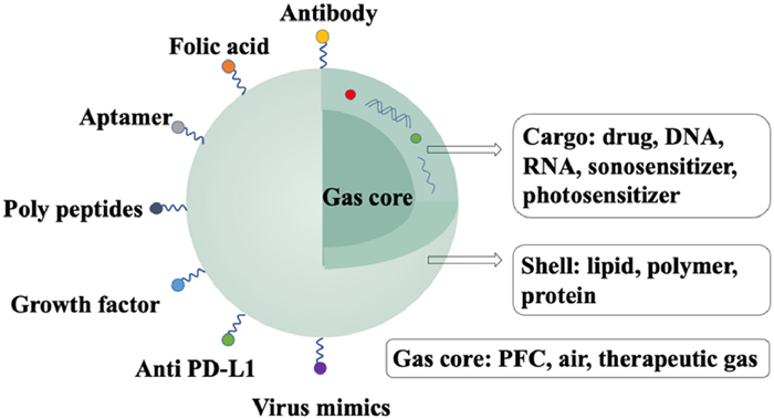
Figure 1.
Illustration of the structure of a MB.
Recent progress in theranostic microbubbles
Ziyao Wang , Ziyan Feng , Fangxue Du , Xi Xiang , Xinyi Tang , Li Qiu , Zhiyong Qian

Ultrasound (US) imaging is an economic, min-invasive diagnostic procedure which has been widely used in clinical fields. Because of the similar water proportion of many soft tissues and tumors, the acoustic impedances between them can be indistinguishable, making the images ambiguous and the diagnosis difficult. To tackle these challenges, US contrast agents (UCAs) were explored to enable US imaging effectiveness. UCAs are mainly gaseous micro-bubbles (MBs), entering the human blood circulation to achieve different goals. Generally, MBs have a more powerful acoustic impedance than that of the surrounding fluids and biological tissue. Consequently, utilizing MBs can efficiently improve the reflection of US, reaching a better image resolution for precise diagnosis [1,2]. Nanosized MBs, designed on the basis of MBs, have the capability to penetrate through blood vessels for extravascular imaging and therapy.
MBs are mostly gas spheres of several µm diameter. Usually, they are composed of three parts: the gas core, the shell enclosing the gas core, and the cargo attached to or encapsulated by the shell, as shown in Fig. 1. The most significant concern in the fabrication of MBs is their stability, which will influence the effects of both imaging and therapy [3].
Stability of MBs is partly determined by the gas core inside. It is reported that gas type with higher water solubility generates larger bubbles [4]. Different solubilities of different gasses, besides, can influence the blood recirculation time of MBs, which is crucial for theranostic efficacy [5]. On the other hand, the gas type influences the zeta potential of MBs, which subsequently has an impact on the stability. According to literature, as for negatively-charged microbubbles, OH− ions accumulate in the gas-water interfaces [6]. As a result, the ability of MBs to absorb OH− ions on the shell surface partly leans on the properties of gas core.
Conventionally, perfluoropropane (C3F8), perfluorobutane (C4F10, PFB), and sulfur hexafluoride (SF6) have been used in clinical fields, as Definity, Sonazoid, Optison and SonoVue.
A unique shell is utilized to support MBs since gas bubbles in aqueous environments are unsteady on their own. Maintaining an MB's unity throughout preparation and utilization is the shell's primary purpose.
The shell's formation controls its elasticity, which in turn affects MBs stability and acoustic response when exposed to a US field [7]. A range of biocompatible substances has been selected as MBs' shells, producing a variety of shell qualities. Both synthetic and natural polymers, which make up the MBs shell by themselves or after adjustment, can enhance the characteristics of MBs, like interacting with particular molecules or tissues.
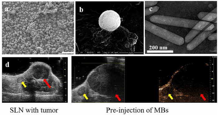
Due to their unique qualities, such as biocompatibility, nano-size, high surface area, and various compositions, MBs are being employed more and more as carriers for both drugs and contrast agents (Fig. 2a) [8,9]. The hydrophobic cargo can either retain covalent or non-covalent connection to the shell of MBs, via specific ligands [10], or they can be enclosed within the MBs (Fig. 2b) [11].
Approaches for preparing MBs involving stochastic techniques, forced extrusion, template layer-by-layer deposition, and microfluidic techniques [12].
Ultrasonication, excimer laser ablation, and high shear emulsification are three main categories of stochastic techniques. Numerous bubbles of a diverse scale range are produced using stochastic techniques [13]. Approaches for forced extrusion comprise electrohydrodynamic atomization, inkjet printing, and membrane emulsification. This creates MBs for medium sizes [14]. Layer-by-layer (LbL) assembling based on different disposable core templates is used in template layer-by-layer deposition to produce hollow microspheres with a homogenous particle size [15]. To improve control of size distribution and, therefore, accuracy and homogeneity, a microfluidic technique is used. Via the utilization of a microfluidic system, MBs are produced by transporting the components through numerous channels [16].
Nanosized microbubbles are bubbles of nanometer-scaled and, as a result, have particular characteristics (Fig. 2c) [17]. The bulk of the construction processes for nanosized microbubbles were developed from MB synthesis methods. Both self-assembled nanosized microbubbles and phase-shift perfluorocarbon droplets are made using corresponding procedures. The main method of creating nanosized microbubbles involves the direct self-assembling of several phospholipids, proteins, and surfactants at the surface of an aqueous dissolving medium [18]. As for nanodroplets, condensation of MBs is a common process using existing MBs [19].
With the air-liquid interface of bubbles, microbubbles are currently the most widely used in clinical diagnosis of diseases, as shown in Fig. 2d [20].
According to the mechanism of contrast enhanced imaging, gas core plays a key role in diagnostic MBs.
New approaches to US imaging have been sparked by the development of MBs. MBs vibrate when exposed to pressure alterations, resulting in a nonlinear acoustic signal. By comparing different signals, MBs can be obviously separated from the surrounding media. This process suggests a number of methods for identifying MBs that take use of nonlinear acoustic signal, including pulse/phase reversal, power modulation, and contrast pulse sequencing [21]. Moreover, in the aqueous medium as well as on solid support, it has been convincingly shown that diagnostic US imaging methods are capable of identifying single MB with high susceptibility, via selective targeting [22].
Traditional MBs typically behave hydrodynamically similarly to red blood cells (RBCs) due to the alike size distribution, in order to travel in the circulation and communicate with the endothelial cells along the vessels [23]. These traits have a significant impact on the targeting property of MBs. The utilizations of classic MB are primarily focused on atherosclerosis, ischemia-reperfusion damage, inflammatory bowel disease, and angiogenesis since only intravascular targets can be addressed by MBs. Contrast-enhanced US (CEUS) with MBs can provide very precise and trustworthy identification in comparison to standard anatomical US, particularly in the early stage of these illnesses [24].
Despite MBs' effectiveness in several intravascular imaging applications, their relatively large particle sizes continue to hinder their ability to identify cell-surface markers extravascularly. For targeting in tissues, necessary altering of MB sizes is needed to accommodate vascular fenestrations. For example, leaky tumor vasculature permit the passage of nanoparticles which are smaller than 700 nm [25], while enhanced vascular permeability in insulitis can cause nanoparticles with a size of 100–300 nm to travel into the islet microenvironment [26]. As a result, nanosized MBs were proposed.
Recent years have seen the development of nanocarriers for their therapeutic efficiency, reduced toxicity and selective accumulation [27]. In comparison with MB, nanosized MBs gain higher collection in extravascular regions, improving the signal of the target areas, especially in tumors with the help of the enhanced permeability and retention (EPR) effect [28,29]. Additionally, the exterior of nanosized MBs may be altered by attaching certain targeting molecules to enhance accumulation in tissues and boost imaging effectiveness. But, there still are drawbacks to employing nanosized MBs, such as weak signal intensity and low stability [30].
Various methods have been tested in order to create nanosized MBs that can maintain their stability throughout circulation while achieving the best imaging efficiency: (1) nanodroplets with acoustic droplet vaporization (ADV); (2) nanoscale gas-filled protein structures produced by cells or bacteria; (3) in situ production of gas via bio/chemical reactions.
Phase-shift MBs, which have an extended recirculation time due to their stability, as well as comparatively easier synthesis procedures, have become popular as MB substitutes in latest days. Most of them are nanoparticles or nanodroplets filled with perfluorocarbon (PFC), which range in size from 250 nm to 500 nm [31,32]. These nanodroplets have a better chance of leaving the vessels and finding the targeted locations because of their reduced sizes [33,34]. The liquid core inside can be transformed into gas and used as conventional MBs at the following stage, which is known as ADV. These ADV-produced bubbles oscillate under acoustic pressure, creating signals that a receiving transducer may pick up [35,36]. Based on spontaneous nucleation, a novel PFBNB nanodroplet with a reduced vaporization threshold was created by Jian An et al. for arterial labeling US subtraction angiography (ALUSA). The outcomes show a color-coded super-resolution ALUSA picture with a resolution of 36 µm in a rabbit kidney, showing the vessels precisely [37]. Similarly, it has been demonstrated that CEUS with phase-shift nanodroplets can identify insulitis before the development of diabetes [38].
The size of the droplets should be thoroughly evaluated in order to assure the stability and phase transition behavior of these nanoagents. Smaller particles have greater surface tension at the border between the droplet and the bulk liquid, which increases the Laplace pressure within the PFC droplet, according to several researchers [39]. They have a larger vaporization threshold, as a result, which might restrict their therapeutic applications [40]. The rise of vaporization threshold can be avoided by employing perfluorocarbon types with lower boiling points according to current study [41]. They have previously proved their effectiveness and qualities as contrast agents assessed in vivo. According to the findings, droplets can offer contrast enhancement comparable to that of traditional MBs and, if constructed appropriately, can stay in circulation for a longer period of time [42]. Another method that concentrated on spontaneous nucleation employed PFCs with boiling temperatures between 37 ℃ and 56 ℃. This approach, which involves saturating a cosolvent with PFC before adding a subpar solvent to lower solvent quality, making droplets to naturally nucleate, is frequently called ouzo method [43].
Aquatic bacteria and archaea produce special gas-filled protein structures called gas vesicles (GVs) that are 200 nm in size, have a 2-nm shell around the air core [44]. GVs serve as floating devices that let aquatic microorganisms live in a habitat at the right water depth for adequate photosynthetic illumination [45]. GVs are structurally stable with nanoscale size because their special shells are permeable to gas but exclude water. GVs provide great US imaging capability and are biocompatible [46]. GVs have been proven to produce stable US contrast that is readily detected in vitro and in vivo [47]. In vitro and in vivo assessments of GVs have been demonstrated. These vesicles' scale makes it possible to mark targets outside the circulation. Gas vesicles may also be produced for bacterial or cell monitoring due to their intracellular detectability, like the activity of enzymes.
However, similar to other common nanoparticles, most of the GVs are often absorbed in non-targeted organs following intravenous injection, such as the liver, spleen, and lungs [48]. GVs were given surface alterations to enhance their pharmacokinetics and address the systemic clearance problem. According to several studies, coating GVs with polyethylene glycol (PEG) or hyaluronic acid (HA) can specifically boost the concentration of GVs at the tumor site [49,50]. By combining PEG and HA in GV shells, PEGylated HA-GVs (pH-GVs) for in-tumor molecular US imaging were designed [17]. This allowed them to bypass clearance from the reticular system (RES) and utilize EPR effects.
Without enclosing any gas precursor, such gas-generating nanoparticles may outperform conventional gas-filled MBs, since they may travel steadily in the circulation and can efficiently concentrate in tumor tissues through EPR effect [51]. For illustration, gas-generating polymeric nanoparticles (GGPNPs) were developed, with the creation of carbon dioxide (CO2) nanobubbles on their surface caused by the hydrolysis reactions. Nanobubbles significantly grow or combine to form MBs, which show resonance [52].
This is the basis for considering the specific microenvironment in situ which may drive gas creation. For example, at the acidic pH of tumor tissues, calcium carbonate (CaCO3) nanocomposites can break down and produce echogenic CO2 bubbles [53]. After mixing it with additional elements, its imaging potential has been investigated in various researches [54,55].
Shell flexibility and stiffness are controlled by shell constitution, which affects MB stability and acoustic response when exposed to ultrasonic pressure [56,57]. MBs have been designed using various typically biocompatible substances, including lipids, inorganic nanoscale polymers, and proteins. Due to the flexible and elastic nature of lipid MB shells, it is simple to produce echoes even in low-acoustic pressure evaluations [1,58]. MB shells made of both polymers and proteins are more robust to contraction and expansion. These kind of MBs have the potential of using in high frequency US imaging [59].
Shells can be established to perform certain tasks based on the unique features of different constituents. By joining HA polymer to the shell, Mary W.N. Burns et al. created targeted and bioresponsive MBs. HA transforms into a hydrogel and muffles the acoustic signal once it cross-linked. Based on this, they created a pH-sensitive MB platform using a reversible pH-sensitive cross-linker [60].
As previously noted, the backscattering capability reduced as the size of the nanosized MBs decreases [61]. One solution to this issue is to change the makeup of the shells. Despite the impact of the bubble size, shell features have an important influence on bubble behaviors [62]. The outcome demonstrates that there is a clear correlation between the nonlinear behavior of the shell under US and its stiffness. The stiffness variation has a significant influence on the pressure threshold. As a result, multiple attempts to employ relatively flexible shells have been made [63,64].
The traditional MB circulates with bloodstream and cannot be employed for tissue-specific applications since it lacks a specialized capacity to react with specific sites. For tackling this challenge, targeted MB is made to promote the accumulation in only certain tissues, organs, etc. Targeted imaging, additionally, can significantly lower the injection volume of MBs and reduce the difficulty of imaging [20,59].
Different ligands, including antibodies, peptides, and carbohydrates, were attached to the MBs to improve their capacity to target. Additionally, the EPR effect is investigated, which also causes nanosized MBs to accumulate more in tumors.
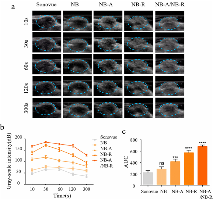
There are various conventional methods for specific ligands coupling to MB membranes [59]. (1) Adding the targeted ligand to the bubble's outer shell directly. This technique works well for ligands that are lipid-bound because they can resist the challenging MBs synthesis conditions (Fig. 3) [65]. (2) Using electrostatic adsorption, self-ionic bonding [66,67], or other techniques to attach antibodies or ligands to the MB shell membrane. However, in vivo environment has an influence on the connections, making the desired targeting action less likely to occur. (3) Coupling agents are a category of compounds with two ends that each has a unique set of characteristics. These various groups may interact with various organic molecules to create a strong connection. Certain ligands or antibodies can link to the MB shell via coupling agents indirectly. (4) The MB shell's formulation includes the bridging agent. The bridging agent's composition is firstly modified by adding the required chemical groups, which will eventually create functional groups. The functional groups are triggered once the MB has formed, and they are then coupled with the ligand. (5) A biotinylated ligand is connected by incorporating biotin into the shell membrane layer and using avidin as a "bridging" agent. The strongest non-covalent interaction currently understood is that between biotin and avidin [68,69].
Another commonly accepted technique to achieve targeting is to use membrane generated from natural cells, such as platelets, erythrocytes, cancer cells, and stem cells. Modern research has focused heavily on cell membrane-camouflaged drug delivery systems (CMCDDs), which are widely recognized for their superior biocompatibility and potential to improve the targeting effect [70]. Additionally, MBs have been included to this targeting strategy. Because the membrane proteins and the original immune components are there, MBs can acquire the cells' innate capacity for multitargeting. This technique can also increase the biocompatibility of MBs and prolonged circulation time [71]. For instance, in the case of sepsis-induced acute renal injury [72] and myocardial ischemia-reperfusion injury [73], platelet membrane-coated hybrid MBs were equipped with a variety of adhesive receptors (such as integrin IIb3), benefiting from selective adherence to injured endothelium. Similar to this, Natacha Jugniot et al. synthesized the first nanosized MBs via triple-negative breast cancer cell membrane as a targeted diagnostic agent [74], taking use of the homotypic recognition of tumor cells.
Multimodal imaging enables the combination of US imaging with other clinical imaging examinations. US imaging is a real-time, practical, and secure imaging option, but it is less accurate in detecting soft tissues than other imaging methods [75]. Combing US with other imaging techniques, say, MRI, can provide more comprehensive understanding to the patients' condition. It is investigated if adding multimodal contrast agents to MBs can address this issue, delivering more precise information [76].
The creation of multimodal MBs involves the use of components with various imaging capabilities. An US-responsive dual-modal US/T1-MRI MB was created by Young Il Yoon et al. for the effective diagnosis of prostate cancer. To identify malignancies using T1-MRI, Fe3+ chelated-melanin nanoparticles (Fe3+MNPs) were created. The MB+Fe3+MNPPs complex was then created via a simple charge interaction between the MBs and Fe3+MNPPs [67]. Furthermore, because of its paramagnetic characteristics and shorter T1 relaxation durations of hydrogens in nearby water molecules, which can provide high contrast on T1 images [77], gadolinium (Gd) has been used for MRI/US dual-modality molecular imaging in breast cancer animal models.
The applications of MBs as UCAs have been highlighted above. MBs have also been thoroughly investigated as a tool to enhance drug delivery [78].
The gas cores of MBs have the potential to improve therapeutic outcomes. Numerous different biological effects are induced by their synthesis, oscillation and collapse [79].
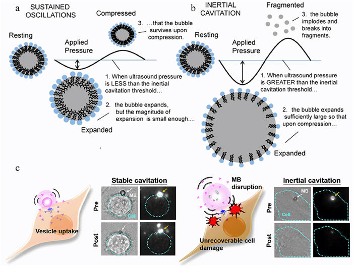
One of the effects US induces is cavitation, or MB oscillation. As mentioned above, when US waves are applied to an aqueous medium, the MBs within will compress and expand in accordance with the waves. This process, in which these tiny bubbles go through growth and collapse sessions or resonance in the medium when exposed to acoustic radiation, is known as the "cavitation process" [80]. Scientists divided them into two types of cavitation bubbles basing on the acoustic pressure and the behavior of MBs. The bubbles in the US field will gradually grow to a crucial size known as resonance size [81]. If the MB exceeds its resonance size within one or several acoustic cycles, becomes unstable and collapses violently, this phenomenon is called inertial cavitation. If, on the contrary, it oscillates for many cycles around its resonance size without collapse, it is called stable cavitation (or non-inertial cavitation) [82], as shown in Figs. 4a and b. Relatively speaking, MBs undergo inertial cavitation under higher acoustic pressure and stable cavitation under lower acoustic pressure [83].
As a mechanical effect, cavitation causes injury to the immediately adjacent tissue, including cell death and thrombolysis [11,84,85]. Its applications in acute myocardial infarction, stroke, and peripheral arterial disease have seen positive outcomes [86]. This effect is also explored in multiple malignant diseases. The treatment with MBs can induce vascular damage and blood flow disruption in tumor, and eventually reduce tumor growth and prolong survival [79].
As mentioned above, during the cavitation process, MB produces great energy to its surroundings. This energy is transmitted to the surrounding cells in the form of mechanical streams or microjets, causing shear stress on their membranes, therefore increases the cell permeability, as shown in Fig. 4c. This is because that it creates transient pores in cellular membranes when bubble dynamics become violent enough. This phenomenon is called sonoporation [78]. In the majority of the experiments, sonoporation was used to increase the uptake of molecules or to penetrate biological barriers [87]. However, different tissues and medications have their own ideal ultrasonic parameters respectively. Thus finding the ideal ultrasonic parameter and maximizing sonoporation effect also worth exploring. According to Qi Liu et al., the optimal parameters of the sonoporation effect for MDA-MB-231 cells were 300 mW/cm2 of US intensity, 2 min of irradiation time, and 20% MBs concentration. In a specific range, the intake of fluorescence staining first increased and then decreased with the increase of US power intensity, irradiation time, and MB concentration [88]. On the other hand, Qu et al. investigated the effectiveness of sonoporation on several breast cancer cell lines and discovered that each cell line has its ideal combination of US parameters to increase the sonoporation efficiency respectively [89]. To accelerate bench-to-bedside translation, Kotopoulis et al. compared three clinically available MBs and summarized that each kind of MBs had the optimal concentration and US parameters to fulfill the sonoporation effect, with SonoVue being best at lower intensities and Sonazoid™ at higher intensities [90].
US-targeted MB destruction (UTMD) is a type of US therapy combining ultrasonic cavitation and sonoporation. Drug-carrying MBs are injected into bloodstream, and when US is applied, the MBs will play as cavitation nuclei as well as sonoporation inducer, thus releasing the drug and enhance its effect [91]. MBs are widely used in targeted drug delivery of drugs that have high systemic side effects or are easily degraded in vivo, ranging from nucleic acids, to proteins, chemotherapies, and even immunotherapy [92]. For example, Liu et al. fabricated chitosan-based injectable hydrogel embedded with SDF-1α-carrying nanodroplets (PFP@NDs-PEG-SDF-1α) that vaporize into MBs under the application of US, and proved them useful in enhancing the cartilage repair in vitro and in vivo [93]. Moreover, a first clinical trial was performed based on UTMD-induced chemotherapy in which researchers combined US, MBs, and chemotherapy in a clinical setting using commercially available equipment. This joint treatment augmented the clinical efficacy of gemcitabine and extend survival time in patients with pancreatic ductal adenocarcinoma [94]. Later, John R. Eisenbrey et al. combined UTMD with transarterial radioembolization in a slightly larger group of 28 patients with hepatocellular carcinoma, and found this approach feasible with an excellent safety profile and an improved hepatocellular carcinoma treatment response [95]. However, there is still a long way to go to introduce it to clinical applications. More and larger trials are needed to fill this gap.
PFC makes up the majority of gas cores, since PFC gas cores are stable, thus can provide longer in vivo circulation time. Aside from these advantages, PFC has high oxygen-carrying capacity, which can be used to alleviate the hypoxic tumor environment and works as a synergistic treatment [96].
MBs are also being explored as gas carriers to delivery therapeutic gasses. This approach has been investigated in alleviating cardiac allograft rejection by Tao Lin et al. who concluded that nitric-oxide (NO) released from MBs significantly suppressed thrombosis and reduced inflammatory cell infiltration [97]. Zhong et al., on the other hand, observed similar anti-inflammatory and thrombolysis effects by administrating H2S-loaded MBs in an ischemia-reperfusion injury model [98].
MBs are usually injected intravenously in clinical practice, and then they circulate throughout the body. This makes the distribution of MBs difficult to predict, and it is possible that the contents of the MBs may be deposited in undesired tissues, causing toxic side effects on the rest of the body. One way to overcome this problem is to combine ligands or receptors to the shells to create "target MBs" that deposit in the target tissues. Glucocorticoids are often the first-line therapy for patients with immune-associated diseases. However, its dose-dependent side effects, such as the increased risk of infection and metabolic disturbances, hamper its clinical use. Kui Fan et al. fabricated a kind of visualized podocyte-targeting and focused US responsive glucocorticoid MBs that delivered dexamethasone (Dex) to podocyte specifically, thus reduced systemic side effects [99]. Except for limiting side effects, targeted MB can also be useful for active drug delivery to target lesions. This approach has been investigated for targeted intracerebral delivery by Xue Mi et al., who fabricated US-responsive NBs to load asparagine endopeptidase (AEP) inhibitor RR-11a, and modified the NB surface with either AEP recognizable peptide AAN or pro-transendothelial transversal motif RGD for AEP-targeted treatment of Alzheimer's disease (AD). They effectively reduced amyloid plaque deposition in the hippocampus and tau cleavage, increased RR-11a molecule accumulation in the AD lesion, and subsequently improved cognitive performance in AD model mouse models [100].
Besides combining targeting ligands to shell, adding environmentally responsive materials to MB shell is another way to produce MBs that deposit cargo at the target spot. By adjusting the fabrication technics of the shells, drugs within can only be released in specific condition. For instance, researchers created a hydroxychloroquine (HCQ)-carrying MB with dual pH and US responsiveness that selectively releases medications in acidic tumor microenvironment or when sufficient ultrasonic pressure is applied. This method prevented tumor lung metastasis and exhibits good tumor-targeting, biocompatibility, and biosafety with a tumor growth suppression value of 80.02% simultaneously (Fig. 5) [101].

Cell membranes can prohibit large molecules (e.g., drugs and genes) from entering cells. Some scientists are practicing lipid nanocarriers for their advantages of improving drug solubility and bioavailability [102]. MBs are one of the commonly used nanocarriers, and drug delivery by targeted use of US to rupture drug-carrying MBs can result in local drug release, thus achieve better therapeutic effect [78].
Chemotherapy has an irreplaceable position in anti-cancer strategies, but conventional chemotherapy has substantial side effects and leads easily to cancer treatment failure [103]. The shell of the MBs can help avoid direct exposure of chemotherapeutic drugs to internal environment, thus reducing their systemic toxicity and enhance the drug deposition in tumorous area. The use of encapsulated chemotherapeutic drugs has proved effective in breast cancer, ovarian cancer and several other kinds of solid tumors by increasing circulation time and tumor drug accumulation, while limiting bioavailability and toxicity in normal tissues, as shown in Fig. 5 [101,104]. To further enhance the efficacy, scientists combined Pt(IV), chloroquine (CQ) and anti-PD-L1 peptide (DPPA-1) by encapsulating all three of the anticancer drugs into the MBs, which achieved a superior synergistic effect of chemoimmunotherapy [105].
A low-intensity US (US) combined with certain chemical agents called sonosensitizers in a process known as sonodynamic therapy (SDT) has been developed as a therapeutic modality since late 1980s [106] and is gaining widespread attention in the treatment of diseases. In SDT, US is used as the stimuli to trigger the production of reactive oxygen species (ROS) by sonosensitizers to affect the survival of target cells [107,108]. The combination of MBs and SDT made it possible for US to treat disease in two different aspects, thus using US in a more efficient way. Logan et al. evaluated the potential to use MBs to deliver Rose Bengal, a sonosensitizer, together with paclitaxel (PTX) and doxorubicin (Dox) to combine SDT and chemotherapy as a potential treatment for breast cancer, and discovered a significant reduction in both the cancer cell viability and tumor volume compared to the ones treated with the drug-carrying MBs alone or a Cremophor EL suspension of PTX and Dox [109]. Since ROS is produced in SDT, combining SDT with other antitumor therapy can effectively alleviate hypoxia in TME, and this approach to encapsulate sonosensitizers into MB has seen applausive results in many malignant as well as benign diseases [110-112].
Gene therapy is a breakthrough in the management of many genetically defect illnesses, including cancer, and this kind of therapy paths the future direction for the treatment of ailments that cannot be cured by modern medicine. Because genes are too large to enter cells passively and easily degraded by enzymes in vivo, reliable and effective carriers are required to deliver them to target cells [112,113]. To date, inducing the expression of a certain gene is realized through the introduction of plasmid DNA (pDNA) or messenger RNA (mRNA) into target cells, while the suppression a certain gene is realized through small interfering RNA (siRNA) or RNA interference (RNAi) technology [114]. The most challenging part for it is to exploit safe and efficient gene delivery vectors to protect genetic cargos as well as facilitate their transfer to the target site of action [115], and the application of US-mediated gene delivery (UMGD) allows for a direct, site-specific transfer of genetic materials into the organ/site of interest. With the help of UTMD and sonoporation, this approach also enhances gene uptake by increasing cell membrane permeability [116]. Wu et al. encapsulated plasmid DNA (pDNA) into a phase-changeable cationic MB, and discovered this kind of DNA-carrying MBs to possess favorable biocompatibility and its possibility to enhance gene transfection efficiency and therapeutic effect [117]. For RNA delivery, Wang et al. synthesized the lipid MBs which were charge-coupled with galectin-7-siRNA, and utilized UTMD to locally enhance gene transfection efficiency and successfully inhibited galectin-7 expression through siRNA-mediated knockdown in a rat abdominal heterotopic heart transplantation acute rejection model [17]. Jennifer C. Wischhusen also announced that UTMD-mediated delivery of microRNA-122 and anti-microRNA-21 modulated the immune microenvironment of Hepa1–6 tumors at the level of cytokine expressions in a murine hepatocellular carcinoma model [118]. However, it is yet to discover the parameters of US transducers and protocols that can achieve efficient gene transfer in large animal models and humans, and optimizing the procedure to avoid potential tissue damage via UMGD.
The emergence of MBs has sparked creative approaches to both diagnosis and treatment. The characteristics of MBs allow them to act as a carrier for drug delivery while imaging. This makes it possible to monitor the lesion and administer drugs simultaneously to avoid any delayed treatment. Besides, the characteristic of MBs can visualize the traces of drugs for more precise treatment, and enhance their intake by UTMD. These methods address the critical clinical requirement for the creation of a more accurate and effective method of therapy in addition to a non-invasive assessment agent.
Combining diagnostic compositions and therapeutic compositions to build one theranostic MB is the most direct way to achieve this goal. It is easy to understand that the integration of functional elements makes a multifunctional MB. Take Wang's HCQ-NDs mentioned above as an example, they fabricated the bubble with HCQ as the cargo for tumor theranostics, which showed excellent tumor-targeting, biocompatibility, biosafety as well as contrast-enhanced ultrasound imaging properties. This approach avoided the insufficient response of traditional MBs to the TME, and paved the way for precise visualization and effective treatment of tumors [101].
The basic structure of a MB—its gas core, is the most favorable elements for diagnosis. After MBs play as imaging contracts for imaging, they explode to release cargos within if US parameters are switched. In this way, the gas core helps not only imaging, but also drug release. As mentioned, scientists have encapsulated a series of drugs [9,78,79,99,103,104,109], even genes [17,114,117,118], into a basic MB, and this approach not only expanded their functions, enhanced their intake, protected irrelevant tissue, but also visualized the administration of the cargos by releasing them at the site they were just imaged. It is reported that gemcitabine MBs not only revealed significantly affect drug activity and efficacy, US contrast activity compared with unmodified MBs, but also provided substantial tumoral image enhancement before and after destructive US pulses [9]. In addition to malignant diseases, combining urokinase (UK) with US and MBs also have a synergistic effect on thrombolysis by remarkably increased the thrombolysis rate deep vein thrombosis dogs [119]. The UTMD effect has also been used to open some biological barriers, say, blood brain barrier, for MBs to enter central nervous system to image and administer drugs [100]. These findings convincingly suggest that the combination of diagnostic compositions and therapeutic compositions in one MB not only fulfill both effects, but may also realize better therapeutic outcomes.
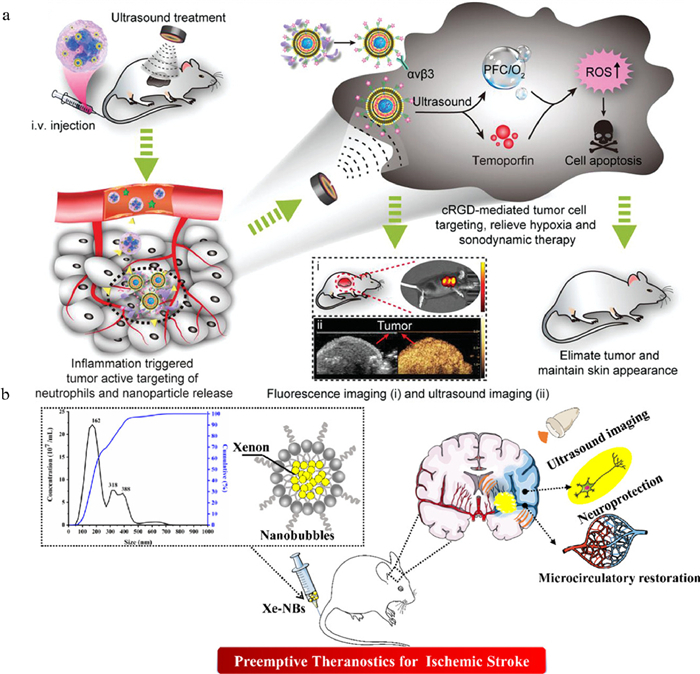
In additional to gas core, contrast agent for other kinds of imaging is another kind of functional composition scientists are loading onto MBs to have a better understanding of a patient's condition. It is reported that the attempt to load thrombus-specific theranostic nanoparticles onto MBs significantly enhanced the photoacoustic contrast in thrombosed vessels specifically and suppressed thrombus formation [120]. Lei Sun et al. chose to load temoporfin, a SDT agent, into their MBs. In this way, the tumors can be monitored in real time by temoporfin-mediated fluorescence imaging and PFC-MB-enhanced ultrasound imaging while reaching complete obliteration of tumors and efficient extension of the survival time of tumor-bearing mice with minimal systemic adverse effects (Fig. 6a) [96]. These loaded contrast agents, especially the ones for photoacoustic imaging, are always adopted to PTT as well. In another research, scientists encapsulated IR780-ND into MBs for photoacoustic and fluorescence imaging, as well as for its SDT effect [121]. Yet, most of the contrast agents loaded are photoacoustic agents, and this method is far from mature.
Concentrating on a single component with several functions is an additional method of creating theranostic MBs. Numerous studies focusing on the gas core have been proposed in light of the structure of MBs.
Inertial cavitation has been extensively exploited in this field due to its potent effects upon remote activation, as was previously indicated. Previous studies have suggested that genetically altered bacteria and cells may heterologously produce designed GVs, allowing for non-invasive gene expression imaging [122]. Additionally, the use of GVs as cavitation sources has been investigated. It was shown that these biomolecules may be transformed into micrometer-scale cavitating bubbles by low-frequency US, which then unleashes powerful local mechanical effects. As a result, in addition to acting as US imaging contrasts, manufactured GVs can operate as distantly actuated cell-eradicating agents [123].
Likely, high intensity focused ultrasound (HIFU) and radiofrequency ablation (RFA) may be improved by bubble-caused cavitation [124]. For instance, l-menthol was used to create a biocompatible "tri-phase transitional" platform [125]. The packed IR-780 would turn light into heat energy when faced an NIR laser, which would cause the enclosed l-menthol to vaporize. The continually created l-menthol bubbles could improve US imaging contrast. l-menthol bubble may also provide a cavitation effect that makes tumor cell membranes more permeable and subsequently enhances the PTT effect.
Gas therapy, which uses physiologically important gasses, is another way to make use of the therapeutic effects of gas core and has inspired novel, inventive ways to construct MBs [126]. The gas can still function as the gas core for imaging in addition to its biological impact. For example, H2O2 is now developing as a novel stimuli-responsive modality, with its capacity to produce O2 bubbles for US imaging, attenuate tumor hypoxia and improve the therapeutic effectiveness of various treatment subsequently [127].
Regarding anti-inflammatory gasses, it has been demonstrated that high levels of NO have a crucial impact on the immune system, with their main effects being anti-inflammatory and immunosuppressive [128]. For the simultaneous detection and precise therapy of antibody-mediated cardiac allograft rejection (AMR) in cardiac allografts, a new C4d-targeted NO-loaded MB was created. MBs are proved to stay in cardiac allografts with AMR, non-invasively sensing C4d deposition and accurately administering NO [97].
Similar work has also been pursued using Xenon (Xe). Xe has a strong therapeutic ability to treat neuronal damage and has been shown to be neuroprotective [129]. For the real-time US image-guided preventive therapy of the early stroke, Xe-enclosed nanosized MBs have been produced [129]. In vivo, the MBs at the site of the ischemic lesion provides the region US contrast imaging effect. Additionally, local neuroprotective Xe administration can restore neurological function with lessened neuron destruction from apoptosis (Fig. 6b) [130].
MBs is proven to be a promising tool for the integration of diagnosis and treatment for a variety of diseases. In this review, we summarized the functions of each component of MBs in diagnosis or treatment, and also proposed several methods to integrate these components to create theranostic MBs. Advances in theranostic MBs have expanded their use mainly to cancers, vascular diseases and CNS diseases. During the past decades, scientists have created more stable MBs, with the ability to circulate longer, target more precisely and carry more therapeutic components. However, there are still some limitations await to be tackled to fully unleash the potential of theranostic MBs. Although it is possible for some kinds of MBs to penetrate to deep tissue, how to balance the bubble size to guarantee both imaging effect and penetration ability is not entirely solved yet. Since imaging and therapeutic US require different US parameters, it will be more convenient if a theranostic US machine is manufactured to image and treat with one US probe. After all, US is the most widely used imaging modality, and the development of theranostic US will be beneficial for all.
The authors declared that they have no financial and personal relationships with other people or organizations that can inappropriately influence their work.
This work was supported by the National Natural Science Foundation of China-Sichuan Joint Fund Key Program (No. NSFCU21A20417), the National Natural Science Foundation of China (Nos. NSFC31930067, 81971622, 82272003), and the 135 Project for Disciplines of Excellence, West China Hospital, Sichuan University (No. ZYGD18002, China).
V. Paefgen, D. Doleschel, F. Kiessling, Front. Pharmacol. 6(2015) 197.
A.S. Hannah, G.P. Luke, S.Y. Emelianov, Theranostics 6(2016) 1866–1876. doi: 10.7150/thno.14961
E.G. Schutt, D.H. Klein, R.M. Mattrey, J.G. Riess, Angew. Chem. Int. Ed. 42(2003) 3218–3235. doi: 10.1002/anie.200200550
J.N. Meegoda, S. Aluthgun Hewage, J.H. Batagoda, Environ. Eng. Sci. 35(2018) 1216–1227. doi: 10.1089/ees.2018.0203
J. Bzyl, W. Lederle, A. Rix, et al., Eur. Radiol. 21(2011) 1988–1995. doi: 10.1007/s00330-011-2138-y
N.F. Bunkin, A.V. Shkirin, N.V. Penkov, et al., Front. Chem. 9(2021) 630074. doi: 10.3389/fchem.2021.630074
E. Stride, T. Segers, G. Lajoinie, et al., Ultrasound Med. Bio. 46(2020) 1326–1343. doi: 10.1016/j.ultrasmedbio.2020.01.027
M.S. Khan, J. Hwang, K. Lee, et al., Cancers 11(2019) 1464. doi: 10.3390/cancers11101464
L.J. Delaney, J.R. Eisenbrey, D. Brown, et al., Acta Biomater. 130(2021) 385–394. doi: 10.1016/j.actbio.2021.05.046
M. Zahiri, S. Taghavi, K. Abnous, et al., J. Control. Release 339(2021) 164–194. doi: 10.1016/j.jconrel.2021.09.032
A.H. Liao, C.R. Hung, H.K. Chen, C.P. Chiang, Sci. Rep. 8(2018) 8327. doi: 10.1038/s41598-018-26702-z
M. Lee, E.Y. Lee, D. Lee, B.J. Park, Soft Matter 11(2015) 2067–2079. doi: 10.1039/C5SM00113G
F. Cavalieri, M. Ashokkumar, F. Grieser, F. Caruso, Langmuir 24(2008) 10078–10083. doi: 10.1021/la801093q
M.R. Böhmer, R. Schroeders, J.A.M. Steenbakkers, et al., Colloid. Surface. A 289(2006) 96–104. doi: 10.1016/j.colsurfa.2006.04.011
M. Motornov, Y. Roiter, I. Tokarev, S. Minko, Prog. Polym. Sci. 35(2010) 174–211. doi: 10.1016/j.progpolymsci.2009.10.004
E. Castro-Hernández, W. van Hoeve, D. Lohse, J.M. Gordillo, Lab Chip 11(2011) 2023–2029. doi: 10.1039/c0lc00731e
G. Wang, L. Song, X. Hou, et al., Biomaterials 236(2020) 119803. doi: 10.1016/j.biomaterials.2020.119803
A.A. Exner, M.C. Kolios, Curr. Opin. Colloid In. 54(2021) 101463. doi: 10.1016/j.cocis.2021.101463
P.S. Sheeran, N. Matsuura, M.A. Borden, et al., IEEE T. Ultrason. Ferr. 64(2017) 252–263. doi: 10.1109/TUFFC.2016.2619685
Z. Hu, S.V. Bachawal, X. Li, et al., Mol. Imaging Biol. 24(2022) 333–340. doi: 10.1007/s11307-021-01680-3
G. Köse, M. Darguzyte, F. Kiessling, Nanomaterials 10(2020) 1935. doi: 10.3390/nano10101935
A.L. Klibanov, P.T. Rasche, M.S. Hughes, et al., Invest. Radiol. 39(2004) 187–195. doi: 10.1097/01.rli.0000115926.96796.75
M.W. Keller, S.S. Segal, S. Kaul, B. Duling, Circ. Res. 65(1989) 458–467. doi: 10.1161/01.RES.65.2.458
S.V. Bachawal, K.C. Jensen, A.M. Lutz, et al., Cancer Res. 73(2013) 1689–1698.
L. Zhang, T. Yin, B. Li, et al., ACS Nano 12(2018) 3449–3460. doi: 10.1021/acsnano.8b00076
D.G. Ramirez, E. Abenojar, C. Hernandez, et al., Nat. Commun. 11(2020) 2238. doi: 10.1038/s41467-020-15957-8
J. Wu, Y. Long, M. Li, Q. He, Acta Pharm. Sin. B 11(2021) 2286–2305. doi: 10.1016/j.apsb.2020.11.024
H. Maeda, Adv. Drug Deliver. Rev. 91(2015) 3–6. doi: 10.1016/j.addr.2015.01.002
X. Huang, W. Liao, Z. Xie, et al., Mat. Sci. Eng. C: Mat. 90(2018) 27–37. doi: 10.1016/j.msec.2018.04.036
J.M. Gorce, M. Arditi, M. Schneider, Invest. Radiol. 35(2000) 661–671. doi: 10.1097/00004424-200011000-00003
P.S. Sheeran, V.P. Wong, S. Luois, et al., Ultrasound Med. Biol. 37(2011) 1518–1530. doi: 10.1016/j.ultrasmedbio.2011.05.021
J. Xu, Y. Chen, L. Deng, et al., Biomaterials 106(2016) 264–275. doi: 10.1016/j.biomaterials.2016.08.034
T.O. Matsunaga, P.S. Sheeran, S. Luois, et al., Theranostics 2(2012) 1185–1198. doi: 10.7150/thno.4846
H. Yang, W. Cai, W. Lv, et al., Int. J. Nanomed. 14(2019) 7079–7093. doi: 10.2147/IJN.S207419
X. Shu, Y. Chen, P. Yan, et al., J. Control. Release 347(2022) 270–281. doi: 10.1016/j.jconrel.2022.05.010
H. Zhu, D. Qin, Y. Wu, et al., ACS Appl. Mater. Interfaces 10(2018) 29251–29259. doi: 10.1021/acsami.8b08190
J. An, J. Zhang, F. Dong, et al., Small 18(2022) 2105989. doi: 10.1002/smll.202105989
D.G. Ramirez, M. Ciccaglione, A.K. Upadhyay, et al., Proc. Natl. Acad. Sci. U. S. A. 118(2021) e2022523118. doi: 10.1073/pnas.2022523118
N.Y. Rapoport, A.M. Kennedy, J.E. Shea, et al., J. Control. Release 138(2009) 268–276. doi: 10.1016/j.jconrel.2009.05.026
S. Park, G. Son, Ultrason. Sonochem. 71(2021) 105361. doi: 10.1016/j.ultsonch.2020.105361
P.S. Sheeran, S.H. Luois, L.B. Mullin, et al., Biomaterials 33(2012) 3262–3269. doi: 10.1016/j.biomaterials.2012.01.021
P.S. Sheeran, J.D. Rojas, C. Puett, et al., Ultrasound Med. Biol. 41(2015) 814–831. doi: 10.1016/j.ultrasmedbio.2014.10.020
D.S. Li, S. Schneewind, M. Bruce, et al., Nano Lett. 19(2019) 173–181. doi: 10.1021/acs.nanolett.8b03585
R.W. Bourdeau, A. Lee-Gosselin, A. Lakshmanan, et al., Nature 553(2018) 86–90. doi: 10.1038/nature25021
F. Pfeifer, Nat. Rev. Microbiol. 10(2012) 705–715. doi: 10.1038/nrmicro2834
D.I. Piraner, A. Farhadi, H.C. Davis, et al., Biochemistry 56(2017) 5202–5209. doi: 10.1021/acs.biochem.7b00443
M.G. Shapiro, P.W. Goodwill, A. Neogy, et al., Nat. Nanotechnol. 9(2014) 311–316. doi: 10.1038/nnano.2014.32
J. Le Floc'h, A. Zlitni, H.A. Bilton, et al., Mol. Imaging Biol. 20(2018) 230–239. doi: 10.1007/s11307-017-1122-6
J. Huang, H. Zhang, Y. Yu, et al., Biomaterials 35(2014) 550–566. doi: 10.1016/j.biomaterials.2013.09.089
L.J. Cruz, P.J. Tacken, R. Fokkink, C.G. Figdor, Biomaterials 32(2011) 6791–6803. doi: 10.1016/j.biomaterials.2011.04.082
Q. Feng, W. Zhang, X. Yang, et al., Adv. Healthc. Mater. 7(2018) 1700957. doi: 10.1002/adhm.201700957
E. Kang, H.S. Min, J. Lee, et al., Angew. Chem. Int. Ed. 49(2010) 524–528. doi: 10.1002/anie.200903841
M. Kim, J.H. Lee, S.E. Kim, et al., ACS Appl. Mater. Interfaces 8(2016) 8409–8418. doi: 10.1021/acsami.6b02115
S. Chen, X.L. Xu, B. Zhou, et al., ACS Appl. Mater. Interfaces 11(2019) 22194–22205. doi: 10.1021/acsami.9b06745
K.H. Min, H.S. Min, H.J. Lee, et al., ACS Nano 9(2015) 134–145. doi: 10.1021/nn506210a
M. Versluis, E. Stride, G. Lajoinie, et al., Ultrasound Med. Biol. 46(2020) 2117–2144. doi: 10.1016/j.ultrasmedbio.2020.04.014
Y. Yang, L. Jing, X. Li, et al., Theranostics 7(2017) 466–481. doi: 10.7150/thno.17411
B. Helfield, Ultrasound Med. Biol. 45(2019) 282–300. doi: 10.1016/j.ultrasmedbio.2018.09.020
Y. Wang, H. Cong, S. Wang, et al., J. Mater. Chem. B 9(2021) 7633–7661. doi: 10.1039/D1TB00850A
M.W.N. Burns, R.F. Mattrey, J. Lux, ACS Appl. Mater. Interfaces 12(2020) 52298–52306. doi: 10.1021/acsami.0c12043
H. Chen, Z. Zhen, T. Todd, et al., Mater. Sci. Eng. R. Rep. 74(2013) 35–69. doi: 10.1016/j.mser.2013.03.001
A. Jafari Sojahrood, A.C. de Leon, R. Lee, et al., ACS Nano 15(2021) 4901–4915. doi: 10.1021/acsnano.0c09701
H. Wu, N.G. Rognin, T.M. Krupka, et al., Ultrasound Med. Biol. 39(2013) 2137–2146. doi: 10.1016/j.ultrasmedbio.2013.05.007
A. de Leon, R. Perera, C. Hernandez, et al., Nanoscale 11(2019) 15647–15658. doi: 10.1039/C9NR04828F
R. Zhao, J. Jiang, H. Li, et al., J. Neuroinflamm. 15(2018) 334. doi: 10.1186/s12974-018-1368-1
C. Wang, S. Yang, X. Chen, et al., Rev. Cardiovasc. Med. 22(2021) 1657–1666. doi: 10.31083/j.rcm2204173
Y.I. Yoon, S.W. Ha, H.J. Lee, J. Magn. Reson. Imaging 48(2018) 1610–1616. doi: 10.1002/jmri.26217
X. Mi, X. Guo, H. Du, et al., Nanomedicine 42(2022) 102533. doi: 10.1016/j.nano.2022.102533
Y. Xie, Y. Chen, L. Zhang, et al., Transplantation 103.8(2019) 1603.
L. Xinlong, Z. Xin, L. Chong, Chin. Chem. Lett. 32(2021) 2347–2358. doi: 10.1016/j.cclet.2021.03.015
M. Zhang, S. Gao, D. Yang, et al., Acta Pharm. Sin. B 11(2021) 2265–2285. doi: 10.1016/j.apsb.2021.03.033
J. Yang, X. Miao, Y. Guan, et al., Adv. Healthc. Mater. 10(2021) 2101628. doi: 10.1002/adhm.202101628
L. Xu, Y. Chen, Q. Jin, et al., Mol. Pharmaceut. 18(2021) 2974–2985. doi: 10.1021/acs.molpharmaceut.1c00145
N. Jugniot, T.F. Massoud, J.J. Dahl, R. Paulmurugan, J. Nanobiotechnol. 20(2022) 267. doi: 10.1186/s12951-022-01484-9
C.K. Kuhl, Annu. Rev. Med. 70(2019) 501–519. doi: 10.1146/annurev-med-121417-100403
M.M. Siddiqui, S. Rais-Bahrami, B. Turkbey, et al., JAMA 313(2015) 390–397. doi: 10.1001/jama.2014.17942
D. Fu, X. Huang, Z. Lv, et al., Bioengineered 13(2022) 7104–7116. doi: 10.1080/21655979.2022.2045832
J. Deprez, G. Lajoinie, Y. Engelen, et al., Adv. Drug Deliv. Rev. 172(2021) 9–36. doi: 10.1016/j.addr.2021.02.015
S. Snipstad, K. Vikedal, M. Maardalen, et al., Adv. Drug Deliv. Rev. 177(2021) 113847. doi: 10.1016/j.addr.2021.113847
S.P. Wrenn, S.M. Dicker, E.F. Small, et al., Theranostics 2(2012) 1140–1159. doi: 10.7150/thno.4305
Z. Izadifar, P. Babyn, D. Chapman, J. Med. Biol. Eng. 39(2019) 259–276. doi: 10.1007/s40846-018-0391-0
Thomas Leong, Ashok kumar, et al., Acoust. Aust. 39.2(2011) 54–63.
C.C. Church, E.L. Carstensen, Ultrasound Med. Biol. 27(2001) 1435–1437. doi: 10.1016/S0301-5629(01)00441-0
Y.J. Ho, H.C. Chang, C.W. Lin, et al., J. Control. Release 333(2021) 316–327. doi: 10.1016/j.jconrel.2021.03.044
J. Tu, A.C.H. Yu, BME Front. 2022(2022) 9807347.
S. El Kadi, T.R. Porter, N.J.W. Verouden, et al., JACC-Cardiovasc. Imag. 15(2022) 345–360.
K.H. Song, B.K. Harvey, M.A. Borden, Theranostics 8(2018) 4393–4408. doi: 10.7150/thno.26869
Q. Liu, J. Jiang, L. Tang, M. Chen, Ann. Transl. Med. 8(2020) 298. doi: 10.21037/atm.2020.02.155
N. Qu, D. Shi, M. Shang, et al., Med. Sci. Monit. 24(2018) 9054–9062. doi: 10.12659/MSM.910790
S. Kotopoulis, M. Popa, M. Mayoral Safont, et al., Pharmaceutics 14(2022) 98. doi: 10.3390/pharmaceutics14010098
J. Wischhusen, F. Padilla, IRBM 40(2019) 10–15. doi: 10.1016/j.irbm.2018.11.005
M. Argenziano, S. Occhipinti, A. Scomparin, et al., Drug Deliv. Transl. Res. 12(2022) 2007–2018. doi: 10.1007/s13346-022-01185-8
L. Hui, X. Xi, H. Jianbo, et al., Chin. Chem. Lett. 32(2021) 1759–1764. doi: 10.1016/j.cclet.2020.12.004
G. Dimcevski, S. Kotopoulis, T. Bjånes, et al., J. Control. Release 243(2016) 172–181. doi: 10.1016/j.jconrel.2016.10.007
J.R. Eisenbrey, F. Forsberg, C.E. Wessner, et al., Radiology 298(2021) 450–457. doi: 10.1148/radiol.2020202321
L. Sun, J.E. Zhou, T. Luo, et al., Adv. Mater. 34(2022) e2109969. doi: 10.1002/adma.202109969
T. Liao, Q. Li, Y. Zhang, et al., J. Heart Lung Transpl. 39(2020) 481–490. doi: 10.1016/j.healun.2020.02.002
J. Zhong, Y. Sun, Y. Han, et al., J. Thromb. Haemost. 19(2021) 738–752. doi: 10.1111/jth.15110
K. Fan, L. Zeng, J. Guo, et al., Theranostics 11(2021) 2670–2690. doi: 10.7150/thno.53083
X. Mi, H. Du, X. Guo, et al., Acta Biomater. 141(2022) 388–397. doi: 10.1016/j.actbio.2022.01.023
X. Wang, M. Shang, X. Sun, et al., J. Control. Release 343(2022) 66–77. doi: 10.1016/j.jconrel.2022.01.009
Z. Liu, H. Wang, Q. Zhao, et al., Mater. Express 11(2021) 240–247. doi: 10.1364/OME.412006
G. Wei, Y. Wang, G. Yang, et al., Theranostics 11(2021) 6370–6392. doi: 10.7150/thno.57828
N. Ingram, L.E. McVeigh, R.H. Abou-Saleh, et al., Theranostics 10(2020) 10973–10992. doi: 10.7150/thno.49670
X. Yang, M. Zhao, Z. Wu, et al., ACS Nano 16(2022) 3417–3431. doi: 10.1021/acsnano.2c00462
N. Yumita, R. Nishigaki, K. Umemura, S. Umemura, Jpn. J. Cancer Res. 80(1989) 219–222. doi: 10.1111/j.1349-7006.1989.tb02295.x
Z. Jiayi, O. Ai, S. Zhuanglin, et al., Chin. Chem. Lett. 33(2022) 1907–1912. doi: 10.1016/j.cclet.2021.11.017
W. Wu, Y. Pu, H. Lin, et al., Research 2021(2021) 9769867.
K. Logan, F. Foglietta, H. Nesbitt, et al., Eur. J. Pharm. Biopharm. 139(2019) 224–231. doi: 10.1016/j.ejpb.2019.04.003
W. Cao, Y. Liu, P. Ran, et al., ACS Appl. Mater. Interfaces 13(2021) 58411–58421. doi: 10.1021/acsami.1c19288
X. Jintao, Z. Nanqian, Y. Yuping, et al., Colloids Surf. B: Biointerfaces 190(2020) 110887. doi: 10.1016/j.colsurfb.2020.110887
R.J. Browning, S. Able, J.L. Ruan, et al., J. Control. Release 337(2021) 371–377. doi: 10.1016/j.jconrel.2021.07.020
G. Sun, J. Zhang, S. Wu, Y. Liu, Mater. Express 11(2021) 1045–1050. doi: 10.1166/mex.2021.2029
X.M. Anguela, K.A. High, Annu. Rev. Med. 70(2019) 273–288. doi: 10.1146/annurev-med-012017-043332
Y. Zhou, M. Yu, C. Tie, et al., Research 2021(2021) 9760398.
A.P.G. Walsh, H.N. Gordon, K. Peter, X. Wang, Adv. Drug Deliv. Rev. 179(2021) 113998. doi: 10.1016/j.addr.2021.113998
M. Wu, H. Xiong, H. Zou, et al., Acta Biomater 70(2018) 211–226. doi: 10.1016/j.actbio.2018.02.006
J.C. Wischhusen, S.M. Chowdhury, T. Lee, et al., J. Control. Release 321(2020) 272–284. doi: 10.1016/j.jconrel.2020.01.051
F. Wang, T. Shi, C. Su, Ultrasound Med. Biol. 45(2019) 859–866. doi: 10.1016/j.ultrasmedbio.2018.10.021
E. Jung, C. Kang, J. Lee, et al., ACS Nano 12(2018) 392–401. doi: 10.1021/acsnano.7b06560
L. Zhang, H. Yi, J. Song, et al., ACS Appl. Mater. Interfaces 11(2019) 9355–9366. doi: 10.1021/acsami.8b21968
A. Farhadi, G.H. Ho, D.P. Sawyer, et al., Science 365(2019) 1469–1475. doi: 10.1126/science.aax4804
A. Bar-Zion, A. Nourmahnad, D.R. Mittelstein, et al., Nat. Nanotechnol. 16(2021) 1403–1412. doi: 10.1038/s41565-021-00971-8
Y. Zhou, X. Han, X. Jing, Y. Chen, Adv. Healthc. Mater. 6(2017) 1700646. doi: 10.1002/adhm.201700646
C. Zhang, J. Liu, H. Guo, et al., Adv. Healthc. Mater. 8(2019) 1900409. doi: 10.1002/adhm.201900409
L. Chen, S.F. Zhou, L. Su, J. Song, ACS Nano 13(2019) 10887–10917. doi: 10.1021/acsnano.9b04954
Y. Luodan, C. Yu, C. Hangrong, Chin. Chem. Lett. 28(2017) 1841–1850. doi: 10.1016/j.cclet.2017.05.023
C. Bogdan, Nat. Immunol. 2(2001) 907–916. doi: 10.1038/ni1001-907
L. Van Hese, L. Al Tmimi, S. Devroe, et al., Brit. J. Anaesth. 121(2018) 1365–1368. doi: 10.1016/j.bja.2018.08.014
J. Jin, M. Li, J. Li, et al., ACS Appl. Mater. Interfaces 13(2021) 43880–43891. doi: 10.1021/acsami.1c06014

Figure 2 Pictures of diagnostic MBs. (a) SEM of air MBs. (Scale bar: 5 µm). Copied with permission [9]. Copyright 2022, Elsevier. (b) SEM images of C3F8 MB. Copied with permission [11]. Copyright 2018, Springer Nature. (c) TEM image of Anabaena flos-aquae derived gas vesicles. Copied with permission [17]. Copyright 2022, Elsevier. (d) CEUS imaging of orthotopic breast tumor and the sentinel lymph node in mice. Copied with permission [20]. Copyright 2021, Springer Nature.

Figure 3 Ultrasound imaging of tumor after injection of biotinylated ligand MBs. (a) Grayscale ultrasound imaging of tumor after injection of SonoVue MB, NB, NB-A, NB-R and NB-A/NB-R at various time points (10, 30, 60, 120 and 300 s). (b, c) Corresponding time-intensity curves and AUC analysis with data extracted from (a). The data represent the mean ± S.E.M. (n = 3 per group). A one-way ANOVA test was used for statistical analysis; ns: no significance, ***P < 0.001, ****P < 0.0001 compared with the Sonovue group. Copied with permission [65]. Copyright 2021, Elsevier.

Figure 4 MB cavitation. (a) Schematic of stable cavitation. Copied with permission [80]. Copyright 2003, Ivyspring International. (b) Schematic of inertial cavitation. Copied with permission [80]. Copyright 2003, Ivyspring International. (c) Impact of cavitation on cells. Copied with permission [84]. Copyright 2021, Elsevier.

Figure 5 US and MB enhance tumor inhibition in vivo. (a) Schematic illustration of the experiment. (b) Photographs of excised tumors at the experiment terminal. (c) The tumor growth curves of different groups in 14 days. (d) The weight of excised tumor in different groups. (e) Body weight changes of mice after different treatments. (f) HE staining, TUNEL and IHC analysis in different groups. Scale bar: 100 µm. All statistical data are expressed as means ± SD (n = 5). Copied with permission [101]. Copyright 2022, Elsevier.

Figure 6 Schematic of the effects of theranostic MBs. (a) Schematic of the MB combining SDT, fluorescence imaging and US imaging. Copied with permission [96]. Copyright 2022, John Wiley and Sons. (b) Schematic of the imaging and treating utilities of Xe-NBs. Copied with permission [130]. Copyright 2018, American Chemical Society.

 扫一扫看文章

扫一扫关注我们
 DownLoad:

 下载:
下载: