Table 1.
Crystallographic data of complexes 1-3

Self-assembly of zero-dimensional cluster {[Cu6(H2tba)6]·2DMF·xSolvent}, 2D sheet {[Sc(H2tba)3(DMF)]·2DMF}n and 3D network {[Fe(H2tba)2(H2O)2]·2DMF}n based on 2-thiobarbituric acid
Chao WANG , Dongmei HE , Yu GUAN , Jiayi XU , Limin ZHENG , Yuejian LIN , Zhenxia CHEN , Kai LIANG


2-Thiobarbituric acid (H3tba) is a substituted mercaptopyrimidine with three mobile H atoms and has many possible tautomeric structures[1-2]. H3tba and its derivatives are not only a kind of barbituric acid drugs, but also a polyfunctional N, N′, O, O′, S-donating ligand with different binding centers, which can form novel complexes of diverse structures in the water phase[3-14]. However, only a few reports have been devoted to the reaction of H3tba with metal ions in the organic phase[15-18]. In this work, we provide three examples of using H3tba and three different metal ions for the assembly of a hexa-copper cluster complex {[Cu6(H2tba)6]·2DMF·xSolvent} (1), 2D sheet {[Sc(H2tba)3(DMF)]·2DMF}n (2), and a 3D network structure coordination polymer {[Fe(H2tba)2(H2O)2]·2DMF}n (3). The luminescence properties of complex 1 were further studied.
All commercially available chemicals and solvents were of reagent grade and used as received without further purification. Sc(ClO4)3·6H2O was prepared by the reaction of Sc2O3 and perchloric acid (70.0%-72.0%) in the fume cupboard. Caution: Perchlorates are potentially explosive and should be handled with great care and in small amounts. Elemental analyses for C, H, and N were carried out with a Vario EL Ⅲ elemental analyzer. IR spectra were recorded on a Nicolet 6700 FT-IR spectrometer with KBr pellets in the 4 000-400 cm-1 region. The thermogravimetric analysis (TGA) measurements were carried out on a TG 209 F1 thermal analyzer heated from room temperature to 900 ℃ under N2 atmosphere with a heating rate of 10 ℃·min-1. Powder X-ray diffraction (PXRD) patterns were taken on a Rigaku D/max-2550 PC X-ray diffractometer (Cu Kα, λ=0.154 184 nm) at room temperature (40 kV, 40 mA, 2θ=5°-70°). The fluorescent spectra were obtained on a PTI QM/TM luminescence spectrometer at room temperature.
A solution of Cu(ClO4)2·6H2O (0.1 mmol, 37 mg) in MeOH (2 mL) was added to a solution of H3tba (0.2 mmol, 29 mg) in DMF (6 mL), and colorless single crystals were obtained several days later. The single crystal diffracted weakly and easily lost some of the occluded solvents. The products were collected, washed with ethanol three times, and dried in air. Yield: 0.009 g (26% based on Cu). Anal. Calcd. for C24H18N12O12S6Cu6·4DMF·5CH3OH(%): C, 29.09; H, 3.93; N, 13.24; Found(%): C, 29.35; H, 3.46; N, 13.63. IR (KBr, cm-1): 3 104 (s), 2 973 (m), 2 976 (m), 1 654 (s), 1 637 (s), 1 526 (s), 1 355 (s), 1 319 (s), 1 161 (s), 1 122 (m), 799 (m), 520 (m), 458 (m).
A solution of Sc(ClO4)3·6H2O (0.25 mmol, 113 mg) in MeOH (2 mL) was added to a solution of H3tba (0.75 mmol, 108 mg) in DMF (3 mL), and red rectangular plates formed. The products were collected, washed with ethanol three times, and dried in air. Yield: 0.058 g (52% based on Sc). Anal. Calcd. for C21H30N9O9S3Sc(%): C, 36.36; H, 4.36; N, 18.17; Found(%): C, 36.59; H, 4.83; N, 18.04. IR (KBr, cm-1): 3 146 (s), 3 071 (s), 2 920 (s), 2 884 (s), 1 645 (s), 1 602 (s), 1 414 (s), 1 386 (s), 1 302(s), 1 187 (s), 1 137 (s), 891 (m), 794 (m), 699 (m), 664 (m).
A solution of Fe(ClO4)3·9H2O (0.1 mmol, 52 mg) in MeOH (1 mL) was added to a solution of H3tba (0.2 mmol, 29 mg) in DMF (0.5 mL), and yellow rectangular plates formed for single-crystal X-ray diffraction. The products were collected, washed with ethanol three times, and dried in air. Yield: 0.021 g (42% based on Fe). Anal. Calcd. for C14H24N6O8S2Fe(%): C, 32.07; H, 4.61; N, 16.03; Found(%): C, 31.94; H, 4.61; N, 16.37. IR (KBr, cm-1): 3 108 (m), 3 078 (m), 2 874(m), 1 645 (s), 1 597 (s), 1 201 (s), 1 182 (s), 890 (m), 815 (m), 539 (s), 485 (m).
Data collection for complexes 1 and 2 were carried out on a Bruker Smart Apex CCD diffractometer with a graphite-monochromatic Mo Kα radiation (λ=0.071 073 nm) at 173 and 273 K, respectively. Data collection for complex 3 was carried out on a Rigaku synergy X-ray single crystal diffractometer with Cu Kα radiation (λ=0.154 184 nm) at 100 K. The structures were solved by direct methods and refined on F 2 with the SHELX-97 program[19]. All nonhydrogen atoms were refined with anisotropic thermal parameters. The positions of hydrogen atoms were generated geometrically. In complex 1, only two of the DMF molecules were refined isotropically with hydrogen atoms included. Due to the disorder, the other solvent molecules were removed using SQUEEZE[20]. The structure of complex 3 is a merohedral twin. The crystallographic data and details of refinements for complexes 1-3 are summarized in Table 1.
 下载:
导出CSV
下载:
导出CSV
| Parameter | 1 | 2 | 3 |
| Empirical formula | C30H32N14O14S6Cu6 | C21H30N9O9S3Sc | C14H24N6O8S2Fe |
| Formula weight | 1 386.36 | 693.71 | 524.36 |
| Temperature / K | 173 | 273(2) | 100(2) |
| Crystal system | Triclinic | Triclinic | Tetragonal |
| Space group | P1 | P1 | I42d |
| a / nm | 1.317 6(3) | 1.132 8(3) | 1.704 54(7) |
| b / nm | 1.326 2(4) | 1.185 4(3) | 1.704 54 |
| c / nm | 1.378 3(4) | 1.327 8(4) | 0.748 58(6) |
| α / (°) | 86.657(5) | 111.959(4) | |
| β / (°) | 85.127(5) | 104.473(4) | |
| γ / (°) | 65.488(5) | 90.099(4) | |
| Volume / nm3 | 2.182 6(10) | 1.592 2(8) | 2.175 0(2) |
| Z | 1 | 2 | 4 |
| Dc / (g·cm-3) | 1.055 | 1.447 | 1.601 |
| μ / mm-1 | 1.620 | 0.488 | 7.850 |
| F(000) | 692 | 720 | 1 088 |
| 2θ range for data collection / (°) | 2.966-53.160 | 3.434-50.484 | 7.34-147.32 |
| Reflection collected | 17 231 | 5 478 | 2 232 |
| Independent reflection | 8 831 | 2 136 | 1 033 |
| Goodness-of-fit on F 2 | 0.97 | 0.672 | 1.177 |
| R1, wR2 [I > 2σ(I)] | 0.057 9, 0.158 1 | 0.050 5, 0.073 3 | 0.039 2, 0.109 8 |
| R1, wR2 (all data) | 0.092 1, 0.178 2 | 0.124 1, 0.082 8 | 0.039 3, 0.110 0 |
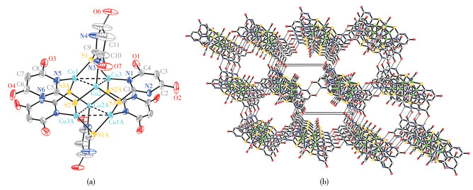
Single-crystal structure analysis of complex 1 reveals that the crystal structure is the triclinic system, space group P1. The six copper atoms adopt a trigonal antiprismatic arrangement as shown in Fig. 1a. Each copper atom is trigonally coordinated to a ligand nitrogen atom and two bridging sulfur atoms with bond lengths: Cu1—N5 0.200 5(4) nm, Cu2—N3 0.199 2(5) nm, Cu3—N1 0.199 8(4) nm, Cu1—S1 0.221 40(14) nm, Cu1—S3A 0.226 92(16) nm, Cu2—S2 0.223 27(14) nm, Cu2—S3 0.224 38(14) nm, Cu3—S1 0.221 52(15) nm, and Cu3—S2A 0.225 86(17) nm. Furthermore, each copper atom exhibits secondary Cu…Cu bonding to two other copper atoms forming a triangle (Cu1…Cu2 0.282 11(12) nm, Cu2…Cu3 0.292 19(12) nm, Cu3…Cu1 0.302 26(10) nm) with bond angles ∠Cu1—Cu2—Cu3 of 63.48(2)°, ∠Cu2—Cu1—Cu3 of 59.88(3)°, ∠Cu2—Cu3—Cu1 of 56.63(3)° indicated by broken lines in Fig. 1a. In complex 1, the C—S bond lengths are in a range of 0.172 6(7)-0.175 3(6) nm, indicating they are single bonds. The ligand loses one proton, and the singly deprotonated ligand bridges the copper atoms through its sulfur and nitrogen donor atoms. Then Cu (Ⅱ) ions were found to result in a reduction to afford Cu (Ⅰ) ions in complex 1. The guest molecules desorb easily within a few minutes when the crystals are taken out from the mother liquor. There are large channels in the structure that contain disorder solvent molecules. The intermolecular hydrogen bonding and non-covalent interactions assemble these aggregates into a distorted face-centered cubic lattice, as illustrated in Fig. 1b
Symmetry code: A: 1-x, 1-y, 1-z.
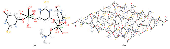
There are two different coordination modes for Sc(Ⅲ) ions in complex 2 as shown in Fig. 2a. The Sc1 atom has six oxygen donors which consist of two O atoms from two DMF molecules, and four O atoms from four bridged H2tba- ligands. The other Sc2 atom has six oxygen donors which consist of two O atoms from two single H2tba- anions, and four O atoms from four bridged H2tba- ligands. The Sc1 atom is bound by six O atoms (O1, O1A, O4C, O4D, O5, and O5A), with bond lengths: Sc1—O1 0.211 1(3) nm, Sc1—O5 0.204 0(3) nm, Sc1—O4C 0.207 9(3) nm, and Sc1—O4D 0.207 9(3) nm, bond angles: O1—Sc1—O1A 180.0°, O4C—Sc1—O4D 180.0°, O5—Sc1—O5A 180.0°, O1—Sc1—O5 88.55(11)°, O1—Sc1—O4C 90.09(11)°, O5—Sc1—O4C 89.34(11)°, O5—Sc1—O4D 90.66(11)°. The Sc2 atom is bound by O2, O2B, O3, O3B, O7, and O7B atoms, with bond lengths: Sc2—O2 0.207 0(4) nm, Sc2—O3 0.204 2(3) nm, Sc2—O7 0.209 6(4) nm, bond angles: O2—Sc2—O2B 180.0°, O3—Sc2—O3B 180.0°, O7—Sc2—O7B 180.0°, O2—Sc2—O7 90.97(14)°, O2—Sc2—O3 91.22(13)°, O3—Sc2—O7 89.52(14)°. The Sc (Ⅲ) ions are bridged by four H2tba- anions to afford a 2D layer network viewed along the crystallographic c-axis, as illustrated in Fig. 2b. The adjacent sheets are observed stacking with an ABC sequence. The 2D layer structure is stabilized by hydrogen bonds and a π-π interaction between the H2tba- ligands and DMF molecules.
Symmetry codes: A: -x, 1-y, 1-z; B: 1-x, -y, 1-z; C: x-1, y, z; D: 1-x, 1-y, 1-z.
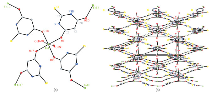
In complex 3, each Fe (Ⅱ) ion has six oxygen donors which consist of two O atoms from two coordinated H2O molecules, and four O atoms from four H2tba- ligands as shown in Fig. 3a. The Fe (Ⅱ) ion is bound by O1, O1A, O1B, O1C, O1W, and O1WB atoms, with bond lengths: Fe1—O1 0.214 8(3) nm, Fe1—O1A 0.214 8(3) nm, Fe1—O1B 0.214 8(3) nm, Fe1—O1C 0.214 8(3) nm, Fe1—O1W 0.206 9(5) nm, and Fe1—O1WB 0.206 9(5) nm, bond angles: O1—Fe1—O1A 173.76(18)°, O1W—Fe1—O1WB 172.6(6)°, O1C—Fe1—O1B 173.76(18)°, O1—Fe1—O1B 90.170(11)°, O1—Fe1—O1C 90.170(10)°, O1W—Fe1—O1C 93.8(10)°, O1A—Fe1—O1C 90.170(11)°. In complex 3, the C3—S1 and C2—O1 distances are 0.166 2(5) and 0.127 1(5) nm, respectively, indicating they are double bonds. The C1—C2 distance is 0.139 5(5) nm, C3—N1 and the C2—N1 bond lengths are in a range of 0.135 7(4)-0.138 7(6) nm, indicating the C1 and N1 atoms are bonded with one H atom respectively. The H3tba ligand loses one proton, and the H2tba- ligand bridges the metal centers through two oxygen donor atoms. Then Fe (Ⅲ) ions were found to result in reduction to afford Fe (Ⅱ) ions in complex 3. There are large channels in the structure that contain the DMF molecules involved in the intermolecular hydrogen bonding that assembles these aggregates into a distorted face- centered cubic lattice, as illustrated in Fig. 3b.
Symmetry codes: A: 1-x, 1-y, z; B: x, 1-y, 1-z; C: 1-x, y, 1-z; D: 0.5-x, y, 0.75-z; E: x, 0.5-y, 1.25-z; F: x, 1.5-y, 1.25-z; G: -0.5+x, 1-y, 0.75-z; H: 0.5+x, 1-y, 0.75-z.
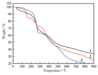
TGA performed for complex 1 as illustrated in Fig. 4 showed a weight loss of about 27.63% at 265 ℃, which is close to the calculated values of 26.74% for the removal of guest DMF molecules and MeOH molecules. Complex 2 showed a weight loss of about 31.60% at 300 ℃, which is close to the calculated values of 28.15% for the removal of guest DMF molecules and coordinated DMF molecules. Complex 3 showed a weight loss of about 26.09% at 280 ℃, which is close to the calculated values of 24.70% for the removal of guest DMF molecules and coordinated H2O molecules. As the temperature increased, the elimination of the organic ligands began for complexes 1-3. The final residues (33.52%) for 1 at 900 ℃ are almost agreed with the calculated values of 34.42% based on CuO. The final residues of about 25.50% for 2 at 900 ℃ and 21.24% for 3 at 800 ℃ are slightly higher than the calculated values of 19.88% and 13.70% based on Sc2O3 and FeO, respectively. The reason could be attributed to the formation of Ln2O2SO4 oxysulfates and oxide of iron.
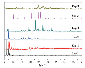
The single crystals of complexes 1 and 3 lost crystallization when the crystals were taken out from the mother liquor a few minutes later. The loss of solvent molecules from the crystals may result in another phase. So, the PXRD analysis was performed for the complexes. The PXRD patterns of complexes 1 and 3 had slight displacement deviation from the single- crystal simulation results. The single-crystal simulation result for complexes 2 agrees well with the experiment PXRD pattern, as illustrated in Fig. 5.
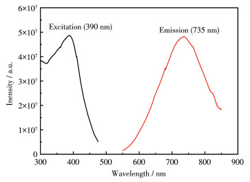
The luminescent spectrum of complex 1 in the solid state is shown in Fig. 6, with a maximum of 735 nm (λex=390 nm). Complex 1 exhibited an emission red shift compared with the free H3tba ligand upon the excitation, indicating that the emission of the complex may be assigned to the ligand-to-metal charge transfer (LMCT)[22].
In summary, three complexes using tautomeric H3tba as ligands, with increasing dimensionality from the 0D cluster, 2D sheet, to the 3D network were synthesized by self-assembly reaction. The results reveal that the dimensionality of this kind of complex can be modulated by using different metal salts. We believe that diverse structure types can be further constructed with H3tba ligands in the future.
Méndez E, Cerdá M F, Gancheff J S, Torres J, Kremer C, Castiglioni J, Kieninger M, Ventura O N. Tautomeric forms of 2-thiobarbituric acid as studied in the solid, in polar solutions, and on gold nanoparticles[J]. J. Phys. Chem. C, 2007, 111: 3369-3383.
Chierotti M R, Ferrero L, Garino N, Gobetto R, Luca Pellegrino, Braga D, Grepioni F, Maini L. The richest collection of tautomeric polymorphs: The case of 2-thiobarbituric acid[J]. Chem.-Eur. J., 2010, 16: 4347-4358. doi: 10.1002/chem.200902485
Dhasmana A, Barthwal J P, Pandey B R, Ali B, Bhargava K P, Parmar S S. Anticonvulsant activity and succinate dehydrogenase inhibitory property of new substituted thiobarbiturates[J]. J. Heterocycl. Chem., 1981, 18(3): 635-637. doi: 10.1002/jhet.5570180341
Balas V I, Hadjikakou S K, Hadjiliadis N, Kourkoumelis N, Light M E, Hursthouse M, Metsios A K, Karkabounas S. Crystal structure and antitumor activity of the novel zwitterionic complex of tri-n-butyltin(Ⅳ) with 2-thiobarbituric acid[J]. Bioinorg. Chem. Appl., 2008, : . doi: 10.1155/2008/654137
Golovnev N N, Molokeev M S. Bridging behaviour of the 2-thiobarbiturate anion in its complexes with Li and Na[J]. Acta Crystallogr. Sect. C, 2013, C69(7): 704-708.
Golovnev N N, Molokeev M S. Crystal structures of cesium and rubidium 2-thiobarbiturates[J]. Russ. J. Inorg. Chem., 2014, 59(9): 943-946. doi: 10.1134/S003602361409006X
Golovnev N N, Molokeev M S. Crystal structures of two barium 2-thiobarbiturate complexes[J]. J. Struct. Chem., 2014, 55(5): 871-878. doi: 10.1134/S0022476614050114
Golovnev N N, Molokeev M S, Vereshchagin S N. Calcium and strontium thiobarbiturates with discrete and polymeric structures[J]. J. Coord. Chem., 2013, 66(23): 4119-4130. doi: 10.1080/00958972.2013.860450
Golovnev N N, Molokeev M S, Vereshchagin S N, Atuchin V V, Sidorenko M Y, Dmitrushkov M S. Crystal structure and properties of the precursor [Ni(H2O)6](TBA)2·2H2O and the complexes M(TBA)2(H2O)2 (M=Ni, Co, Fe)[J]. Polyhedron, 2014, 70: 71-76. doi: 10.1016/j.poly.2013.12.021
Golovnev N N, Molokeev M S. Crystal structure of catena-bis (2-thiobarbiturato-O, S)diaquacadmium[J]. Russ. J. Inorg. Chem., 2013, 58(10): 1193-1196. doi: 10.1134/S0036023613100094
Golovnev N N, Molokeev M S. Crystal structure of catena-di(2-thiobarbiturato-O, S)aqualead(Ⅱ)[J]. J. Struct. Chem., 2017, 54(5): 968-971.
Golovnev N N, Molokeev M S. Crystal structure of polymer hexaaqua-hexakis-(2-thiobarbiturato)dieuropium(Ⅲ)[J]. Russ. J. Coord. Chem., 2014, 40(9): 648-652. doi: 10.1134/S1070328414090036
Golovnev N N, Molokeev M S, Sterkhova I V, Vereshchagin S N, Golovneva I I. Crystal structure and properties of polymeric hexaaqua-hexakis-(2-thiobarbiturato)disamarium(Ⅲ)[J]. J. Struct. Chem., 2017, 58(3): 539-543. doi: 10.1134/S0022476617030155
Golovnev N N, Molokeev M S, Sterkhova I V. Structure and thermal decomposition of Nd(Ⅲ), Gd(Ⅲ) and Tb(Ⅲ) 2-thiobarbiturates[J]. Russ. J. Inorg. Chem., 2019, 64(9): 1146-1151. doi: 10.1134/S0036023619090134
Golovnev N N, Molokeev M S, Lesnikov M K. Hydrates of lanthanide (Ⅲ) 2-thiobarbiturates: Synthesis, structure and thermal decomposition[J]. Russ. J. Inorg. Chem., 2020, 65(7): 999-1005. doi: 10.1134/S0036023620070098
Hunks W J, Jennings M C, Puddephatt R J. Supramolecular gold (Ⅰ) thiobarbiturate chemistry: Combining aurophilicity and hydrogen bonding to make polymers, sheets, and networks[J]. Inorg. Chem., 2002, 41: 4590-4598. doi: 10.1021/ic020178h
Pan Z R, Zhang Y C, Song Y L, Zhuo X, Li Y Z, Zheng H G. Synthesis, structure and nonlinear optical properties of three dimensional compounds[J]. J. Coord. Chem., 2008, 61: 3189-3199. doi: 10.1080/00958970802020673
Gong Y, Hao Z, Li J H, Wu T, Lin J H. Mg(Ⅱ)-induced second-harmonic generation based on bis-monodentate coordination mode of thiobarbiturate[J]. Dalton Trans., 2013, 42: 6489-6494. doi: 10.1039/c3dt32380c
Wa ng, C , Zh ou, M S, Ya ng, L J, Liang , K , Z hu, Z J, Ch en, Z X. Synthesis, structure and luminescent properties of a 2D cobalt (Ⅱ) coordination polymer based on 2-thiobarbituric acid[J]. Chin. J. Struct. Chem., 2017, 36(7): 1210-1215.
Sheldrick G M. SHELXTL 97, Program for the refinement of crystal structure. University of Göttingen, Germany, 1997.
Spek A L. Platon squeeze: A tool for the calculation of the disordered solvent contribution to the calculated structure factors[J]. Acta Crystallogr. Sect. C, 2015, C71: 9-18.
Yam V W W, Lo K K W. Luminescent polynuclear d10 metal complexes[J]. Chem. Soc. Rev., 1999, 28: 323-334. doi: 10.1039/a804249g

Figure 1 Coordination environments of the Cu (Ⅰ) ions of complex 1 with thermal ellipsoid at 50% probability levels; (b) Packing diagram of 1 viewed along the a-axis
Symmetry code: A: 1-x, 1-y, 1-z.

Figure 2 (a) Coordination environments of the Sc (Ⅲ) ions of complex 2 with thermal ellipsoid at 50% probability levels; (b) 2D network of one sheet in 2 viewed along the c-axis
Symmetry codes: A: -x, 1-y, 1-z; B: 1-x, -y, 1-z; C: x-1, y, z; D: 1-x, 1-y, 1-z.

Figure 3 (a) Coordination environments of the Fe (Ⅱ) ions complex 3 with thermal ellipsoid at 30% probability levels; (b) Packing diagram of 3 viewed along the c-axis
Symmetry codes: A: 1-x, 1-y, z; B: x, 1-y, 1-z; C: 1-x, y, 1-z; D: 0.5-x, y, 0.75-z; E: x, 0.5-y, 1.25-z; F: x, 1.5-y, 1.25-z; G: -0.5+x, 1-y, 0.75-z; H: 0.5+x, 1-y, 0.75-z.
Table 1. Crystallographic data of complexes 1-3
| Parameter | 1 | 2 | 3 |
| Empirical formula | C30H32N14O14S6Cu6 | C21H30N9O9S3Sc | C14H24N6O8S2Fe |
| Formula weight | 1 386.36 | 693.71 | 524.36 |
| Temperature / K | 173 | 273(2) | 100(2) |
| Crystal system | Triclinic | Triclinic | Tetragonal |
| Space group | P1 | P1 | I42d |
| a / nm | 1.317 6(3) | 1.132 8(3) | 1.704 54(7) |
| b / nm | 1.326 2(4) | 1.185 4(3) | 1.704 54 |
| c / nm | 1.378 3(4) | 1.327 8(4) | 0.748 58(6) |
| α / (°) | 86.657(5) | 111.959(4) | |
| β / (°) | 85.127(5) | 104.473(4) | |
| γ / (°) | 65.488(5) | 90.099(4) | |
| Volume / nm3 | 2.182 6(10) | 1.592 2(8) | 2.175 0(2) |
| Z | 1 | 2 | 4 |
| Dc / (g·cm-3) | 1.055 | 1.447 | 1.601 |
| μ / mm-1 | 1.620 | 0.488 | 7.850 |
| F(000) | 692 | 720 | 1 088 |
| 2θ range for data collection / (°) | 2.966-53.160 | 3.434-50.484 | 7.34-147.32 |
| Reflection collected | 17 231 | 5 478 | 2 232 |
| Independent reflection | 8 831 | 2 136 | 1 033 |
| Goodness-of-fit on F 2 | 0.97 | 0.672 | 1.177 |
| R1, wR2 [I > 2σ(I)] | 0.057 9, 0.158 1 | 0.050 5, 0.073 3 | 0.039 2, 0.109 8 |
| R1, wR2 (all data) | 0.092 1, 0.178 2 | 0.124 1, 0.082 8 | 0.039 3, 0.110 0 |
 下载: 导出CSV

 扫一扫看文章


扫一扫关注我们