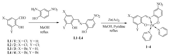
Scheme 1.
Syntheses of complexes 1~4

Zinc is a very important trace element in the normal growth and development of the human body[1]..The content of zinc ions in the living body is the second only to iron, and it ranks second in the total trace elements. Zinc is known as the 'spark of life'. Zinc is a constituent of more than 200 enzymes in the human body, mainly in the form of metalloenzymes, or as an activator of enzymes. Therefore, zinc has a wide range of distribution and action in the body. It is also a component of insulin in the body, which can prolong the physiological effect of insulin and have different effects on the metabolism of body cells[2]. Therefore, the intelligent design of Zn(Ⅱ) complex metal drugs has important research significance for human health. Zinc is a transition metal element, which is well coordinated with a molecule containing lone-pair electron to form a complex[3-5]. Zinc ions have the following characteristics: good Lewis acidity; no redox activity; good solubility; low toxicity, and so on[6, 7]. Since the coordination bonds have a certain directionality and intensity, the researchers can regulate molecular structure through a coordination bond, so the ligand and zinc ion unit having a specific structure and function are designed to obtain a new zinc complex. In recent years, more and more zinc complexes have been synthesized, and their structures have been continuously published[8, 9]. These zinc complexes exhibit diverse properties due to the different ligand structures and coordination configuration, such as catalysis[10, 11], fluorescence[12-15], biological activity[16, 17], and so on. In this paper, four pyridine zinc(Ⅱ) complexes based on halogenated salicylaldehyde Schiff base are synthesized (Scheme 1), and the interaction between complexes and ct-DNA were studied to screen the biologically active drug structure, which can provide an important theoretical basis for developing new metal drugs.
Infrared spectrum (KBr) was recorded by the Prestige-21 infrared spectrometer (Japan Shimadzu, 4000~400 cm–1). 1H NMR spectra were measured with a Bruker AVANCE-400 NMR spectrometer. The elemental analysis was determined by PE-2400(Ⅱ) elemental analyzer. Crystallographic data of the complexes were collected on a Bruker SMART APEX Ⅱ CCD diffractometer. Melting points were determined using an X4 digital microscopic melting point apparatus without correction (Beijing Tektronix Instrument Co. Ltd.). Thermogravimetric analyses (TGA) were recorded on a NETZSCH TG 209 F3 instrument at a heating rate of 20 ℃·min-1 from 40 to 800 ℃ under air. The UV spectra were determined with the UV-2550 spectrophotometer (Shimazu). Fluorescence spectra were obtained with a Hitachi F-7000 spectrophotometer with quartz cuvette (path length = 1 cm).
The reagents used in experiment were all analytical reagent, and used directly without further purification.
A mixture of 4-NAP (2-amino-4-nitrophenol) (10 mmol), halogenated salicylaldehyde (10 mmol) and CH3OH (50 mL) was added in a round-bottomed flask (100 mL), and refluxed with agitating for 4.0 h. The solution was cooled, and then the insoluble matter is removed by filtration. Four ligands L1~L4 were obtained.
1 mmol Schiff base ligand (L1/L2/L3/L4) and 1 mmol zinc acetate were added in CH3OH (50 mL) and refluxed with agitating for 4 h. Then, most of the methanol was evaporated, followed by adding pyridine dropwise until it just dissolved. The reaction solution continued to reflux for 2.5 h, filtered. Finally, the crystals were precipitated by controlling solvent volatilization.
The crystals of complexes were mounted on a diffractometer equipped with graphite-monochromated MoKα radiation at a φ-ω mode. All the data were corrected by Lp factors and empirical absorbance. Structure has been solved by direct methods. All non-hydrogen atoms were defined in successive difference Fourier synthesis, and H atoms were added according to theoretical models or located from the Fouriermaps. Non-hydrogen atoms were refined by their isotropic and anisotropic thermal parameters. All calculations were completed by the SHELXTL-97[18] program. Crystallographic data are listed in Table S1, and the bond data are summarized in Table S2.
ct-DNA (30 μM), EB (3 μM) and different concentration complex solution (0~50 μM) were placed in a 5 mL volumetric flask in tris-HCl (0.01 mol·L–1) buffer solution[19]. After 3 h, the fluorescence spectra were acquired at 25 ℃. The excitation wavelength was 258 nm, and the emission wavelength is shown in the spectrum. The slit scanning width of emission and excitation is 5.0 nm.
In the synthesis of ligands L1~L4, their solubility in CH3OH is relatively small. After cooling, they will be precipitated from the reaction mixture. The raw material will be dissolved in CH3OH, so the crude products of L1~L4 have been obtained by Vacuum filtration. After that, these ligands were recrystallized with CH3OH to obtain pure product with the yields of 68%~78%. This is a general way to synthesize Schiff base compounds. Complexes 1~4 have been obtained by self-assembly reactions. Pyridine participates in coordination as an electron donor. Finally, the complex crystals were obtained by solvent evaporation method. All complexes are orange yellow transparent crystals with their yields all around 80%.
The infrared peak shapes of the four ligands are the same, and the infrared peak shapes of the four complexes are also very similar, which preliminarily shows that they all have similar main structures. By comparing the infrared-spectra of ligands L1~L4 and complexes 1~4, we can see that the latter do not show O–H bond stretching vibration in the high-frequency region, indicating that the ligand may participate in coordination of new complexes through hydrogen protons[20-24]. The characteristic absorption peaks of -C=N and -NO2 are basically the same in ligands L1~L4 and complexes 1~4.
In the 1H NMR spectrum, the peak positions and integrated areas appearing in the spectrum match the number of hydrogen protons of the predicted complexes[25]. The other peaks are all in the low-field position, and no peak signal is found in the high-field position, indicating that all the complexes contain unsaturated hydrogen atoms without saturated ones. In the complex, the two -OH hydrogen proton peaks disappeared, indicating that the metal atom is coordinated with the Schiff base ligand. The characterization results of the complexes are consistent with those of X-ray single-crystal diffraction.
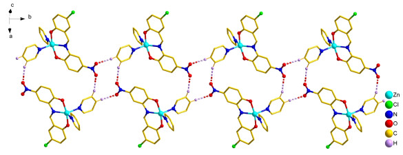
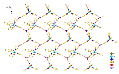
The structures of complexes 1~4 in solid state are shown in Fig. 1. The complexes are all single-metal nuclear molecules, and the coordination mode of the central Zn atom is similar. Take complex 1 as an example, Zn(1) forms a five-coordinated triangular bipyramidal configuration by two oxygen atoms (O(1) or O(2)) and one imino nitrogen atom (N(1)) from the ligand, and N(3) or N(4) from the coordination pyridine, respectively. N(1), N(3), and N(4) occupy three positions on the equatorial plane, on both sides of which the two oxygen atoms O(1) and O(2) are located. The axial angle O(1)–Zn(1)–O(2) is 167.08°, which obviously deviates from a line (180°). The angle and distance of three atoms of complex 1 in the equatorial-plane and Zn(1) atom are different: dZn(1)–N(1) = 2.0846(17) Å, dZn(1)–N(3) = 2.1060(17) Å, dZn(1)–N(4) = 2.0711(17) Å, bond angle N(4)–Zn(1)–N(3) = 100.87(7)°, bond angle N(1)–Zn(1)–N(3) = 134.23(7)°, and bond angle N(4)–Zn(1)–N(1) = 124.77(6)°. Therefore, the center zinc atom adopts a distorted five-coordinate triangular bipyramidal-configuration. 2~4 are similar to 1 in crystal structure, but the bond parameters are different, and the center Zn atom also shows a distorted five-coordinate triangular bipyramidal configuration.

Complexes 1 and 3 form respectively a one-dimensional chain structure and a two-dimensional grid structure by lots of hydrogen bonds (Figs. 2 and 3). In 1 and 3, the C–H···O hydrogen bonds are also mainly constructed by hydrogen atoms from phenyl group and oxygen atoms from -NO2. Interestingly, despite all complexes have similar structures, the hydrogen bonds are different. There are no hydrogen bonds in complexes 2 and 4. In a basic unit, the number of hydrogen bonds in 1 and 3 is also 2 maybe due to the too large steric hindrance of the phenyl group in complexes 2 and 4. For 1, dC(21)–H(21)...O(3) = 2.410 Å, dC(22)–H(22)...O(4) = 2.535 Å, C(21)–H(21)···O(3) = 176.21°, C(22)–H(22)···O(4) = 161.46°; For 3, dC(16)–H(16)...O(3) = 2.670 Å, dC(22)–H(22)...O(4) = 2.594 Å, C(16)–H(16)···O(3) = 150.65°, C(22)–H(22)···O(4) = 125.90°. The weak action of the complexes is listed in Table S3 (SI).
Thermal stability was performed under air atmosphere with the gas-flow rate of 20.0 mL·min–1 from 40 to 800 ℃ at a heating rate of 20 ℃·min–1. As depicted in Fig. 4, complexes 1~4 show a similar weight loss process, which can be roughly divided into two weight loss stages. All four complexes display weight loss at 150~250 ℃, corresponding to the departure of pyridine molecule. In Fig. 4, there is a short and small platform at the first weight loss stage (150~250 ℃) in complexes 2 and 4, while 1 and 3 don not have, so the two pyridine molecules in 1 and 3 are lost at the same time, while in 2 and 4 they are lost one by one. In the next stages, complexes 1~4 undergo the second decomposition until 800 ℃, which corresponds to the loss of Schiff base. The remaining weight indicates the final products are ZnO[26-29]. In summary, complexes 1~4 can exist stably below 150 ℃.
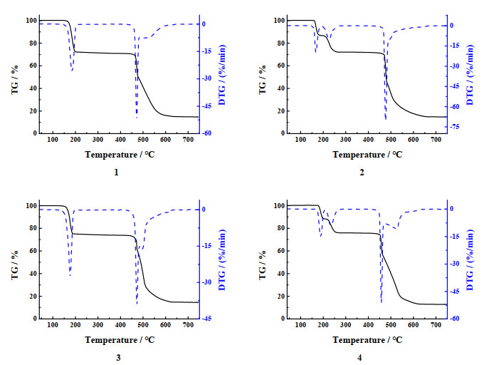
Fig. 5 shows the fluorescent curve of complexes 1~4 in EB-DNA system with different concentrations. With increasing the concentration, the fluorescence intensity of EB-DNA-complex declines. Therefore, it speculated the complexes can coordinate with the bases of DNA, resulting in the extrusion of EB out of DNA. To analyze quantificationally the binding capacity of complexes and DNA, we employed Stern-Volmer equation[30] I0/I = 1 + KSVccomplex to obtain the quenching constants KSV of complexes replacing EB and DNA with 6.27 × 10⁴ L·mol–1 (1), 4.69 × 10⁴ L·mol–1 (2), 5.95 × 10⁴ L·mol–1 (3), and 2.99 × 10⁴ L·mol–1 (4). This value suggests the complexes insert in DNA to a certain degree. Furthermore, the quenching constant is consistent with the reported zinc complex. For example, in 2020, Marzieh Daryanavard' groups have reported a new mononuclear Zn(Ⅱ) complex, with its KSV value to be 7.57 × 10⁴[31]. Tapan Kumar Mondal' groups have reported a Zn(Ⅱ) complex with hex-adentate N4S2 donor thioether ligand, and the KSV value of Zn(Ⅱ) complexes is 2.6 × 10⁴[32].
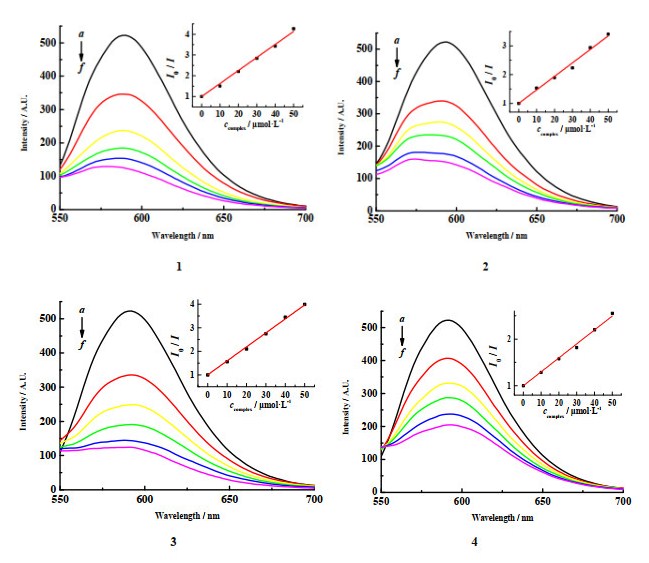
By comparing the KSV values of the four complexes, it is not difficult to find that they are all in the same order of magnitude, and the difference is not very large. In general, the interactions between complexes 1~4 and DNA are stronger than EB, and they all can squeeze EB from DNA, but complexes 1 and 3 are slightly stronger than 2 and 4. It is speculated that the aromatic ring of the ligand in the complex is inserted into the base pair of DNA, which competes for the binding of EB to DNA, squeezing EB out of the base pair of the DNA, and the steric hindrance effect of 1 and 3 is relatively small, so that it is the relatively strongest interaction with DNA.
Schiff base pyridine Zn(Ⅱ) complexes 1~4 have been synthesized and characterized. Structural analysis results show that they have the same coordination mode and similar steric structures. Through intermolecular hydrogen bonds, complex 1 constitutes a one-dimensional chain structure, and complex 3 constitutes a two-dimensional network structure, respectively. Thermogravimetric analysis shows complexes 1~4 can exist stably before 150 ℃. The result of the interaction with ct-DNA shows that it Is intercalation, and the insertion effect of complex 1 is the strongest. It will provide an important theoretical basis for developing new metal drugs.
Stefanidou, M.; Maravelias, C.; Dona, A.; Spiliopoulou, C. Zinc: a multipurpose trace element. Arch. Toxicol. 2006, 80, 1–9. doi: 10.1007/s00204-005-0009-5
Chasapis, C. T.; Loutsidou, A. C.; Spiliopoulou, C. A.; Stefanidou, M. E. Zinc and human health: an update. Arch. Toxicol. 2012, 86, 521–534. doi: 10.1007/s00204-011-0775-1
Han, A. Y.; Su, H.; Xu, G. H.; Khan, M. A.; Li, H. Synthesis, crystal structures, and luminescent properties of Zn(Ⅱ), Cd(Ⅱ), Eu(Ⅲ) complexes and detection of Fe(Ⅲ) ions based on a diacylhydrazone Schiff base. RSC Adv. 2020, 10, 23372–23378. doi: 10.1039/D0RA03642K
Ríos Gómez, M. L.; Lampronti, G. I.; Yang, Y.; Mauro, J. C.; Bennett, T. D. Relating structural disorder and melting in complex mixed ligand zeolitic imidazolate framework glasses. Dalton T. 2020, 49, 850–857. doi: 10.1039/C9DT03559A
Guo, K.; Wang, P.; Tan, W.; Li, Y.; Gao, X.; Wang, Q.; Pu, L. Structure of a dimeric BINOL-imine-Zn(Ⅱ) complex and its role in enantioselective fluorescent recognition. Inorg. Chem. 2020, 59, 17992–17998. doi: 10.1021/acs.inorgchem.0c02330
Jedrzejas, M. J.; Setlow, P. Comparison of the binuclear metalloenzymes diphosphoglycerate-independent phosphoglycerate mutase and alkaline phosphatase: their mechanism of catalysis via a phosphoserine intermediate. Chem. Rev. 2001, 101, 607–618. doi: 10.1021/cr000253a
Lipscomb, W. N.; Sträter, N. Recent advances in zinc enzymology. Chem. Rev. 1996, 96, 2375–2434. doi: 10.1021/cr950042j
Sanatkar, T. H.; Khorshidi, A.; Janczak, J. Dinuclear Zn(Ⅱ) and tetranuclear Co(Ⅱ) complexes of a tetradentate N2O2 Schiff base ligand: synthesis, crystal structure, characterization, DFT studies, cytotoxicity evaluation, and catalytic activity toward benzyl alcohol oxidation. Appl. Organomet. Chem. 2020, 34, e5493.
Huang, H. S.; Zhang, T. L.; Zhang, S. T.; Zhang, J. G.; Wu, X. F.; Xu, J. H. Theoretical study of the structure, mechanism of detonation initiation and stability of transition metal carbohydrazide nitrates. Chin. J. Struct. Chem. 2013, 32, 1491-1496.
Opalade, A. A.; Karmakar, A.; Rúbio, G. M. D. M.; Pombeiro, A. J. L.; Gerasimchuk, N. Zinc complexes with cyanoxime: structural, spectroscopic, and catalysis studies in the pivaloylcyanoxime–Zn system. Inorg. Chem. 2017, 56, 13962–13974. doi: 10.1021/acs.inorgchem.7b01891
Mohamed, M. F.; Brown, R. S. Cleavage of an RNA model catalyzed by dinuclear Zn(Ⅱ) complexes containing rate-accelerating pendants. Comparison of the catalytic benefits of H-bonding and hydrophobic substituents. J. Org. Chem. 2010, 75, 8471–8477. doi: 10.1021/jo1017316
Pandey, R.; Kumar, A.; Xu, Q.; Pandey, D. S. Zinc(Ⅱ), copper(Ⅱ) and cadmium(Ⅱ) complexes as fluorescent chemosensors for cations. Dalton T. 2020, 49, 542–568. doi: 10.1039/C9DT03017D
Bazany-Rodríguez, I. J.; Salomón-Flores, M. K.; Viviano-Posadas, A. O.; García-Eleno, M. A.; Barroso-Flores, J.; Martínez-Otero, D.; Dorazco-González, A. Chemosensing of neurotransmitters with selectivity and naked eye detection of l-DOPA based on fluorescent Zn(Ⅱ)-terpyridine bearing boronic acid complexes. Dalton T. 2021, 50, 4255–4269. doi: 10.1039/D0DT04228E
Mikata, Y.; Yamashita, A.; Kawamura, A.; Konno, H.; Miyamoto, Y.; Tamotsu, S. Bisquinoline-based fluorescent zinc sensors. Dalton T. 2009, 3800–3806.
Majumdar, D.; Das, D.; Sreejith, S. S.; Das, S.; Kumar Biswas, J.; Mondal, M.; Ghosh, D.; Bankura, K.; Mishra, D. Dicyanamide-interlaced assembly of Zn(Ⅱ)-Schiff-base complexes derived from salicylaldimino type compartmental ligands: syntheses, crystal structures, FMO, ESP, TD-DFT, fluorescence lifetime, in vitro antibacterial and anti-biofilm properties. Inorg. Chim. Acta 2019, 489, 244–254. doi: 10.1016/j.ica.2019.02.022
Martínez, V. R.; Aguirre, M. V.; Todaro, J. S.; Ferrer, E. G.; Williams, P. A. M. Candesartan and valsartan Zn(Ⅱ) complexes as inducing agents of reductive stress: mitochondrial dysfunction and apoptosis. New J. Chem. 2021, 45, 939–951. doi: 10.1039/D0NJ02937H
Aleshin, G. Y.; Egorova, B. V.; Priselkova, A. B.; Zamurueva, L. S.; Khabirova, S. Y.; Zubenko, A. D.; Karnoukhova, V. A.; Fedorova, O. A.; Kalmykov, S. N. Zinc and copper complexes with azacrown ethers and their comparative stability in vitro and in vivo. Dalton T. 2020, 49, 6249–6258. doi: 10.1039/D0DT00645A
Sheldrick, G. M. SHELXL-97, A Program for Crystal Structure Refinement. Germany Geöttingen: University of Geöttingen 1997.
Wang, M.; Yu, F.; Jiang, W. J.; Tan, Y. X.; Zhang, F. X.; Kuang, D. Z. Syntheses, crystal structures and biological activity of the diorganotin 2-(2-(4-methoxybenzoyl)hydrazono)-3-phenylpropanoic carboxylate complexes. Chin. J. Struct. Chem. 2020, 39, 1965-1972.
Zhang, S. Z.; Guo, G.; Ding, W. M.; Li, J.; Wu, Y.; Zhang, H. J.; Guo, J. Q.; Sun, Y. X. Synthesis and spectroscopic properties of two different structural Schiff base Zn(Ⅱ) complexes constructed with/without auxiliary ligands. J. Mol. Struct. 2021, 1230, 129627. doi: 10.1016/j.molstruc.2020.129627
Uddin, M. N.; Ferdous, T.; Islam, Z.; Jahan, M. S.; Quaiyyum, M. A. Development of chemometric model for characterization of non-wood by FT-NIR data. J. Bioresour. Bioprod. 2020, 5, 196–203. doi: 10.1016/j.jobab.2020.07.005
Chen, S.; Jiang, S.; Jiang, H. A review on conversion of crayfish-shell derivatives to functional materials and their environmental applications. J. Bioresour. Bioprod. 2020, 5, 238–247. doi: 10.1016/j.jobab.2020.10.002
Cao, Y.; Wang, X. Z.; Li, Y. J.; Shen, D. H.; Dai, Y. P.; Zhang, S. Z.; Zhang, W. G. Effect of high temperature oil heat treatment on the stsrch content and mold-resistant property of bamboo. J. For. Eng. 2020, 5, 109–115.
Chen, Z. J.; Gao, H.; Li, W.; Li, S. J.; Liu, S. X.; Li, J. Research progree of biomass-based optical materials. J. For. Eng. 2020, 5, 1–12. doi: 10.31186/jenggano.5.1.1-10
Pretsch, E.; Bühlmann, P.; Badertscher, M. Structure Determination of Organic Compounds. Fourth ed. Berlin Heidelberg: Springer-Verlag 2009, p69-242.
Ashok, B.; Hariram, N.; Siengchin, S.; Rajulu, A. V. Modification of tamarind fruit shell powder with in situ generated copper nanoparticles by single step hydrothermal method. J. Bioresour. Bioprod. 2020, 5, 180–185. doi: 10.1016/j.jobab.2020.07.003
Tan, W.; Hao, X. L.; Wang, Q. W.; Ou, R. X. Mechanical and thermal properties of bamboo plastic composites reinforced by thermotropic liquid crystal copolyesters. J. For. Eng. 2020, 5, 97–103.
Zheng, C. X.; Zhu, S.; Lu, Y.; Mei, C. T.; Xu, X. W.; Yue, Y. Y.; Han. J. Q. Synthesis and characterization of cellulose nanofibers/polyacrylic acid-polyacrylamide double network electroconductive hydrogel. J. For. Eng. 2020, 5, 93–100.
Zhu, L. T.; Gu, Y. F.; Wu, G. S. Biomass thermal conductivity measurement system design. J. For. Eng. 2020, 5, 97–102.
Yan, C.; Zhang, J.; Liang, T.; Li, Q. Diorganotin (Ⅳ) complexes with 4-nitro-N-phthaloyl-glycine: synthesis, characterization, antitumor activity and DNA-binding studies. Biomed. Pharmacother. 2015, 71, 119–127. doi: 10.1016/j.biopha.2015.02.027
Daryanavard, M.; Jannesari, Z.; Javeri, M.; Abyar, F. A new mononuclear zinc(Ⅱ) complex: crystal structure, DNA- and BSA-binding, and molecular modeling; in vitro cytotoxicity of the Zn(Ⅱ) complex and its nanocomplex. Spectrochim. Acta A 2020, 233, 118175. doi: 10.1016/j.saa.2020.118175
Mondal, A. S.; Jana, M. S.; Manna, C. K.; Naskar, R.; Mondal, T. K. Synthesis of a zinc(Ⅱ) complex with hexadentate N4S2 donor thioether ligand: X-ray structure, DNA binding study and DFT computation. J. Mol. Struct. 2018, 1164, 94–99. doi: 10.1016/j.molstruc.2018.03.038

 扫一扫看文章

扫一扫关注我们
 DownLoad:

 下载:
下载:


