表 1
无机配合物光热试剂的主要特征
Table 1.
Main characteristics of inorganic complex‑based photothermal reagents*

癌症是严重威胁人类健康的重大疾病之一,目前临床上常使用的非手术治疗方式以化疗、放疗为主,存在细胞耐药和副作用大等问题。作为一种新型的癌症治疗策略,光热治疗(photothermal therapy,PTT)通过光热试剂将近红外(NIR)光能转化为热能,导致肿瘤部位快速升温至46 ℃以上,从而实现癌细胞的定点消除,具有治疗时间短、毒性小和副作用低等优点。与化疗、放疗等治疗方式相比,PTT因其在肿瘤治疗方面的微创性和有效性而备受关注,在临床上已开始应用于乳腺癌和前列腺癌等癌症的治疗[1-2]。因此,开发新型高效的光热试剂对于推动PTT的发展至关重要。
在过去的几年中,人们开发了性能各异的NIR光热试剂,极大地促进了高性能光热试剂的设计合成及其在PTT领域的应用。迄今为止,基于无机材料和有机小分子的光热试剂得到了较好的发展。无机材料主要包括量子点、二维纳米材料、贵金属纳米材料和金属硫属纳米材料等[3-6],虽然其因光热稳定性高的优势获得广泛的关注和研究,纳米材料合成可重复性差和生物毒性高的问题限制了它们在生物医学领域的应用。近年来,有机小分子光热材料由于其生物毒性低、结构易修饰和光物理性质可调等优势,已被应用于生物医学领域[7-11]。例如,吲哚菁绿和亚甲蓝已经被美国食品和药物管理局批准用于临床手术中的荧光引导肿瘤切除术。然而,该类材料容易被光漂白的问题阻碍了其进一步发展。
无机金属配合物(简称无机配合物)是一类由金属和有机配体通过配位键形成的化合物。无机配合物的组成易于确定,中心的金属离子与有机配体之间可以协同优化,在光物理性质方面,可以通过配体修饰调节其共轭结构或D-A(donor-acceptor)电子结构,使其吸收波长红移至NIR区,同时改善生物兼容性和水溶性。另一方面,可以选择不同的中心金属调控其吸光能力。最为重要的是,利用金属与配体螯合或者配体的聚集可以实现激发态能量耗散的非辐射衰变路径,有助于将光能转化为热能并得到较高的光热转化效率。因此,无机配合物作为一类重要的NIR吸收型光热材料可被应用于PTT领域。此类材料不仅具有无机纳米材料热稳定性高、抗光漂白能力强的优势,而且又具备有机小分子结构易修饰、光物理性质可调的特点,近些年来取得了较快的发展。
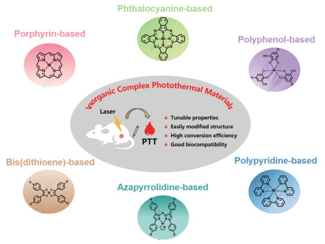
为了提高无机配合物材料的PTT效果,一般从以下3个方面进行调节:(1) 通过不同的金属中心或对配体的修饰提高其吸光能力和吸收波长;(2) 通过配合物自组装成纳米颗粒(nanoparticals,NPs)或将配合物聚集体包覆成纳米颗粒以提高配合物的光热转化效率、水溶性及其对肿瘤部位的靶向能力;(3) 利用对配体分子的修饰调控其分子内运动或调节金属和配体之间的电荷转移实现光热转换效率的提高。除了需要考虑PTT效果,还要求配合物材料具有良好的生物兼容性。鉴于此,人们投入了大量精力开发利用NIR光高效产热并将热量精准传递到目标部位的先进配合物光热试剂。本综述旨在总结近些年的研究在以上方面的努力。本文中讨论的无机配合物光热试剂的主要特征总结在表 1中,其分类见图 1。
 下载:
导出CSV
下载:
导出CSV
| Ligand | Metal | Probe construction | Laser irradiation | Photothermal effect | Therapeutic strategy | Ref. |
| Porphyrin | Zn | HIV-Tat/DSPE-mPEG 2000-Mal/PorCP NPs | 808 nm (0.75 W·cm-2, 10 min) | 63.8%, ΔT=42 ℃ (H2O, 50 μg·mL-1) | Passive targeting (EPR)/Tat mediated cell uptake | [12] |
| Porphyrin | Zn | Por-DPP NPs | 808 nm (1.5 W·cm-2, 10 min) | 62.5%, ΔT=29.6 ℃ (tumor tissue) | Passive targeting (EPR) | [13] |
| Porphyrin | Zn | DC-CHOL/DSPC/DEP-BH NPs | 808 nm (1.0 W·cm-2, 6 min) | 39.7%, ΔT=23 ℃ (tumor tissue) | Passive targeting (EPR)/electrostatic interaction | [14] |
| Porphyrin | Zn | (Zn)Por NPs | 635 nm (1.8 W·cm-2, 10 min) | 16.3%, n.d. | Passive targeting (EPR) | [15] |
| Porphyrin | Zn | PEG/DOX NPs | 808 nm (3 W·cm-2, 10 min) | 28.4%, Tmax=47 ℃ (tumor tissue) | Passive targeting (EPR) | [16] |
| Porphyrin | Zn | NI-(Zn)Por NPs | 635 nm (1.4 W·cm-2, 10 min) | 35.8%, n.d. | Passive targeting (EPR) | [17] |
| Porphyrin | Zn | BDE/ZnATPP NPs | 635 nm (0.6 W·cm-2, 6 min) | 54.1%, ΔT=18 ℃ (tumor tissue) | Passive targeting (EPR) | [18] |
| Porphyrin | Zn | P1/ZnP1/ZnP2 NPs | 660 nm (0.75 W·cm-2, 15 min) | ca. 33.4%, ΔT=26 ℃ (H2O, 20 μg·mL-1) | Passive targeting (EPR) | [19] |
| Porphyrin | Zn | P-4 NPs | 660 nm (0.8 W·cm-2, 8 min) | 30.0%, ΔT=15.4 ℃ (tumor tissue) | Passive targeting (EPR) | [20] |
| Porphyrin | Au | AuPNSs | 635 nm (0.8 W·cm-2, 5 min) | ca. 48.2%, Tmax=50.8 ℃ (tumor tissue) | Passive targeting (EPR)/cRGD mediated cell uptake | [21] |
| Porphyrin | Ag | AgTMPyP@GO | 808 nm (1.0 W·cm-2, 10 min) | 32.7%, ΔT=25.8 ℃ (tumor tissue) | n.d. | [22] |
| Porphyrin | Cu | Cu-TCPP MOF | 808 nm (1.0 W·cm-2, 10 min) | 36.8%, ΔT=14 ℃ (tumor tissue) | Passive targeting (EPR) | [23] |
| Porphyrin | Ir | DSPE-PEG-MAL/HIV-1 Tat/4-Ir NPs | 635 nm (0.4 W·cm-2, 5 min) | 49.5%, Tmax=61.9 ℃ (tumor tissue) | Passive targeting (EPR)/HIV-Tat mediated cell uptake | [24] |
| Porphyrin | Ni | DSPE-PEG2000/NGP-1-NPs | 808 nm (1.0 W·cm-2, 10 min) | 60.0%, Tmax=49.3 ℃ (H2O, 30 μg·mL-1) | Passive targeting (EPR) | [25] |
| Porphyrin | Ni | DSPE-PEG2000/NGP-2-NPs | 1064 nm (1.0 W·cm-2, 10 min) | 69.0%, Tmax=52.5 ℃ (tumor tissue) | Passive targeting (EPR) | [25] |
| Porphyrin | Fe | PEG/siRNA/Zr-FeP MOF | 635 nm (1.9 W·cm-2, 5 min) | 33.7%, ΔT=17 ℃ (PBS, 50 μg·mL-1) | Passive targeting (EPR) | [26] |
| Porphyrin | Mn | S-nitrosothiol/Zr2+/Mn-TCPP MOF | 808 nm (1.0 W·cm-2, 10 min) | 48.3%, Tmax=54.0 ℃ (H2O, 400 μg·mL-1) | Passive targeting (EPR) | [27] |
| Porphyrin | Mn | Mn-TEPP/HA/HAuCl4 | 1064 nm (1 W·cm-2, 10 min) | 41.0%, Tmax=56 ℃ (tumor tissue) | Passive targeting (EPR)/active targeting (HA) | [28] |
| Phthalocyanine | Zn | PcA | 730 nm (1.0 W·cm-2, 10 min) | n.d., ΔT=15 ℃ (H2O, 4.7 μg·mL-1) | n.d. | [31] |
| Phthalocyanine | Zn | Biotin/TEG/Pc | 655 nm (0.22 W·cm-2, 5 min) | n.d., Tmax=67 ℃ (H2O, 14 μg·mL-1) | Passive targeting (EPR)/Active targeting (Biotin) | [32] |
| Phthalocyanine | Zn | ZnPc | 650 nm (0.7 W·cm-2, 10 min) | 31.3%, Tmax=53.2 ℃ (tumor tissue) | Passive targeting (EPR) | [33] |
| Phthalocyanine | Cu | PcC1 | 685 nm (0.2 W·cm-2, 10 min) | n.d., Tmax=53 ℃ (tumor tissue) | n.d. | [31] |
| Phthalocyanine | Fe | PcC2 | 630 nm (1.0 W·cm-2, 5 min) | n.d., n.d. | n.d. | [31] |
| Phthalocyanine | Fe | FePc/HSA | 671 nm (0.5 W·cm-2, 10 min) | 44.4%, Tmax=55.4 ℃ (tumor tissue) | Passive targeting (EPR)/Active targeting (HSA) | [34] |
| Phthalocyanine | Mn | Curcumin/HA/MnPc nanosheet | 730 nm (0.9 W·cm-2, 5 min) | 72.3%, Tmax=56 ℃ (tumor tissue) | Passive targeting (EPR)/Active targeting (HA) | [35] |
| Polyphenol | Fe | Fe3+-TA | 808 nm (6 W·cm-2, 10 min) | 77.3%, ΔT=15 ℃ (H2O, 125 μg·mL-1) | Passive targeting (EPR) | [36] |
| Polyphenol | Fe | PNV@FeⅢTA | 808 nm (6 W·cm-2, 10 min) | 45.4%, Tmax=44.5 ℃ (H2O, 50 μg·mL-1) | Passive targeting (EPR) | [37] |
| Polyphenol | Fe | Fe3+-TA/Agarose | 808 nm (1.0 W·cm-2, 10 min) | n.d., ΔT=58 ℃ (H2O, n.d.) | n.d. | [38] |
| Polyphenol | Fe | BSA-Fe-GA nanonetwork | 808 nm (2 W·cm-2, 10 min) | 20.33%, ΔT=37.2 ℃ (tumor tissue) | n.d. | [39] |
| Polyphenol | Fe | Fe3+-HA/BAI | 808 nm (1.0 W·cm-2, 5 min) | 29.44%, n.d. | Passive targeting (EPR)/Active targeting (HA) | [40] |
| Polyphenol | Fe | Cro-Jul/Fe-Qu/DSPE-PEG2000 | 808 nm (1.0 W·cm-2, 5 min) | 48.96%, Tmax=45.0 ℃ (H2O, 200 μg·mL-1) | Passive targeting (EPR) | [41] |
| Bis(dithiolene) | Ni | nickel-bis(dithiolene) derivatives | 940 nm (1.0 W·cm-2, 10 min) | 12%, ΔT=59.2 ℃ (H2O, 500 μg·mL-1) | n.d. | [42] |
| Bis(dithiolene) | Ni | F127/NiBD-Cz NPs | 1064 nm (1.0 W·cm-2, 6 min) | 63.6%, ΔT=50.1 ℃ (H2O, 100 μg·mL-1) | Passive targeting (EPR) | [43] |
| Bis(dithiolene) | Ni | DSPE-PEG2000/DPPC/OPYO/TPE-Ni NPs | 940 nm (1.0 W·cm-2, 10 min) | 28.9%, Tmax=54 ℃ (tumor tissue) | Passive targeting (EPR) | [44] |
| Aza-BODIPY | Pt | DSPE-mPEG5000/PtDP-N NPs | 730 nm (0.5 W·cm-2, 5 min) | 27.9%, Tmax=54 ℃ (tumor tissue) | Passive targeting (EPR) | [45] |
| Polypyridine | Ir | PEG/IrDAD-NPs | 808 nm (0.7 W·cm-2, 10 min) | 34.9%, ΔT=26.6 ℃ (tumor tissue) | Passive targeting (EPR) | [46] |
| Polypyridine | Ru | PEG/HL-PEG2K | 808 nm (1.0 W·cm-2, 5 min) | 41.77%, ΔT=28.2 ℃ (tumor tissue) | Passive targeting (EPR) | [47] |
| *n.d.: not described, EPR: enhanced permeability and retention, PEG: polyethylene glycol, HIV: human immunodeficiency virus, Tat: transactivator of transcription, DSPE-PEG2000: 1,2-distearoyl-sn-glycero-3-phosphoethanolamine-N-[methoxy(polyethylene glycol)-2000], BSA: bovine serum albumin, DEP-BH: 5,15-bis{2,5-bis(2-ethyl-hexyl)-3,6-di-thienyl-2-yl-2,5-dihydro-pyrrolo[3,4-c]pyrrole-1,4-dione-5′-yl-ethynyl}-10,20-bis(2-butylheptyl)-porphyrin zinc, DC-CHOL: 3β[N-(N′, N′-dimethylaminoethane)carbamoyl]cholesterol, DSPC: 1,2-distearoyl-sn-glycero-3-phosphocholine, DOX: doxorubicin, ZnATPP: zinc(Ⅱ) 5-(4-aminophenyl)-10,15,20-triphenylporphyrin, BDE: 1,4-butanediol diglycidylether, AuPNSs: Au based porphyrin nanospheres, TCPP: meso-tetra(4-carboxyphenyl)porphine, MOF: metal-organic framework, DSPE: 1,2-distearoyl-sn-glycero-3-phospho-ethanolamine, MAL: maleic anhydride-acrylic acid copolymer, siRNA: small interfering RNA, PBS: phosphate buffer saline, TEPP: 5,10,15,20-tetrakis(4′-ethynylphenyl) porphyrin, HA: hyaluronic acid, Pc: phthalocyanine, TEG: triethylene glycol, HSA: human serum albumin, TA: tannic acid, BSA: bovine serum albumin, GA: gallic acid, PNV: polymeric nanovesicles, BAI: baicalein, Cro-Jul: croconic acid-julolidine, Qu: quercetin, BD: bis(dithiolene), Cz: carbazolyl, DPPC: 1,2-dipalmitoyl-sn-glycero-3-phosphocholine, TPE-Ni: nickel-bis(dithiolene)-containing photothermal agent, BODIPY: boron-dipyrromethene. | ||||||
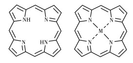
卟啉由4个吡咯环通过亚甲基相互连接而成,具有复杂的环状结构(图 2)。其衍生物是一类重要的有机发色团,具有消光系数大和生物相容性好等优势,被广泛应用于PTT。作为一种常见的四齿配体,卟啉可以与众多金属配位形成金属配合物,也称为金属卟啉。事实上,多种金属卟啉存在于自然界中,如构成血红素的铁卟啉和构成叶绿素的镁卟啉,在生物体中承担着吸收光的重要作用。作为一类潜在的光热试剂,人们对于金属卟啉类配合物的研究主要集中在生物体所需微量金属元素-卟啉配合物,如Zn、Fe、Ni、Cu、Mn等。此外,还包括非必需但有益的金属元素-卟啉配合物,如Au、Ag、Ir等。金属卟啉母核不仅吸收波长短(一般低于700 nm),而且光热转化效率较低,水溶性和肿瘤靶向效果较差,不能实现理想的PTT效果。因此,针对这些问题,人们主要从4个方面进行了改进:(1) 通过分子工程策略,以金属卟啉作为电子给体,在卟啉的亚甲基位置连接电子受体或金属卟啉增大共轭来调节分子的吸收波长;(2) 在卟啉的亚甲基上引入亲水性基团调节配合物的水溶性,同时促进配合物分子的自组装聚集和提高配合物的光热转化效率;(3) 通过嵌段聚合物包覆配合物聚集体得到纳米颗粒或者肿瘤微环境,利用EPR效应或响应系统实现肿瘤靶向;(4) 除了卟啉中心与金属离子配位外,卟啉的外围也可以结合金属离子,通过形成金属有机骨架(MOF)增加金属卟啉单元的个数,从而提高配合物的摩尔吸光度。
锌元素是人体必需的微量元素之一,与卟啉形成的卟啉配合物具有高的生物安全性。基于这个原因,在金属卟啉类的配合物光热试剂研究中,关于锌卟啉类的报道最多。
Guo等[12]通过炔基桥连接交替的锌卟啉供体(D)和苯并噻二唑(BT)受体(A)获得了共轭聚合物PorCP(图 3A)。这种D-A结构能够有效地引起分子内电荷转移(intramolecular charge transfer,ICT),导致吸收波长红移并增强NIR吸收,延长的共轭结构可以进一步导致基于每个锌卟啉单元的平均摩尔吸光度的提高。用808 nm激光照射被聚合物封装后的PorCP NPs,其光热转换效率高达63.8%。在斑马鱼体内实验中,PorCP NPs表现出对斑马鱼肝脏增生模型显著的PTT效果。Wu等[13]以锌卟啉作为电子给体单元,二酮吡咯并吡咯用作电子受体单元,设计合成了一种具有D-A结构的新型锌卟啉类的配合物Por-DPP(图 3B)。Por-DPP可以在不添加任何辅助试剂的情况下自组装成纳米颗粒,并在800 nm附近表现出有效的吸收。Por-DPP NPs的形成导致荧光猝灭并促进非辐射热产生。在808 nm激光照射下,Por-DPP NPs的光热转化效率(PCE)可达62.5%。在体外具有明显的PTT效果,并且可以在体内有效损伤癌细胞,而光疗后没有明显的副作用。Duan等[14]也是以二酮吡咯并吡咯为电子受体将其与锌卟啉通过炔基连接构成稳定的A-D-A结构,扩展了π共轭体系,开发了一种可重复使用的NIR响应二酮吡咯修饰的锌卟啉DEP-BH(图 3C)用于PTT。另外,利用卟啉的疏水性,作者将DEP-BH用热敏的、带正电荷的脂质体包裹得到DEP-BH@TSLs,其PCE为39.7%。DEP-BH@TSLs可以通过静电作用靶向带负电荷的细菌。由于DEP-BH的光稳定性,1个纳米治疗剂量可以重复进行2次光治疗,从而避免了资源的浪费和操作的复杂性。以上文献报道中,作者主要在调控配合物的吸收波长方面做出了努力,水溶性的调节基本是靠嵌段聚合物的封装来实现。
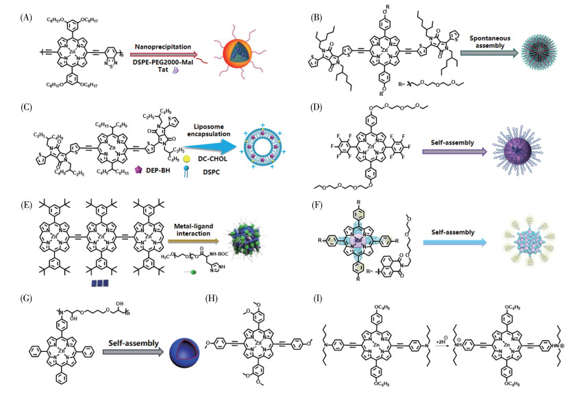
通过在卟啉结构上修饰聚乙二醇(PEG)也可以改善金属卟啉的水溶性。Yang等[15]通过在母体卟啉结构上引入亲水性PEG链和五氟苯部分,合成了新的锌卟啉配合物ZnPor(图 3D)。自组装成纳米颗粒后,与配合物分子相比,ZnPor NPs荧光被显著猝灭,呈现出红移的NIR吸收和更高的光热转化效应。体外实验结果表明,ZnPor NPs具有良好的生物相容性,并表现出较高的光细胞毒性。另外,其体内癌症的PTT效果表现优异。Zhang等[16]利用L-组氨酸改性的PEG与共轭三锌卟啉的超分子相互作用,设计获得了仿生水分散性共轭三卟啉基纳米颗粒(图 3E)。该纳米粒子溶液的最大吸收达到了NIR区,在808 nm激光照射下,其光热转换效率为28.4%。体内实验中,在808 nm激光照射下,肿瘤小鼠的肿瘤温度在5 min内迅速达到47℃,表明了该纳米颗粒在PTT方面具有良好的应用潜力。Yang等[17]以PEG修饰的1,8-萘酰亚胺为电子受体,与锌卟啉核形成D-A结构(NI-ZnPor),其吸收波长红移至NIR区,通过自组装得到纳米颗粒NI-ZnPor NPs(图 3F)。由于PEG的存在,NI-ZnPor NPs具有良好的水溶性。此外,在635 nm激光照射下,NI-ZnPor NPs具有良好的PTT效果。
长波长吸收和良好水溶性是配合物光热材料需要改善之处,此外,光热转换效率和肿瘤靶向能力的提高也是实现高效PTT的必要条件。Chen等[18]基于锌卟啉(ZnATPP)与环氧树脂(BDE)的点击反应设计合成了一种交替卟啉共聚物TPP-a-BDE,并自组装形成聚合物囊泡TPPBVs(图 3G)。在交替共聚物独特的链折叠机制作用下。囊泡的光热转换效率高达54.1%,在635 nm激发下,TPPBV对革兰氏阳性耐甲氧西林金黄色葡萄球菌(MRSA)和革兰氏阴性超广谱β-内酰胺酶大肠杆菌(ESBL)均表现出很强的光热抗菌活性。此外,TPPBVs在MRSA感染的小鼠模型中也具有显著的体内治疗效果。Ding等[19]通过纳米沉淀法合成了卟啉衍生物ZnP2(图 3H)纳米颗粒,该纳米颗粒具有良好的生物相容性和肿瘤靶向性。成像结果显示ZnP2 NPs在肿瘤部位停留了24 h,证明了ZnP2 NPs优异的高通透性和滞留效应。在注射ZnP2 NPs后,小鼠的肿瘤体积显著减小,表明ZnP2 NPs可以成为潜在的光热治疗剂。缺氧条件下糖酵解会触发酸性肿瘤微环境,从而激发pH响应系统建立某些触发器,以有效地进行肿瘤靶向光疗。Liang等[20]设计并合成了一种pH响应性锌(Ⅱ)卟啉(P-4)用于癌症PTT。在P-4自组装纳米粒子中,具有pH响应性的二丁基氨基苯基基团可以在酸性肿瘤微环境中进行质子化(图 3I),光热转换效率为30.0%。体外实验证明了P-4 NPs具有较强的稳定性、低暗毒性和理想的光毒性。体内实验中,用660 nm激光照射12 h后,肿瘤部位的温度逐渐上升到了51.6 ℃,表明了其良好的肿瘤靶向积累和光热效果。
金、银和铜作为元素周期表的第Ⅰ副族元素,能够与卟啉配位,并表现出良好的光热转换效率。其中,金纳米颗粒在NIR区域表现出显著的表面等离子体共振,能够在激光照射下实现有效的光热转换,从而提高PTT治疗效果。由于金与硫氧还蛋白还原酶活性中心的半胱氨酸残基之间的高亲和力,金卟啉可以通过抑制其活性来根除癌细胞。Wang等[21]以四(4-吡啶基)卟吩金(Ⅲ)为骨架合成了不含响应载体的自组装金卟啉纳米球AuPNSs(图 4A)。在635 nm激光照射下,AuPNSs溶液温度迅速增加到78 ℃,PCE约为48.2%。在体内实验中,经肿瘤靶向肽cRGD表面功能化后,cRGD-AuPNSs能够对HeLa细胞显示出显著的靶向和抗增殖作用,照射5 min后,肿瘤部位的温度迅速升高到50.8 ℃,具有明显的光热转化能力。
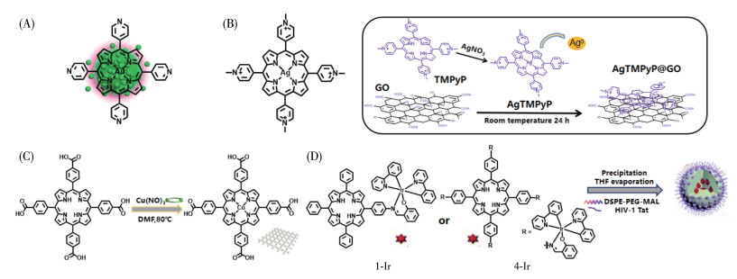
传统的含银抗菌剂因其广谱活性而备受关注,与Ag+相比,高价银离子如Ag2+和Ag3+具有更好的抗菌能力。然而,它们的极度不稳定性严重限制了其作为抗菌剂的发展。作为首例将高价银设计为抗菌素的报道,Lin等[22]制备了一种水溶性高价银(Ag2+和Ag3+)-卟啉配合物AgTMPyP(图 4B),其相应的复合物AgTMPyP@GO在808 nm NIR激光激发下具有优异的PCE(32.17%)及酶活性。溶血、血常规和血液生化实验均表明AgTMPyP@GO具有良好的生物相容性。当被用于PTT时,AgTMPyP@GO可以有效地破坏生物膜、杀死MRSA并促进体内伤口快速愈合。
铜离子的d-d能级跃迁使其具有改善卟啉MOF在NIR区吸收不足的潜力。Li等[23]利用溶剂法制备了超薄铜-四酮基(4-羧基苯基)卟啉MOF纳米片Cu-TCPP MOF(图 4C)。超薄Cu-TCPP MOF纳米片的厚度能够产生较强的NIR吸收和优异的光热性能,在808 nm激光照射下的光热转换效率为36.8%。体内外实验研究表明了Cu-TCPP MOF具有良好的生物相容性。在小鼠肿瘤模型中,Cu-TCPP MOF组的治疗小鼠在激光照射下表现出了明显的恶性细胞收缩,表明Cu-TCPP MOF纳米片能够应用于体内癌症PTT。
作为第Ⅷ族元素,铁、铱和镍也能与卟啉配位并应用于PTT。Zhang等[24]设计合成了具有远红外吸收、NIR发射的单核(1-Ir)和四核(4-Ir)铱卟啉纳米颗粒1-Ir NPs和4-Ir NPs(图 4D)。与化合物1-Ir相比,4-Ir具有更小的单三态能级差、更大的能级能隙、丰富有效的光激发态和高自旋轨道耦合常数,从而增加其长波长下的摩尔吸收系数和光热活性。因此,4-Ir NPs是治疗癌症的理想光敏剂,PCE达到了49.5%。在对U14荷瘤小鼠静脉注射4-Ir NPs并照射10 min后,肿瘤温度升高到了53.0 ℃且肿瘤体积明显减少,有效地抑制了肿瘤生长,表现出了优异的治疗效果。Zhao等[25]合成了5,15-(二甲基苯基)卟啉镍(DMP)并将其与多个苯并冠烯结合得到了NGP-1和NGP-2,水分散性纳米粒子NGP-1-NPs和NGP-2-NPs(图 5A)分别显示了在NIR-Ⅰ和NIR-Ⅱ区域的吸收(约1 000和1 400 nm)。在808和1 064 nm激光的照射下,两者的PCE高达60%和69%。其中,NGP-1-NPs在NIR-Ⅰ表现出了强大的癌细胞杀伤能力,但位于NIR-Ⅱ的NGP-2-NPs具有更优异的光声响应和肿瘤消融能力。对4T1肿瘤小鼠注射NGP-2-NPs 6 h后以1 064 nm激光照射肿瘤部位5 min,肿瘤温度迅速升高至52.5℃,达到了诱导肿瘤热疗温度45 ℃。该治疗完全消除了小鼠体内肿瘤,且14 d内无复发,显示了NGP-2-NPs良好的PTT能力。Zhang[26]等将热休克蛋白70(Hsp70)抑制剂siRNA负载到经PEG修饰的多孔Zr-FeP MOF中(图 5B),实现了低温PTT。在635 nm激光激发下,siRNA/Zr-FeP MOF溶液温度可从20.2 ℃升到45.5 ℃,光热转换效率为33.7%。在4个激光照射周期下,轻微的温度变化也显示了其良好的光热转换稳定性。体内实验中,在635 nm激光照射下,siRNA/Zr-FeP MOF有效地抑制了小鼠体内的肿瘤生长,表现出了明显的治疗效果。
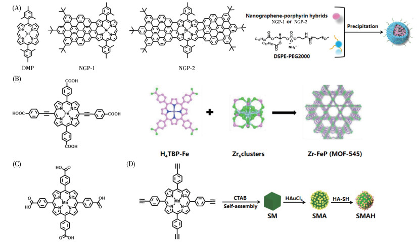
Zhang等[27]通过将具有生物相容性的Zr4+离子与Mn-TCPP自组装制备了锰卟啉NMOFs(图 5C)。在TCPP中引入锰离子同时提供了NMOFs的光热和磁共振成像对比能力,然后用NMOFs与S-亚硝基硫醇(SNO,一种热不稳定的一氧化氮供体)结合得到NMOF-SNO,可实现磁共振成像引导下的一氧化氮释放和光热协同治疗。通过记录808 nm激光激发下的5次循环,作者验证了NMOFs对光激发的光热稳定性。同时,在光照射8 min后,NMOF-SNO迅速升温到54 ℃,是盐水对照组的3倍,PCE为48.3%,证明了NMOF-SNO的光热转化能力,在细胞和小鼠模型中也验证了这一点。此外,通过耐药模型的设计,作者也证明了NMOF-SNO复合材料比DOX化疗具有更高的肿瘤抑制效率。因此,将锰离子并入卟啉环中,能够有效提高锰卟啉的光热转换能力。Xu等[28]以锰卟啉(MnTEPP)作为结构单元合成了自组装有序的金属纳米颗粒SM,通过透明质酸(hyaluronic acid,HA)的修饰使其具有靶向性并提高其水溶性,由此得到了等离子体异质结构纳米复合材料SMAH(图 5D)。体外实验中,用1 064 nm激光照射10 min并循环5次后,SMAH的温度仍可升高至56 ℃,PCE值达到了41.0%。体内实验中,4T1小鼠在1 064 nm激光照射5 min后,肿瘤温度升高到56 ℃左右,且无明显副作用,证实了SMAH良好的光热效应,在体内具有巨大的PTT潜力。
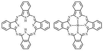
酞菁(phthalocyanine,Pc)可看作是卟啉的衍生物,是一类由8个N原子、8个C原子组成的16中心18π电子的芳香共轭体系的大环分子(图 6)。高度的共轭结构使其在NIR区(670~850 nm)具有强吸收,同时,在400~600 nm范围吸收较弱,相对于卟啉在该范围的强吸收,酞菁能够更好地降低皮肤光敏性[29]。此外,酞菁比卟啉具有更高的摩尔消光系数[30]。以上特点使得金属酞菁成为一类具有研究价值的光热试剂。为了减弱其荧光发射,人们采取了各种有利的措施进行改进。比如,在酞菁骨架上修饰供电子基团,利用光诱导电子转移(PET)抑制其荧光发射;选择顺磁性且具有开壳电子结构的金属离子作为中心离子与之配位,通过金属螯合后的荧光猝灭来提高配合物的光热转化效率;利用分子的堆积或自组装聚集增强分子间的相互作用从而减弱激发能量的辐射衰减。
PET是与电子供体/受体连接的发色团中一种常见的光物理过程。引入电子供体可以猝灭发色团的荧光,从而提高其光热转化效率。Li等[31]首次系统地研究了酞菁的不同结构在PTT中的应用。在锌酞菁的外围引入氨基(PcA,图 7A),氨基对Pc产生的PET效应降低了其荧光发射,从而使其光热转化效率升高。在730 nm激光照射下,PcA溶液的温度提高了20 ℃,光热转换效率达到了30%。为了评估PcA的体外PTT效果,作者将HepG2细胞与PcA孵化,用730 nm激光照射10 min后,发现有90%的HepG2细胞被杀死,表现出了PcA良好的PTT潜力。
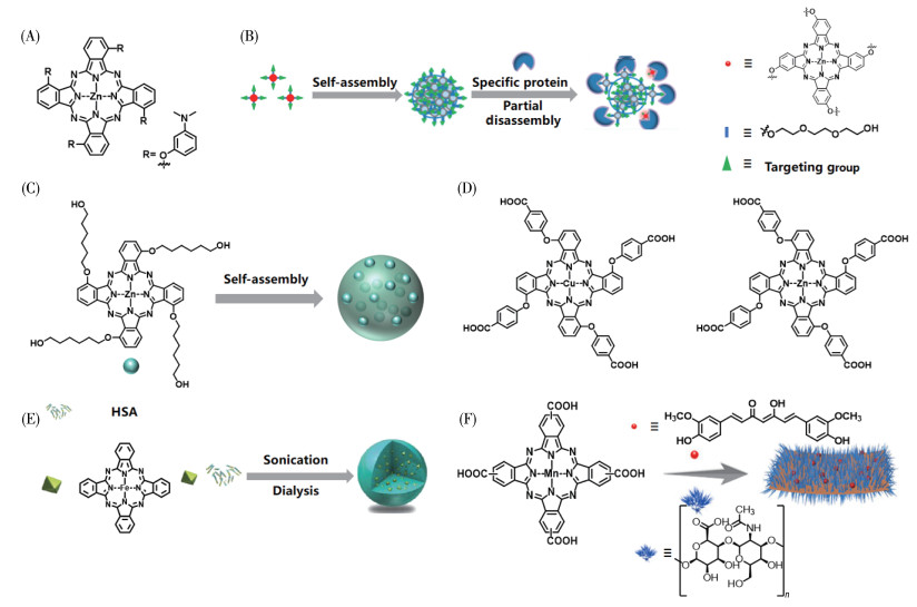
分子的堆积能够引起分子间有效的电子或能量转移,使分子的荧光发射被抑制,从而导致非辐射跃迁过程。Li等[32]将具有亲水性的三乙醇连接锌酞菁骨架与肿瘤靶向分子生物素,设计合成了一种多功能的锌酞菁衍生物Pc-4TEG-B。Pc-4TEG-B可以在水溶液中自组装成均匀的纳米颗粒(NanoPcTB,图 7B),其面对面堆积模式猝灭了Pc-4TEG-B的荧光。在655 nm激光照射下,NanoPcTB在水溶液中的温度可以升至67 ℃,说明其具有良好的光热效应。全身给药后,体内成像结果显示NanoPcTB对肿瘤具有良好的靶向能力。此外,激光照射16 d后能够抑制40%的肿瘤生长。Wang等[33]合成了1,8,15,22-四[3-(6-羟基)己氧基]酞菁锌(Ⅱ)(ZnPc),并通过酞菁分子的聚集构建了ZnPc NPs(图 7C)。制备的ZnPc NPs不仅表现出良好的稳定性、优异的生物相容性和在适当的NIR区域的吸收,而且还表现出高光热转换效率(高达31.3%)和卓越的PTT性能。
除了通过引入不同取代基或分子堆积进行光热性质调控之外,对其中心的金属离子进行选择配位可使其具有不同的光学性质。Li等[31]研究了铜和锌对酞菁光物理性质的影响,与四羧酸锌酞菁(PcC2,图 7D)相比,四羧酸铜酞菁(PcC1,图 7D)的荧光由于金属铜的引入而猝灭。在685 nm激光照射下,PcC1溶液的温度升高了40 ℃,但是PcC2在不同波长的激光照射下没有明显的PTT效应。在连续630 nm激光照射5min后,PcC1对肝癌细胞的抑制率约为92.1%,且在黑暗条件下未观察到明显的细胞毒性。Jia等[34]制备了人血清白蛋白(HSA)-铁(Ⅱ)酞菁纳米颗粒(FePc NPs,图 7E),由于顺磁性铁(Ⅱ)金属的影响,FePc的荧光被猝灭。在671 nm激光照射下,HSA-FePc NPs的温度可提高25.3 ℃,光热转换效率高达44.4%。在体内实验中,通过光声成像,HSA-FePc NPs表现出优异的肿瘤靶向能力和体内光热效应。Zeng等[35]利用四羧酸锰酞菁(MnPc)开发了一种纳米片,然后将其负载姜黄素以提高其治疗效率,并用HA进行修饰以提高其分散性和靶向能力(图 7F)。在730 nm激光照射下,MnPc纳米片溶液的温度升高了48.3 ℃,具有高达72.3%的光热转换效率。与ICG相比,MnPc纳米片在5次光照循环后仍能保持良好的光热效应。小鼠实验表明,MnPc纳米片表现出显著的肿瘤热消除能力。
多酚是植物中天然存在的化合物,具有抗氧化、抗炎等用途。其化学结构中含有多个邻位酚羟基,可以作为多齿配体与多种金属离子螯合形成稳定的金属配合物,而且与一些金属离子配位之后,能有效地拓展其光吸收范围,同时促进激发态电子以非辐射跃迁的形式返回基态,大幅提高其光热转换效率。铁是人体必需的微量元素,Fe(Ⅲ)可以通过配位模式与多酚形成配合物,已被证实具有细胞相容性。利用多酚配体和金属离子的配位作用构建多功能纳米治疗平台,已成为光热消融肿瘤的研究热点。
Liu等[36]首次利用单宁酸(tannic acid,TA)与Fe(Ⅲ)配位制备了可以用于杀死癌细胞的Fe3+-TA配合物(Fe3+-TA)。该配合物具有较强的NIR光吸收、良好的光热稳定性和高达77.3%的光热转换效率(图 8A)。在激光照射下,暴露在Fe3+-TA溶液中的细胞死亡率达到了85%。此外,即使质量浓度高达0.500 mg·mL-1,细胞活力仍在95%以上,说明Fe3+-TA配合物具有较低的细胞毒性。该研究中作者并未继续探究Fe3+-TA在生物过程中的光热特性,但Fe3+-TA已显示出应用于癌症治疗的潜力。同年,Liu等[37]在生物应用中证明了单宁酸-Fe(Ⅲ)的PTT能力,作者以聚(乳酸共乙醇酸)基聚合物纳米球、纳米囊泡和纳米颗粒为模板制备了3种模版支撑的Fe(Ⅲ)-单宁酸纳米颗粒(FeⅢTA,图 8B),光热转换效率为77.3%。在808 nm激光照射10 min后,溶液的温度升高了44.5 ℃,表明FeⅢTA能够快速有效地将NIR能量转化为热能。在体内实验中,作者通过尾静脉注射将FeⅢTA纳米颗粒(PNV@FeⅢTA)注射至小鼠体内,照射4 h后发现小鼠肿瘤逐渐缩小至完全消融,且14 d内未见肿瘤复发,显示了PNV@FeⅢTA在NIR光消融中的应用潜力。Deng等[38]制备了单宁酸-Fe(Ⅲ)光热材料水凝胶ATF用于伤口的光热杀菌,促进伤口愈合(图 8C)。在808 nm NIR光照射下,ATF水凝胶表现出良好的光热转换能力,对其抗菌活性起着重要作用。体外抗菌实验表明ATF水凝胶在NIR光照射下具有良好的灭菌效果。对于动物模型实验,在治疗5 d后,NIR+ATF水凝胶组中的细菌数量明显少于其他组,说明ATF水凝胶在NIR光照射下能有效杀灭细菌。
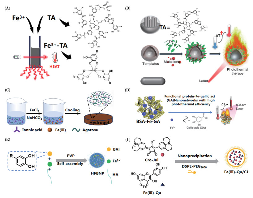
Wang等[39]将Fe(Ⅲ)与没食子酸(gallic acid,GA)配位构建了功能蛋白辅助的BSA-Fe-GA纳米网络,用于局部PTT,该纳米网络具有良好的NIR吸光度和优异的光热性能,同时独特的网络结构(大小为193.5 nm)促进了其在肿瘤部位的保留,可减少局部给药后向周围正常组织的渗透(图 8D)。为评价BSA-Fe-GA纳米网络的光热性能,作者将纯水和BSA-Fe-GA纳米网络分别用808 nm激光照射10 min,可以看到BSA-Fe-GA纳米网络的温度升高了23.6 ℃,光热转换效率为20.33%,而纯水组的温度只增加了4.7 ℃。体内实验中,作者在小鼠瘤内注射BSA-Fe-GA纳米网络,激光照射后,肿瘤表面温度可达65.8 ℃,足以杀死肿瘤细胞。相比之下,PBS作为对照组,温度仅升高了8.4 ℃。同时,4T1荷瘤小鼠在不同时间点的数码照片直观地证实了BSA-Fe-GA纳米网络的PTT能够完全根除肿瘤,30 d内平均相对肿瘤体积的变化也证明了BSA-Fe-GA纳米网络优异的光热肿瘤消融效果。
Wang等[40]采用一种简单的物理自组装方法将HA嵌入金属多酚纳米颗粒中,首次成功制备了一种新型HA修饰的黄芩素亚铁纳米颗粒(HFBNP,图 8E),HFBNP具有恰当的尺寸分布和良好的生物相容性,在5次激光照射后仍能达到几乎相同的最高温度,表现出良好的光热稳定性,能有效地将NIR光能量转化为热能,光热转换效率为(29.44±1.13)%。HA介导的主动靶向能力显著增加了肿瘤细胞对纳米颗粒的摄取,而HFBNP在NIR激光照射下对肿瘤细胞具有较强的生长抑制作用。将PTT与化疗联合使用后,HFBNP可显著抑制荷瘤小鼠的肿瘤生长。
Chen等[41]设计合成了一种新的克罗尼酸-久洛尼定分子(Cro-Jul),该分子在NIR-Ⅱ窗口具有宽吸收、较高的光热转换效率和可控的光热容量(图 8F)。另外,作者利用槲皮素(Qu)和Fe(Ⅲ)之间的配位键得到了Fe(Ⅲ)-Qu配合物,继续将Cro-Jul和Fe(Ⅲ)-Qu与DSPE-PEG2000结合后形成了多功能Fe(Ⅲ)-Qu/CJ纳米体系,能够精准控制高温诱导肿瘤细胞坏死,PCE为48.96%。在808 nm激光照射下,Fe(Ⅲ)-Qu/CJ溶液温度达到了50.8 ℃,证明了其显著的光热转换能力。在体内实验中,Fe(Ⅲ)-Qu/CJ组在照射后肿瘤温度升至44.98 ℃,表明Fe(Ⅲ)-Qu/CJ在体内聚集后的光热效应增强。此外,每2 d观察小鼠的肿瘤状态发现小鼠肿瘤体积明显减少,治疗13 d后的肿瘤重量结果也证实了这一点。
金属双(二硫代烯)配合物是一类具有强NIR吸收的重要材料。通过改变金属中心或不同的二硫代烯取代基,它们的最大吸收区域可以在从可见光到近红外光的很宽范围内进行调整。另外,这些配合物是不发光的,意味着被吸收的能量几乎全部以热的形式释放出来。在金属双(二硫代烯)配合物中,镍(Ⅱ)二硫代烯配合物因其良好的光热稳定性和强的NIR吸收而备受关注。近年来,人们主要通过修饰二硫代烯外围对其吸收波长进行了调控,利用水溶性基团的修饰或者嵌段聚合物的封装进行水溶性生物相容性的改善。
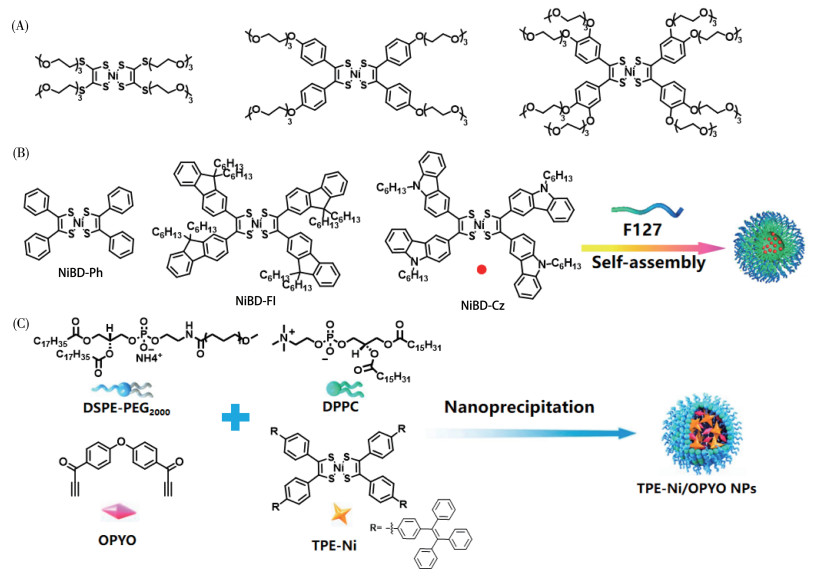
Mebrouk等[42]制备了3种在水中溶解度较高的聚乙二醇化镍双(二硫烯)衍生物(图 9A),并首次将其作为光热剂诱导细胞凋亡。之后,他们又用生物相容性脂质体将其封装,通过光热控制释放药物。Chen等[43]通过调整配体设计并合成了3种NIR-Ⅱ吸收的镍(Ⅱ)双(二硫烯)配合物(NiBD-Ph、NiBD-Fl和NiBD-Cz,图 9B),这些配合物表现出配体依赖的NIR吸收性能。其中,NiBD-Cz的吸收峰可达1 010 nm,通过亲水-疏水相互作用,作者将其包裹在两亲性嵌段共聚物F-127中,从而形成水溶性NiBD-Cz/F-127 NPs,吸收峰也随之红移至1 036 nm。NiBD-Cz/F-127 NPs在水中具有良好的分散性及光稳定性,其PCE高达63.6%。高的PCE使NiBD-Cz/F-127 NPs在低浓度和低功率激光照射下也能达到很好的光热处理效果。Zhang等[44]将点击反应和光热疗法结合,设计并开发了具有强NIR吸收的含镍双(二硫烯)纳米颗粒(TPE-Ni/OPYO NPs,图 9C)。该纳米颗粒具有良好的生物相容性,在940 nm激光照射下,温度可达65 ℃,并有良好的热稳定性。该纳米颗粒在热响应触发下释放出活性OPYO,与癌细胞中的氨基蛋白发生反应,从而实现化疗。此外,基于实体瘤的高通透性和滞留效应,在940 nm激光照射下静脉注射纳米颗粒后,肿瘤能够得到有效消融。
氮杂吡咯烷类化合物具有丰富的可修饰位点、高摩尔消光系数、长波长光吸收和优异的光稳定性,被广泛用于光疗试剂的设计。由于氮杂吡咯烷分子结构中2个半裸露的氮原子具有合适的空间距离,其可以与金属离子进行螯合,形成结构与功能更丰富的配合物分子。金属配位的氮杂吡咯烷类化合物可以通过电子自旋-轨道耦合作用并充分利用重原子效应使系间穿跃速率提升,促使吸收峰红移至NIR生物窗口,因此光激发过程产生的PTT效果可以发挥到深层生物组织中。Li等[45]制备了一系列铂氮杂吡咯烷配合物纳米粒子PtDP-X(X=N、C、S),并探究了氮杂吡咯烷与Pt(Ⅱ)配位后的光热性能。用690 nm激光照射3种PtDP-X溶液,PtDP-N的温度升高了20 ℃,分别比PtDP-C和PtDP-S高2.7和3.4 ℃,表明PtDP-N具有最佳的光热效应。基于此,为了提高PtDP-N的水溶性、生物相容性和光稳定性,作者利用两亲性聚合物1,2-二硬脂酰-SN-3-磷酰乙醇胺-甲氧基聚乙二醇5000(DSPE-mPEG5000)封装PtDP-N,得到了亲水纳米颗粒PtDP-N NPs(图 10A)。在700 nm激光连续照射5 min后,PtDP-N NPs的温度快速升高,光热转换效率达27.9%。另外,PtDP-N NPs在5次循环重复照射后的温度变化可忽略不计,表明PtDP-N NPs具有良好的光热稳定性。在体内实验中,将PtDP-N NPs注射至HeLa荷瘤小鼠体内,随着照射时间的延长,肿瘤部位温度迅速升高,5 min后达到了54 ℃,可有效消融肿瘤,表明PtDP-N NPs在体内具有良好的光热效应。
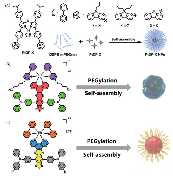
研究发现PTT治疗方式与金属药物发挥协同作用化疗,可促进整体治疗效果,同时最大限度地减少副作用。因此,具有多种光物理特性和生物活性的Ir(Ⅲ)、Ru(Ⅱ)多吡啶类配合物是结合这些治疗方式的理想候选者之一。Zhao等[46]基于D-A-D策略开发了一种掺杂上转化纳米颗粒的铱配合物(IrDAD)的聚合物胶束(IrDAD NPs,图 10B)。在808 nm激光照射下,IrDAD NPs和IrDAD溶液的温度迅速升高,在浓度为100 μmol·L-1时,IrDAD NPs水溶液的温度升高了45 ℃,高于同条件下的IrDAD溶液的温度,两者的光热转换效率分别为34.9%和27.5%,表明IrDAD NPs具有优异的光热转换能力。在体内实验中,作者通过尾静脉注射IrDAD和IrDAD NPs,发现IrDAD NPs导致温度显著升高了26.6 ℃,远高于IrDAD的16.1 ℃,这可能是由于IrDAD NPs具有更好的肿瘤积累。总之,研究表明IrDAD NPs可以有效地将光能转化为热能并应用于癌症体内热疗。Liu等[47]构建了NIR-Ⅱ的聚吡啶配合物纳米平台HL-PEG2K(图 10C),用于皮下和原位U87MG胶质瘤肿瘤的PTT,其最大吸收波长达到了822 nm。在808 nm激光照射下,HL-PEG2K的温度迅速从31.4 ℃升至62.5 ℃,光热转换效率为41.77%。在体内实验中,作者用808 nm激光照射肿瘤小鼠中的神经胶质瘤,治疗的肿瘤温度从29.2 ℃升高到57.4 ℃。作者每4 d监测一次小鼠的肿瘤生长大小,发现在NIR激光照射下HL-PEG2K处理的小鼠肿瘤的生长被有效抑制,且22 d后肿瘤完全消失,无复发,同时HL-PEG2K具有优越的生物相容性,使其成为一种潜在的有效抑制肿瘤生长和复发的治疗平台。
近年来,无机配合物光热材料的研究领域取得了相当大的进展。许多有机配体如卟啉、酞菁、多酚、双(二硫烯)以及氮杂吡咯烷等能够与金属配位并产生良好的光热效应。综述发现,为了提高无机配合物的水溶性及靶向性,大多研究选择引入PEG等水溶性基团或者基于自身的自组装纳米结构,又或者通过嵌段聚合物的封装等手段,这也同时降低了无机纳米材料会带来的生物毒性问题等。为了获得吸收波长更长的配合物,主要是通过推拉电子结构或者增加共轭范围的策略来实现。总之,作为近红外吸收的重要光热材料,无机配合物既具有无机材料光、热稳定性好等优势,又具有有机小分子生物相容性好、光物理性质可调等优势。因此,基于无机配合物的光热试剂的开发对于推动临床上光热治疗的有效实施起着重要作用。
然而,尽管已有不少研究报道了高效的近红外无机配合物光热材料用于疾病治疗,但事实上,近红外激光对人体组织的穿透深度有限,同时人体对高温的承受范围也是导致一些光热剂无法进一步应用于临床的一大原因。针对这一点,开发具有深层组织穿透能力的光热材料仍是目前光热研究需要克服的难题。此外,设计开发可进行多模态协同治疗的药物不失为一种良策。仅靠单一成像或单一光热治疗难以精确彻底地检测和根除实体肿瘤,而利用多种成像技术如光声成像和磁共振成像,以及将光热治疗与包括光动力治疗、电子计算机断层扫描和免疫疗法在内的其他治疗方法相结合,可以充分利用每种技术的优点,降低甚至抵消它们各自的缺点,能够产生协同治疗效果,实现诊疗一体化。
Li X S, Lovell J F, Yoon J, Chen X Y. Clinical development and potential of photothermal and photodynamic therapies for cancer[J]. Nat. Rev. Clin. Oncol., 2020, 17(11): 657-674. doi: 10.1038/s41571-020-0410-2
An D, Fu J Y, Zhang B, Xie N, Nie G H, Agren H, Qiu M, Zhang H. NIR-Ⅱ responsive inorganic 2D nanomaterials for cancer photothermal therapy: Recent advances and future challenges[J]. Adv. Funct. Mater., 2021, 31(32): 2101625. doi: 10.1002/adfm.202101625
Ghaffarkhah A, Hosseini E, Kamkar M, Sehat A A, Dordanihaghighi S, Allahbakhsh A, van der Kuur C, Arjmand M. Synthesis, applications, and prospects of graphene quantum dots: A comprehensive review[J]. Small, 2022, 18(2): 2102683. doi: 10.1002/smll.202102683
Li J, Zhang W, Ji W H, Wang J Q, Wang N X, Wu W X, Wu Q, Hou X Y, Hu W B, Li L. Near infrared photothermal conversion materials: Mechanism, preparation, and photothermal cancer therapy applications[J]. J. Mater. Chem. B, 2021, 9(38): 7909-7926. doi: 10.1039/D1TB01310F
Yu Q, Zhou J, Wang H, Liu Y, Zhou H, Kang B, Chen H Y, Xu J J. A multiple-response cascade nanoreactor for starvation and deep catalysis chemodynamic assisted near-infrared-Ⅱ mild photothermal therapy[J]. Chem. Biomed. Imaging, 2023, 1(3): 242-250. doi: 10.1021/cbmi.2c00003
Ding Z, Gu Y, Zheng C, Gu Y, Yang J, Li D, Xu Y, Wang P. Organic small molecule-based photothermal agents for cancer therapy: Design strategies from single-molecule optimization to synergistic enhancement[J]. Coord. Chem. Rev., 2022, 464: 214564. doi: 10.1016/j.ccr.2022.214564
Jung H S, Verwilst P, Sharma A, Shin J, Sessler J L, Kim J S. Organic molecule-based photothermal agents: an expanding photothermal therapy universe[J]. Chem. Soc. Rev., 2018, 47(7): 2280-2297. doi: 10.1039/C7CS00522A
Lan M H, Zhao S J, Liu W M, Lee C S, Zhang W J, Wang P F. Photosensitizers for photodynamic therapy[J]. Adv. Healthc. Mater., 2019, 8(13): 1900132. doi: 10.1002/adhm.201900132
Li L, Han X, Wang M F, Li C L, Jia T, Zhao X H. Recent advances in the development of near-infrared organic photothermal agents[J]. Chem. Eng. J., 2021, 417: 128844. doi: 10.1016/j.cej.2021.128844
Mu X, Wu F P, Tang Y, Wang R, Li Y J, Li K X, Li C F, Lu Y X, Zhou X F, Li Z B. Boost photothermal theranostics via self-assembly-induced crystallization (SAIC)[J]. Aggregate, 2022, 3(5): e170. doi: 10.1002/agt2.170
Guo B, Feng G X, Manghnani P N, Cai X L, Liu J, Wu W B, Xu S D, Cheng X M, Teh C, Liu B. A porphyrin-based conjugated polymer for highly efficient in vitro and in vivo photothermal therapy[J]. Small, 2016, 12(45): 6243-6254. doi: 10.1002/smll.201602293
Wu F S, Chen L, Yue L L, Wang K, Cheng K, Chen J, Luo X G, Zhang T. Small-molecule porphyrin-based organic nanoparticles with remarkable photothermal conversion efficiency for in vivo photoacoustic imaging and photothermal therapy[J]. ACS Appl. Mater. Interfaces, 2019, 11(24): 21408-21416. doi: 10.1021/acsami.9b06866
Duan X Z, Li J, Huang S Y, Li A R, Zhang Y F, Xue Y, Song X H, Zhang Y, Hong S H, Gao H H, Wu Z M, Zhang X E. Reusable and near-infrared light-activated Zinc(Ⅱ) metalated porphyrin with synergetic PDT/PTT for eradicating bacterial pneumonia[J]. Chem. Eng. J., 2023, 477: 146937. doi: 10.1016/j.cej.2023.146937
Yang L X, Li H L, Liu D, Su H F, Wang K, Liu G Y, Luo X G, Wu F S. Organic small molecular nanoparticles based on self-assembly of amphiphilic fluoroporphyrins for photodynamic and photothermal synergistic cancer therapy[J]. Colloid Surf. B-Biointerfaces, 2019, 182: 110345. doi: 10.1016/j.colsurfb.2019.110345
Zhang Z, Tang W W, Li Y F, Cao Y, Shang Y H. Bioinspired conjugated tri-porphyrin-based intracellular pH-sensitive metallo-supramolecular nanoparticles for near-infrared photoacoustic imaging-guided chemo- and photothermal combined therapy[J]. ACS Biomater. Sci. Eng., 2021, 7(9): 4503-4508. doi: 10.1021/acsbiomaterials.1c00597
Yang M Q, Cao S, Sun X Z, Su H F, Li H L, Liu G Y, Luo X G, Wu F S. Self-assembled naphthalimide conjugated porphyrin nanomaterials with D-A structure for PDT/PTT synergistic therapy[J]. Bioconjugate Chem., 2020, 31(3): 663-672. doi: 10.1021/acs.bioconjchem.9b00819
Chen C S, Chu G Y, Qi M W, Liu Y N, Huang P, Pan H, Wang Y L, Chen Y F, Zhou Y F. Porphyrin alternating copolymer vesicles for photothermal drug-resistant bacterial ablation and wound disinfection[J]. ACS Appl. Bio Mater., 2020, 3(12): 9117-9125. doi: 10.1021/acsabm.0c01343
Ding K K, Zhang Y W, Si W L, Zhong X M, Cai Y, Zou J H, Shao J J, Yang Z, Dong X C. Zinc(Ⅱ) metalated porphyrins as photothermogenic photosensitizers for cancer photodynamic/photothermal synergistic therapy[J]. ACS Appl. Mater. Interfaces, 2018, 10(1): 238-247. doi: 10.1021/acsami.7b15583
Liang P P, Tang H, Gu R, Xue L, Chen D P, Wang W J, Yang Z, Si W L, Dong X C. A pH-responsive zinc(Ⅱ) metalated porphyrin for enhanced photodynamic/photothermal combined cancer therapy[J]. Sci. China Mater., 2019, 62(8): 1199-1209. doi: 10.1007/s40843-019-9423-5
Wang X, Wang J F, Wang J H, Zhong Y, Han L L, Yan J L, Duan P C, Shi B Y, Bai F. Noncovalent self-assembled smart gold(Ⅲ) porphyrin nanodrug for synergistic chemo-photothermal therapy[J]. Nano Lett., 2021, 21(8): 3418-3425. doi: 10.1021/acs.nanolett.0c04915
Lin Y L, He X J, Huang T, Zhao J, Liu L Y, He J Q, Shen J L, Ren Q Z. High-valent silver-porphyrin complex hybrid graphene oxide nanoplatform promotes MRSA-infected wound healing[J]. Chem. Eng. J., 2024, 483: 149279. doi: 10.1016/j.cej.2024.149279
Li B, Wang X Y, Chen L, Zhou Y L, Dang W T, Chang J, Wu C T. Ultrathin Cu-TCPP MOF nanosheets: A new theragnostic nanoplatform with magnetic resonance/near-infrared thermal imaging for synergistic phototherapy of cancers[J]. Theranostics, 2018, 8(15): 4086-4096. doi: 10.7150/thno.25433
Zhang L P, Geng Y, Li L J, Tong X F, Liu S, Liu X M, Su Z M, Xie Z G, Zhu D X, Bryce M R. Rational design of iridium-porphyrin conjugates for novel synergistic photodynamic and photothermal therapy anticancer agents[J]. Chem. Sci., 2021, 12(16): 5918-5925. doi: 10.1039/D1SC00126D
Zhao H, Wang Y, Chen Q, Liu Y, Gao Y J, Muellen K, Li S L, Narita A. A nanographene-porphyrin hybrid for near-infrared-Ⅱ phototheranostics[J]. Adv. Sci., 2024, 11(18): 2309131. doi: 10.1002/advs.202309131
Zhang K, Meng X D, Cao Y, Yang Z, Dong H F, Zhang Y D, Lu H T, Shi Z J, Zhang X J. Metal-organic framework nanoshuttle for synergistic photodynamic and low-temperature photothermal therapy[J]. Adv. Funct. Mater., 2018, 28(42): 1804634. doi: 10.1002/adfm.201804634
Zhang H, Tian X T, Shang Y, Li Y H, Yin X B. Theranostic Mn-porphyrin metal-organic frameworks for magnetic resonance imaging-guided nitric oxide and photothermal synergistic therapy[J]. ACS Appl. Mater. Interfaces, 2018, 10(34): 28390-28398. doi: 10.1021/acsami.8b09680
Xu P J, Wen C C, Gao C J, Liu H H, Li Y S, Guo X L, Shen X C, Liang H. Near-infrared-Ⅱ-activatable self-assembled manganese porphyrin-gold heterostructures for photoacoustic imaging-guided sonodynamic-augmented photothermal/photodynamic therapy[J]. ACS Nano, 2023, 18(1): 713-727.
Lo P C, Rodríguez Morgade M S, Pandey R K, Ng D K P, Torres T, Dumoulin F. The unique features and promises of phthalocyanines as advanced photosensitisers for photodynamic therapy of cancer[J]. Chem. Soc. Rev., 2020, 49(4): 1041-1056. doi: 10.1039/C9CS00129H
Zheng B D, He Q X, Li X S, Yoon J, Huang J D. Phthalocyanines as contrast agents for photothermal therapy[J]. Coord. Chem. Rev., 2021, 426: 213548. doi: 10.1016/j.ccr.2020.213548
Li X S, Peng X H, Zheng B D, Tang J L, Zhao Y Y, Zheng B Y, Ke M R, Huang J D. New application of phthalocyanine molecules: From photodynamic therapy to photothermal therapy by means of structural regulation rather than formation of aggregates[J]. Chem. Sci., 2018, 9(8): 2098-2104. doi: 10.1039/C7SC05115H
Li X, Kim C Y, Lee S, Lee D, Chung H M, Kim G, Heo S H, Kim C, Hong K S, Yoon J. Nanostructured phthalocyanine assemblies with protein-driven switchable photoactivities for biophotonic imaging and therapy[J]. J. Am. Chem. Soc., 2017, 139(31): 10880-10886. doi: 10.1021/jacs.7b05916
Wang Z, Gai S L, Wang C Q, Yang G X, Zhong C N, Dai Y L, He F, Yang D, Yang P P. Self-assembled zinc phthalocyanine nanoparticles as excellent photothermal/photodynamic synergistic agent for antitumor treatment[J]. Chem. Eng. J., 2019, 361: 117-128. doi: 10.1016/j.cej.2018.12.007
Jia Q Y, Ge J C, Liu W M, Zheng X L, Wang M Q, Zhang H Y, Wang P F. Biocompatible iron phthalocyanine-albumin assemblies as photoacoustic and thermal theranostics in living mice[J]. ACS Appl. Mater. Interfaces, 2017, 9(25): 21124-21132. doi: 10.1021/acsami.7b04360
Zeng K, Xu Q F, Ouyang J, Han Y J, Sheng J P, Wen M, Chen W S, Liu Y N. Coordination nanosheets of phthalocyanine as multifunctional platform for imaging-guided synergistic therapy of cancer[J]. ACS Appl. Mater. Interfaces, 2019, 11(7): 6840-6849. doi: 10.1021/acsami.8b22008
Liu P Y, Miao Z H, Li K, Yang H J, Zhen L, Xu C Y. Biocompatible Fe3+-TA coordination complex with high photothermal conversion efficiency for ablation of cancer cells[J]. Colloid Surf. B-Biointerfaces, 2018, 167: 183-190. doi: 10.1016/j.colsurfb.2018.03.030
Liu T, Zhang M K, Liu W L, Zeng X, Song X L, Yang X Q, Zhang X, Feng J. Metal ion/tannic acid assembly as a versatile photothermal platform in engineering multimodal nanotheranostics for advanced applications[J]. ACS Nano, 2018, 12(4): 3917-3927. doi: 10.1021/acsnano.8b01456
Deng H L, Yu Z P, Chen S G, Fei L T, Sha Q Y, Zhou N, Chen Z T, Xu C. Facile and eco-friendly fabrication of polysaccharides-based nanocomposite hydrogel for photothermal treatment of wound infection[J]. Carbohydr. Polym., 2020, 230: 115565. doi: 10.1016/j.carbpol.2019.115565
Wang Y Q, Zhang J, Zhang C Y, Li B J, Wang J J, Zhang X J, Li D, Sun S K. Functional-protein-assisted fabrication of Fe-gallic acid coordination polymer nanonetworks for localized photothermal therapy[J]. ACS Sustain. Chem. Eng., 2019, 7(1): 994-1005. doi: 10.1021/acssuschemeng.8b04656
Wang T B, Yang J H, Kang H M, Zhang L K, Chen H. Facile preparation of a novel hyaluronic acid-modified metal-polyphenol photothermal nanoformulation for tumor therapy[J]. Int. J. Biol. Macromol., 2022, 222: 3066-3076. doi: 10.1016/j.ijbiomac.2022.10.081
Chen Y, Liu L C, Yu L Y, Kang Y H, Yao S J, Wu D P, Xu J J, Mou X Z, Cai Y. Succinct NIR-Ⅱ absorbed croconic acid-julolidine molecule uniting Fe(Ⅲ)-quercetin complex for efficient mild photothermal therapy of oropharyngeal carcinoma[J]. Chem. Eng. J., 2024, 488: 150907. doi: 10.1016/j.cej.2024.150907
Mebrouk K, Chotard F, Le Goff-Gaillard C, Arlot-Bonnemains Y, Fourmigue M, Camerel F. Water-soluble nickel-bis(dithiolene) complexes as photothermal agents[J]. Chem. Commun., 2015, 51(25): 5268-5270. doi: 10.1039/C4CC08231A
Chen K, Fang W J, Zhang Q Y, Jiang X Y, Chen Y, Xu W J, Shen Q M, Sun P F, Huang W. Tunable NIR absorption property of a dithiolene nickel complex: A promising NIR-Ⅱ absorption material for photothermal therapy[J]. ACS Appl. Bio Mater., 2021, 4(5): 4406-4412. doi: 10.1021/acsabm.1c00168
Zhang G Q, Chen X M, Chen X, Du K H, Ding K K, He D, Ding D, Hu R, Qin A J, Tang B Z. Click-reaction-mediated chemotherapy and photothermal therapy synergistically inhibit breast cancer in mice[J]. ACS Nano, 2023, 17(15): 14800-14813. doi: 10.1021/acsnano.3c03005
Li M D, Xu Y J, Zhao M L, Li F Y, Feng W, Feng T, Liu S J, Zhao Q. Rational design of near-infrared-absorbing Pt(Ⅱ)-chelated azadipyrromethene dyes as a new generation of photosensitizers for synergistic phototherapy[J]. Inorg. Chem., 2020, 59(24): 17826-17833. doi: 10.1021/acs.inorgchem.0c02631
Zhao J, Yan K W, Xu G, Liu X, Zhao Q, Xu C J, Gou S H. An iridium(Ⅲ) complex bearing a donor-acceptor-donor type ligand for NIR-triggered dual phototherapy[J]. Adv. Funct. Mater., 2021, 31(11): 2008325. doi: 10.1002/adfm.202008325
Liu Y S, Li Q Q, Gu M J, Lu D S, Xiong X X, Zhang Z Y, Pan Y N, Liao Y Q, Ding Q H, Gong W X, Chen D S, Guan M T, Wu J Z, Tian Z Q, Deng H, Gu L J, Hong X C, Xiao Y L. A second near-infrared Ru(Ⅱ) polypyridyl complex for synergistic chemo-photothermal therapy[J]. J. Med. Chem., 2022, 65(3): 2225-2237. doi: 10.1021/acs.jmedchem.1c01736

图 1 无机配合物光热材料的分类
Figure 1 Classification of photothermal materials based on inorganic complexes

图 3 (A) PorCP的分子结构及其纳米颗粒[12]; (B) Por-DPP的分子结构及其纳米颗粒[13]; (C) DEP-BH的分子结构及其纳米颗粒DEP-BH@TSLs[14]; (D) ZnPor的分子结构及其纳米颗粒[15]; (E) 共轭三卟啉基纳米颗粒的分子结构及其形成过程[16]; (F) NI-ZnPor的分子结构及其纳米颗粒[17]; (G) TPP-a-BDE的分子结构及其纳米颗粒TPPBVs[18]; (H) ZnP2的分子结构[19]; (I) P-4的分子结构及其自组装纳米粒子在酸性肿瘤微环境中的质子化过程[20]
Figure 3 (A) Molecular structure of PorCP and its nanoparticles[12] (reprinted with permission from Ref.[12], Copyright 2016, WILEY-VCH Verlag GmbH & Co. KGaA, Weinheim); (B) Molecular structure of Por-DPP and its nanoparticles[13] (reprinted with permission from Ref.[13], Copyright 2019, American Chemical Society); (C) Molecular structure of DEP-BH and its nanoparticles[14]; (D) Molecular structure of ZnPor and its nanoparticles[15]; (E) Molecular structure of conjugated triporphyrin-based nanoparticles and its formation process[16] (reprinted with permission from Ref.[16], Copyright 2021, American Chemical Society); (F) Molecular structure of NI-ZnPor and its nanoparticles[17] (reprinted with permission from Ref.[17], Copyright 2020, American Chemical Society); (G) Molecular structure of TPP-a-BDE and its nanoparticles TPPBVs[18] (reprinted with permission from Ref.[18], Copyright 2020, American Chemical Society); (H) Molecular structure of ZnP2[19]; (I) Molecular structure of P-4 and its protonation process of self-assembled nanoparticles in an acidic tumor microenvironment[20]

图 4 (A) AuPNSs的分子结构[21]; (B) AgTMPyP的分子结构及其复合物AgTMPyP@GO[22]; (C) Cu-TCPP MOF的分子结构及其制备过程[23]; (D) 1-Ir和4-Ir的分子结构及其纳米颗粒[24]
Figure 4 (A) Molecular structure of AuPNSs[21] (reprinted with permission from Ref.[21], Copyright 2021, American Chemical Society); (B) Molecular structure of AgTMPyP and its composite AgTMPyP@GO[22]; (C) Molecular structure of Cu-TCPP MOF and its preparation process[23]; (D) Molecular structures of 1-Ir and 4-Ir and their nanoparticles[24]

图 5 (A) DMP、NGP-1和NGP-2的分子结构及NGP-1和NGP-2的纳米颗粒[25]; (B) Zr-FeP MOF的分子结构及其组成[26]; (C) NMOFs的分子结构[27]; (D) MnTEPP的分子结构及其纳米颗粒[28]
Figure 5 (A) Molecular structures of DMP, NGP-1, and NGP-2 and nanoparticles of NGP-1 and NGP-2[25]; (B) Molecular structures of Zr-FeP MOF and its components[26] (reprinted with permission from Ref.[26], Copyright 2018, WILEY-VCH Verlag GmbH & Co. KGaA, Weinheim); (C) Molecular structure of NMOFs[27]; (D) Molecular structure of MnTEPP and its nanoparticles[28] (reprinted with permission from Ref.[28], Copyright 2023, American Chemical Society)

图 7 (A) PcA的分子结构[31]; (B) Pc-4TEG-B的分子结构及其纳米颗粒NanoPcTB[32]; (C) ZnPc的分子结构及其纳米颗粒[33]; (D) PcC1和PcC2的分子结构[31]; (E) FePc的分子结构及其纳米颗粒[34]; (F) MnPc的分子结构及其纳米片[35]
Figure 7 (A) Molecular structure of PcA[31]; (B) Molecular structure of Pc-4TEG-B and its nanoparticles[32] (reprinted with permission from Ref.[32], Copyright 2017, American Chemical Society); (C) Molecular structure of ZnPc and its nanoparticles[33]; (D) Molecular structures of PcC1 and PcC2[31]; (E) Molecular structure of FePc and its nanoparticles[34] (reprinted with permission from Ref.[34], Copyright 2017, American Chemical Society); (F) Molecular structure of MnPc and its nanosheets[35] (reprinted with permission from Ref.[35], Copyright 2019, American Chemical Society)

图 8 (A) 具有显著光热效应的Fe3+-TA配合物的合成路线示意图[36]; (B) 金属离子/单宁酸组装体与不同模板的合成示意图[37]; (C) ATF水凝胶的合成路线示意图[38]; (D) 用于局部PTT的BSA-Fe-GA纳米网络的示意图[39]; (E) HFBNP的制备过程[40]; (F) Fe(Ⅲ)-Qu/CJ的制备示意图[41]
Figure 8 (A) Schematic diagram of the synthetic route of Fe3+-TA complex with significant photothermal effect[36]; (B) Schematic diagram of the synthesis of adhesive MITAs and different templates[37] (reprinted with permission from Ref.[37], Copyright 2018, American Chemical Society); (C) Schematic diagram of the synthetic route of ATF hydrogel[38]; (D) Schematic diagram of the BSA-Fe-GA nanonetwork for local PTT[39] (reprinted with permission from Ref.[39], Copyright 2019, American Chemical Society); (E) Preparation process of HFBNP[40]; (F) Schematic diagram of the preparation process for Fe(Ⅲ)-Qu/CJ[41]

图 9 (A) 三种聚乙二醇化镍双(二硫烯)衍生物的分子结构[42]; (B) NiBD-Ph、NiBD-Fl和NiBD-Cz的分子结构及纳米组装的NiBD-Cz/F-127 NPs[43]; (C) TPE-Ni/OPYO NPs的制备示意图[44]
Figure 9 (A) Molecular structures of three polyethylene glycol nickel bis(disulfide) derivatives[42]; (B) Molecular structures of NiBD-Ph, NiBD-Fl and NiBD-Cz and nanoassembly nanoparticles NiBD-Cz/F-127 NPs[43] (reprinted with permission from Ref.[43], Copyright 2021, American Chemical Society); (C) Schematic diagram of the preparation of TPE-Ni/OPYO NPs[44] (reprinted with permission from Ref.[44], Copyright 2023, American Chemical Society)

图 10 (A) PtDP-X的分子结构及其纳米颗粒的制备示意图[45]; (B) IrDAD的分子结构及其纳米颗粒制备过程[46]; (C) HL-PEG2K的分子结构及其纳米颗粒制备过程[47]
Figure 10 (A) Molecular structure of PtDP-X and schematic diagram of its nanoparticle preparation[45] (reprinted with permission from Ref.[45], Copyright 2020, American Chemical Society); (B) Molecular structure of IrDAD and schematic diagram of its nanoparticle preparation process[46] (reprinted with permission from Ref.[46], Copyright 2021, WILEY-VCH GmbH); (C) Molecular structure of HL-PEG2K and schematic diagram of its nanoparticle preparation process[47] (reprinted with permission from Ref.[47], Copyright 2022, American Chemical Society)
表 1 无机配合物光热试剂的主要特征
Table 1. Main characteristics of inorganic complex‑based photothermal reagents*
| Ligand | Metal | Probe construction | Laser irradiation | Photothermal effect | Therapeutic strategy | Ref. |
| Porphyrin | Zn | HIV-Tat/DSPE-mPEG 2000-Mal/PorCP NPs | 808 nm (0.75 W·cm-2, 10 min) | 63.8%, ΔT=42 ℃ (H2O, 50 μg·mL-1) | Passive targeting (EPR)/Tat mediated cell uptake | [12] |
| Porphyrin | Zn | Por-DPP NPs | 808 nm (1.5 W·cm-2, 10 min) | 62.5%, ΔT=29.6 ℃ (tumor tissue) | Passive targeting (EPR) | [13] |
| Porphyrin | Zn | DC-CHOL/DSPC/DEP-BH NPs | 808 nm (1.0 W·cm-2, 6 min) | 39.7%, ΔT=23 ℃ (tumor tissue) | Passive targeting (EPR)/electrostatic interaction | [14] |
| Porphyrin | Zn | (Zn)Por NPs | 635 nm (1.8 W·cm-2, 10 min) | 16.3%, n.d. | Passive targeting (EPR) | [15] |
| Porphyrin | Zn | PEG/DOX NPs | 808 nm (3 W·cm-2, 10 min) | 28.4%, Tmax=47 ℃ (tumor tissue) | Passive targeting (EPR) | [16] |
| Porphyrin | Zn | NI-(Zn)Por NPs | 635 nm (1.4 W·cm-2, 10 min) | 35.8%, n.d. | Passive targeting (EPR) | [17] |
| Porphyrin | Zn | BDE/ZnATPP NPs | 635 nm (0.6 W·cm-2, 6 min) | 54.1%, ΔT=18 ℃ (tumor tissue) | Passive targeting (EPR) | [18] |
| Porphyrin | Zn | P1/ZnP1/ZnP2 NPs | 660 nm (0.75 W·cm-2, 15 min) | ca. 33.4%, ΔT=26 ℃ (H2O, 20 μg·mL-1) | Passive targeting (EPR) | [19] |
| Porphyrin | Zn | P-4 NPs | 660 nm (0.8 W·cm-2, 8 min) | 30.0%, ΔT=15.4 ℃ (tumor tissue) | Passive targeting (EPR) | [20] |
| Porphyrin | Au | AuPNSs | 635 nm (0.8 W·cm-2, 5 min) | ca. 48.2%, Tmax=50.8 ℃ (tumor tissue) | Passive targeting (EPR)/cRGD mediated cell uptake | [21] |
| Porphyrin | Ag | AgTMPyP@GO | 808 nm (1.0 W·cm-2, 10 min) | 32.7%, ΔT=25.8 ℃ (tumor tissue) | n.d. | [22] |
| Porphyrin | Cu | Cu-TCPP MOF | 808 nm (1.0 W·cm-2, 10 min) | 36.8%, ΔT=14 ℃ (tumor tissue) | Passive targeting (EPR) | [23] |
| Porphyrin | Ir | DSPE-PEG-MAL/HIV-1 Tat/4-Ir NPs | 635 nm (0.4 W·cm-2, 5 min) | 49.5%, Tmax=61.9 ℃ (tumor tissue) | Passive targeting (EPR)/HIV-Tat mediated cell uptake | [24] |
| Porphyrin | Ni | DSPE-PEG2000/NGP-1-NPs | 808 nm (1.0 W·cm-2, 10 min) | 60.0%, Tmax=49.3 ℃ (H2O, 30 μg·mL-1) | Passive targeting (EPR) | [25] |
| Porphyrin | Ni | DSPE-PEG2000/NGP-2-NPs | 1064 nm (1.0 W·cm-2, 10 min) | 69.0%, Tmax=52.5 ℃ (tumor tissue) | Passive targeting (EPR) | [25] |
| Porphyrin | Fe | PEG/siRNA/Zr-FeP MOF | 635 nm (1.9 W·cm-2, 5 min) | 33.7%, ΔT=17 ℃ (PBS, 50 μg·mL-1) | Passive targeting (EPR) | [26] |
| Porphyrin | Mn | S-nitrosothiol/Zr2+/Mn-TCPP MOF | 808 nm (1.0 W·cm-2, 10 min) | 48.3%, Tmax=54.0 ℃ (H2O, 400 μg·mL-1) | Passive targeting (EPR) | [27] |
| Porphyrin | Mn | Mn-TEPP/HA/HAuCl4 | 1064 nm (1 W·cm-2, 10 min) | 41.0%, Tmax=56 ℃ (tumor tissue) | Passive targeting (EPR)/active targeting (HA) | [28] |
| Phthalocyanine | Zn | PcA | 730 nm (1.0 W·cm-2, 10 min) | n.d., ΔT=15 ℃ (H2O, 4.7 μg·mL-1) | n.d. | [31] |
| Phthalocyanine | Zn | Biotin/TEG/Pc | 655 nm (0.22 W·cm-2, 5 min) | n.d., Tmax=67 ℃ (H2O, 14 μg·mL-1) | Passive targeting (EPR)/Active targeting (Biotin) | [32] |
| Phthalocyanine | Zn | ZnPc | 650 nm (0.7 W·cm-2, 10 min) | 31.3%, Tmax=53.2 ℃ (tumor tissue) | Passive targeting (EPR) | [33] |
| Phthalocyanine | Cu | PcC1 | 685 nm (0.2 W·cm-2, 10 min) | n.d., Tmax=53 ℃ (tumor tissue) | n.d. | [31] |
| Phthalocyanine | Fe | PcC2 | 630 nm (1.0 W·cm-2, 5 min) | n.d., n.d. | n.d. | [31] |
| Phthalocyanine | Fe | FePc/HSA | 671 nm (0.5 W·cm-2, 10 min) | 44.4%, Tmax=55.4 ℃ (tumor tissue) | Passive targeting (EPR)/Active targeting (HSA) | [34] |
| Phthalocyanine | Mn | Curcumin/HA/MnPc nanosheet | 730 nm (0.9 W·cm-2, 5 min) | 72.3%, Tmax=56 ℃ (tumor tissue) | Passive targeting (EPR)/Active targeting (HA) | [35] |
| Polyphenol | Fe | Fe3+-TA | 808 nm (6 W·cm-2, 10 min) | 77.3%, ΔT=15 ℃ (H2O, 125 μg·mL-1) | Passive targeting (EPR) | [36] |
| Polyphenol | Fe | PNV@FeⅢTA | 808 nm (6 W·cm-2, 10 min) | 45.4%, Tmax=44.5 ℃ (H2O, 50 μg·mL-1) | Passive targeting (EPR) | [37] |
| Polyphenol | Fe | Fe3+-TA/Agarose | 808 nm (1.0 W·cm-2, 10 min) | n.d., ΔT=58 ℃ (H2O, n.d.) | n.d. | [38] |
| Polyphenol | Fe | BSA-Fe-GA nanonetwork | 808 nm (2 W·cm-2, 10 min) | 20.33%, ΔT=37.2 ℃ (tumor tissue) | n.d. | [39] |
| Polyphenol | Fe | Fe3+-HA/BAI | 808 nm (1.0 W·cm-2, 5 min) | 29.44%, n.d. | Passive targeting (EPR)/Active targeting (HA) | [40] |
| Polyphenol | Fe | Cro-Jul/Fe-Qu/DSPE-PEG2000 | 808 nm (1.0 W·cm-2, 5 min) | 48.96%, Tmax=45.0 ℃ (H2O, 200 μg·mL-1) | Passive targeting (EPR) | [41] |
| Bis(dithiolene) | Ni | nickel-bis(dithiolene) derivatives | 940 nm (1.0 W·cm-2, 10 min) | 12%, ΔT=59.2 ℃ (H2O, 500 μg·mL-1) | n.d. | [42] |
| Bis(dithiolene) | Ni | F127/NiBD-Cz NPs | 1064 nm (1.0 W·cm-2, 6 min) | 63.6%, ΔT=50.1 ℃ (H2O, 100 μg·mL-1) | Passive targeting (EPR) | [43] |
| Bis(dithiolene) | Ni | DSPE-PEG2000/DPPC/OPYO/TPE-Ni NPs | 940 nm (1.0 W·cm-2, 10 min) | 28.9%, Tmax=54 ℃ (tumor tissue) | Passive targeting (EPR) | [44] |
| Aza-BODIPY | Pt | DSPE-mPEG5000/PtDP-N NPs | 730 nm (0.5 W·cm-2, 5 min) | 27.9%, Tmax=54 ℃ (tumor tissue) | Passive targeting (EPR) | [45] |
| Polypyridine | Ir | PEG/IrDAD-NPs | 808 nm (0.7 W·cm-2, 10 min) | 34.9%, ΔT=26.6 ℃ (tumor tissue) | Passive targeting (EPR) | [46] |
| Polypyridine | Ru | PEG/HL-PEG2K | 808 nm (1.0 W·cm-2, 5 min) | 41.77%, ΔT=28.2 ℃ (tumor tissue) | Passive targeting (EPR) | [47] |
| *n.d.: not described, EPR: enhanced permeability and retention, PEG: polyethylene glycol, HIV: human immunodeficiency virus, Tat: transactivator of transcription, DSPE-PEG2000: 1,2-distearoyl-sn-glycero-3-phosphoethanolamine-N-[methoxy(polyethylene glycol)-2000], BSA: bovine serum albumin, DEP-BH: 5,15-bis{2,5-bis(2-ethyl-hexyl)-3,6-di-thienyl-2-yl-2,5-dihydro-pyrrolo[3,4-c]pyrrole-1,4-dione-5′-yl-ethynyl}-10,20-bis(2-butylheptyl)-porphyrin zinc, DC-CHOL: 3β[N-(N′, N′-dimethylaminoethane)carbamoyl]cholesterol, DSPC: 1,2-distearoyl-sn-glycero-3-phosphocholine, DOX: doxorubicin, ZnATPP: zinc(Ⅱ) 5-(4-aminophenyl)-10,15,20-triphenylporphyrin, BDE: 1,4-butanediol diglycidylether, AuPNSs: Au based porphyrin nanospheres, TCPP: meso-tetra(4-carboxyphenyl)porphine, MOF: metal-organic framework, DSPE: 1,2-distearoyl-sn-glycero-3-phospho-ethanolamine, MAL: maleic anhydride-acrylic acid copolymer, siRNA: small interfering RNA, PBS: phosphate buffer saline, TEPP: 5,10,15,20-tetrakis(4′-ethynylphenyl) porphyrin, HA: hyaluronic acid, Pc: phthalocyanine, TEG: triethylene glycol, HSA: human serum albumin, TA: tannic acid, BSA: bovine serum albumin, GA: gallic acid, PNV: polymeric nanovesicles, BAI: baicalein, Cro-Jul: croconic acid-julolidine, Qu: quercetin, BD: bis(dithiolene), Cz: carbazolyl, DPPC: 1,2-dipalmitoyl-sn-glycero-3-phosphocholine, TPE-Ni: nickel-bis(dithiolene)-containing photothermal agent, BODIPY: boron-dipyrromethene. | ||||||
 下载: 导出CSV

 扫一扫看文章

扫一扫关注我们