Table 1.
Crystal data and structure refinement of CP 1

A fluorescence sensor based on Cd(Ⅱ) coordination polymer for recognition of nitrofurantoin
Kai-Min WANG , Xiong ZHAO , Xu-Ling BAI , Yan-Qiu DONG , Rui-Feng FAN , Hong-Mei YU , Huai-Jun TANG , Yu-Lu MA


With the development of the economy and the improvement of people′s living standards, people have higher and higher requirements for physical health and environmental protection. In recent years, antibiotics, as an important drug, have been widely used in the treatment and prevention of infectious diseases in humans and animals[1]. However, if a large number of antibiotics are misused and abused, antibiotic residues will bring many problems to humans, animals, and the ecological environment, increase bacterial resistance, and cause tumors, cancer, serious drinking water, or ecological pollution[2-4]. Therefore, the detection of antibiotic residues is very necessary and urgent. At present, Methods commonly used to detect antibiotic residues include liquid chromatography-tandem mass spectrometry (LC-MS)[5-6], high-performance liquid chromatography (HPLC)[7-8], capillary electrophoresis (CE)[9-10] and so on[11-12]. However, these methods are limited by long time, complex operation, high cost, and high repair costs. Therefore, it is necessary to develop a simple, efficient, and low-cost method to detect antibiotic residues.
Recently, coordination polymers (CPs), as a kind of emerging inorganic-organic hybrid crystalline material, have been widely concerned by many researchers because of their varied, rich and adjustable porous crystal structure. CPs have many applications because of their attractive structure, such as gas adsorption and desorption[13-15], catalysis[16-18], and sensing[19-21]. In addition, CPs can also be used as a fluorescence sensor to measure small molecules such as anions[22-23], cations[24-25], and antibiotics[26-27]. Among them, the research on antibiotics is more and more popular. Antibiotic nitrofurantoin (NFT) belongs to the nitrofuran class of antibacterial drugs, which mainly has relatively good antibacterial activity against Escherichia coli and other Enterobacteriaceae. Excessive or insufficient residual levels of NFT in human or animal bodies will produce adverse effects, so it is important to detect the content of antibiotic residue of NFT.
In this work, a stable and interesting 2D coordination polymer [Cd2(L)(dbpy)2(HCOO)(H2O)]·3H2O (1), where H3L=5-((4-carboxybenzyl)amino)isophthalic acid and dbpy=5, 5′-dimethyl-2, 2′-bipyridine, was successfully synthesized using Cd(ClO4)2·6H2O, H3L, and dbpy under hydrothermal conditions. CP 1 can be used as a fluorescence sensor to selectively and sensitively identify NFT in an aqueous medium among eight antibiotics, namely nitrofurantoin (NFT), ofloxacin (OFX), norfloxacin (NFX), cefixime (CFX), cefradine (CFA), amoxicillin (AMO), gentamycin (GEM), penicillium (PCL) (Scheme S1, Supporting information), with a low detection limit (LOD) of 0.19 μmol·L-1. The fluorescence quenching mechanism has also been explored and proposed, which may be the fluorescence resonance energy transfer (FRET). These results indicate that CP 1 can be used as a novel fluorescence sensor for the detection of NFT and has a potential application prospect in the detection of antibiotics.
All chemical reagents and solvents were purchased commercially from reagent companies and used directly without further purification. Powder X-ray diffraction (PXRD) patterns were determined on a Bruker D8 ADVANCE A25X diffractometer equipped (Cu Kα, λ=0.154 056 nm) at a voltage of 40 kV and a current of 40 mA with a scan speed of 8 (°)·min-1 and from 5° to 50° (2θ). Elemental analysis (C, H, and N) were obtained by an Elementar Vario EL Ⅲ analyzer. The UV-Vis absorption spectrum was attained at ambient temperature by a Beijing Puxi TU-1901 double-beam UV-Vis spectrophotometer. Thermogravimetric analyses (TGA) were performed on a STA449F31 thermogravimetric analyzer from room temperature to 800 ℃ under an N2 atmosphere with a heating rate of 15 ℃·min-1. The solid and liquid fluorescence spectra were recorded on a Hitachi F-7000 fluorescence spectrophotometer at room temperature.
Cd(ClO4)2·6H2O (63.0 mg, 0.15 mmol), H3L (16.0 mg, 0.05 mmol), dbpy (9.3 mg, 0.05 mmol), NaOH (6.0 mg, 0.15 mmol), DMF (2.0 mL), and H2O (4.0 mL) were added in a 15 mL hydrothermal reactor, covered and sonicated for 10 min, and then heated at 150 ℃ for 48 h. After cooling to room temperature, light yellow granular crystals were obtained, with a yield of 17.9% (based on H3L). Elemental analysis Cacld. for C41H43Cd2N5O12(%): C, 48.15; H, 4.24; N, 6.85. Found(%): C, 48.43; H, 4.20; N, 6.89. Main IR peaks(KBr, cm-1): 3 410, 1 663, 1 552, 1 382, 1 249, 1 162, 1 042, 779.
The luminescence responses to antibiotics were investigated according to literature approaches[28]. 4.0 mg finely ground powder of CP 1 was separately added into 4 mL aqueous solutions of 100 μmol·L-1 antibiotics to obtain uniform suspensions by sonicated for 30 min, including NFT, OFX, NFX, CFX, CFA, AMO, GEM, and PCL. Like the above method, the blank experiments were performed by the suspension of CP 1 in deionized water. The titration tests were conducted by collecting the emission spectra of CP 1 in the presence of NFT with different concentrations, respectively.
Single-crystal X-ray diffraction data of CP 1 were collected on a Bruker APEX-Ⅱ CCD single X-ray diffractometer with Mo Kα radiation (λ=0.071 073 nm) at 293 K. The crystal structure of CP 1 was solved by direct methods and refined by the full-matrix least-squares method based on F2 using Olex2 and SHELXTL-2018/3[29]. Anisotropic thermal parameters were used to refine all the non-hydrogen atoms of CP 1, and all the hydrogens of CP 1 were obtained by geometric calculation and placed in appropriate positions. A part of the solvent molecules was disordered in the pores of the structural framework, and it is difficult to fix and refine solvent peaks. So the disordered guest molecules need to be removed through step SQUEEZE. The final chemical formula was determined by combining the results of the crystal structure, TGA, elemental analysis, and SQUEEZE results. Crystallographic data and structure refinements are summarized in Table 1, and some selected lengths and bond angles are listed in Table 2.
 下载:
导出CSV
下载:
导出CSV
| Parameter | 1 | Parameter | 1 | |
| Empirical formula | C41H43Cd2N5O12 | Z | 2 | |
| Formula weight | 1 022.65 | μ / mm-1 | 0.922 | |
| Crystal system | Triclinic | F(000) | 972 | |
| Space group | P1 | Reflection collected | 22 326 | |
| a / nm | 1.250 16(13) | Independent reflection | 10 302 (Rint=0.027 6) | |
| b / nm | 1.470 19(15) | Goodness-of-fit on F2 | 1.071 | |
| c / nm | 1.477 81(15) | Data, restraint, parameter | 10 302, 0, 518 | |
| α / (°) | 66.571 0(10) | Final R indices* [I > 2σ(I)] | R1=0.033 4, wR2=0.086 3 | |
| β / (°) | 89.373 0(10) | R indices (all data) | R1=0.046 0, wR2=0.099 6 | |
| γ / (°) | 78.957 0(10) | |||
下载:
导出CSV
| Cd1—O4 | 0.225 3(2) | Cd2—O1A | 0.234 3(2) | N1—C18 | 0.132 9(4) |
| Cd1—O2 | 0.231 9(3) | Cd2—O6B | 0.236 3(2) | C2—C3 | 0.149 9(4) |
| Cd1—N2 | 0.233 8(3) | Cd2—N3 | 0.238 7(3) | ||
| Cd2—O7 | 0.233 6(2) | O1—C1 | 0.123 9(4) | ||
| O4—Cd1—O9 | 81.10(9) | O7—Cd2—O1A | 91.45(8) | C9—O5—Cd2D | 93.72(18) |
| O9—Cd1—O2 | 106.87(11) | N4—Cd2—O5B | 106.88(9) | C18—N1—Cd1 | 124.2(2) |
| O4—Cd1—N2 | 88.75(9) | O7—Cd2—N3 | 84.88(9) | ||
| N2—Cd1—N1 | 70.62(10) | C1—O2—Cd1C | 125.8(2) | ||
| Symmetry codes: A: x-1, y+1, z; B: x-1, y, z; C: x+1, y-1, z; D: x+1, y, z. | |||||
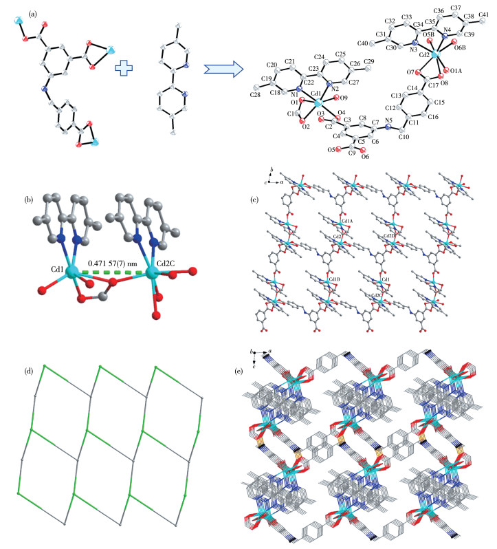
Single crystal X-ray diffraction study reveals that CP 1 conforms to the triclinic P1 space group and each asymmetric unit contains two crystallographically independent Cd2+ cations, one completely deprotonated L3- anion, two dbpy ligands, one HCOO- anion coming from the decompose of DMF, one coordinated water molecule and three lattice water molecules (Fig. 1a). The Cd1 ion is six-coordinated surrounded by one O atom from one μ1-η1 carboxylate group in L3- ligand (O4), two O atoms from one HCOO- anion (O1 and O2), O atom from the coordinated water molecule (O9), and two N atoms from one dbpy ligand (N1 and N2). Cd2 center is encircled by four O atoms originating from two μ1-η1η1 carboxylate groups of two separated L3- anions (O7, O8, O5B, and O6B), one O atom coming from HCOO- (O1A), and two N atoms deriving from dbpy ligand(N3 and N4), presenting a seven-coordinated environment. The Cd—O bond lengths range from 0.225 3(2) to 0.256 6(2) nm, and the minimum and maximum Cd—N bond distances are 0.233 8(3) and 0.238 7(3) nm, respectively, which are within the normal ranges. Two adjacent Cd2+ cations share one HCOO- anion adopting a μ2-η2η1 bridging mode to construct a dinuclear secondary building unit (SBU) [Cd2(HCOO)(dbpy)2], in which the Cd1…Cd2C non-bonding distance is 0.471 57(7) nm and pyridine rings in the two dbpy molecules are almost parallel (Fig. 1b). Neighboring SBUs are firstly linked by isophthalate groups in L3- linkers to form a 1D chain along the b-axis (Fig.S1a). Adjacent 1D chains are further connected through the 4-carboxybenzylamino groups to generate the 2D layer of 1 (Fig. 1c). In this 2D network, the bridging L3- ligands are located in the anti-conformation with the dbpy ligand (Fig.S1b). From the view of topology, the whole structure can be simplified into a 3-connected 2D hcb layer with the point symbol of (63), in which every L3- ligand and SBU can be viewed as three-connected nodes (Fig. 1d). Moreover, 2D layers of 1 are held together via abundant intermolecular interactions, such as the π…π stacking interactions existing between the phenyl rings N1-C18-C19-C20-C21-C22 and N2-C23-C24-C25-C26-C27, or between rings N3-C30-C31-C32-C33-C34 and N4-C35-C36-C37-C38-C39 of dbpy ligands (centroid-to-centroid distance falling in a range of 0.365 97(3) to 0.367 97(3) nm, and dihedral angles of π…π planes are 2.273(131)° and 2.315(139)°, respectively), and the N—H…O hydrogen bonds between amino and carboxylate oxygen atoms in two L3- (Fig.S1c), generating a 3D supramolecular structure (Fig. 1e). The hydrogen bond distance (H5A…O8) of N5—H5A…O8 is 0.291 nm, and N5…O8 distance is 0.302 7(4) nm. Detailed hydrogen bonds are shown in Table S1.
The guest species are omitted for clarity; Symmetry codes: A: x-1, y+1, z; B: x-1, y, z; C: x+1, y-1, z; D: x+1, y, z.
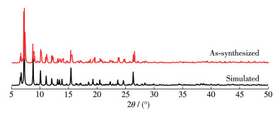
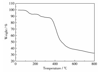
To determine the purity and stability of CP 1, PXRD and TGA experiments were explored. As shown in Fig. 2, the diffraction peaks of as-synthesized CP 1 were similar to the simulated one, which indicates that the synthesized CP 1 is relatively pure. Moreover, the thermal stability of CP 1 was also investigated under a nitrogen atmosphere from room temperature to 800 ℃. As shown in Fig. 3, firstly, the actual weight loss of CP 1 from 80 to 164 ℃ was 5.76%, corresponding to the loss of three free water molecules (Calcd. 5.28%). Secondly, the actual weight loss of 6.05% between 214 and 335 ℃ corresponds to the loss of a coordination H2O molecule and an HCOO- ion (Calcd. 6.16%). Finally, the rapid weight loss at 346 ℃ indicates that the skeleton of CP 1 has collapsed. The above results indicate that the synthesized CP 1 is a crystal material with high purity and stability.
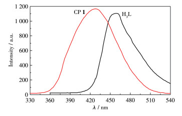
To explore the luminescence properties of CP 1. The solid-state luminescence properties of the H3L ligand and CP 1 were investigated and measured at room temperature. As shown in Fig. 4, the H3L ligand showed a strong maximum emission peak at 456 nm (λex=276 nm), which is due to the typical and common π-π* transition of ligands[30-31]. At the same time, CP 1 performed a similar and broad emission peak at 427 nm (λex=305 nm). The maximum emission peak of CP 1 exhibited a slight blue shift compared with the H3L ligand, which may be due to the ligand-to-metal charge transfer (LMCT)[32-34]. The aqueous suspensions of CP 1 were found to be highly luminescent, with peak maxima at 416 nm (Fig.S2). The curves remained almost similar to that observed in the case of solid samples. The slight blue shift in the emission maxima may be due to the solvent effect. As shown in Fig.S3 of the supplementary information, to verify the fluorescence stability of the suspension, we tested the fluorescence emission spectra for 0, 0.5, 1, 2, 4, and 6 h, respectively. By comparing the spectra, it was found that the fluorescence emission spectra of the complex suspension were relatively stable within at least 6 h. At the same time, after placing the suspension for 24 h, we recovered the CP 1 material through centrifugation. After drying, PXRD identification showed that the main diffraction peaks did not change (Fig.S4). The results illustrate that the structure of CP 1 is stable in the aqueous phase and is not damaged. The above interesting fluorescent properties of CP 1 arouse our great interest in the study of antibiotic identification with CP 1.
Because CP 1 has strong stability and fluorescence properties, we conducted a series of fluorescence sensing experiments for eight antibiotics (NFT, OFX, NFX, CFX, CFA, AMO, GEM, and PCL). Firstly, the selective sensing ability of CP 1 toward different antibiotics was explored by adding 100 μmol·L-1 aqueous solutions of NFT, OFX, NFX, CFX, CFA, AMO, GEM, and PCL to the suspensions of CP 1 (Fig. 5a). The fluorescence intensity of CP 1 was the weakest when 100 μmol·L-1 NFT was added. Meanwhile, the corresponding fluorescence quenching efficiency (QE) was also investigated according to the formula: QE=[(I0-I)/I0]×100%, where I0 and I are the fluorescence intensity of CP 1 suspension without and with antibiotics, respectively. The quenching efficiencies of PCL, AMO, GEM, CFA, NFX, CFX, OFX, and NFT were 1.47%, 1.95%, 1.17%, 2.44%, 4.29%, 4.39%, 7.02%, and 95.12%, respectively (Fig. 5b). These results indicate that CP1 can selectively detect NFT.
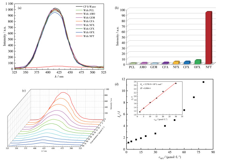
To explore the sensitivity of CP 1 to detect NFT, a fluorescence titration experiment was performed. After adding different concentrations of NFT into the suspension of CP 1, the fluorescence intensity under different concentrations was measured. As shown in Fig. 5c, when the concentration of NFT gradually increased to 80 μmol·L-1, the fluorescence intensity of CP 1 suspension was quenched by more than 90%. To better describe the results of fluorescence titration, the fluorescence quenching efficiency can be quantitatively analyzed using the Stern-Volmer equation: I0/I=1+KsvcNFT, where Ksv is the quenching constant, cNFT is the molar concentration of NFT, and I0 and I are the fluorescence intensity of CP 1 suspension before and after adding NFT[35-36]. According to the Stern-Volmer equation, after adding different concentrations of NFT, the fluorescence intensity showed a good linear relationship at a low concentration of 2 to 30 μmol·L-1 with a high correlation coefficient (R2=0.994 4), and the Ksv of NFT was 5.576 9×10⁴ L·mol-1 (Fig. 5d). In addition, according to the detection limit formula: LOD=3σ/Ksv (σ is the standard deviation of ten standard blank samples)[37-38], the LOD of NFT was calculated to be 0.19 μmol·L-1, which is better than previously reported CP-based sensors performance (Table 3).
下载:
导出CSV
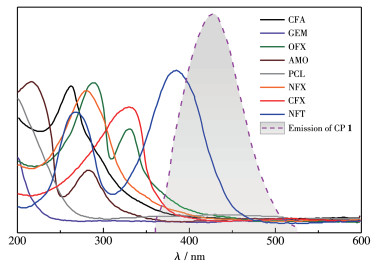
In addition, to explore the possible reason for CP 1 fluorescence quenching caused by NFT, the UV-Vis spectra of eight antibiotics were gained. As shown in Fig. 6, the fluorescence emission spectra of CP 1 overlapped with the UV-Vis absorption spectra of NFT, OFX, CFX, and NFX antibiotics (size order of overlapping areas: NFT > OFX > NFX > CFX), and there was almost no overlap with the other antibiotics. This indicates that CP 1 fluorescence quenching may be caused by the fluorescence resonance energy transfer (FRET), in which NFT can readily absorb the emission light of CP 1[44-46]. These results indicate that CP 1 can be used as a novel fluorescence sensor to selectively and sensitively identify NFT.
In conclusion, a new stable coordination polymer (CP) [Cd2(L)(dbpy)2(HCOO)(H2O)]·3H2O (1) was successfully synthesized by hydrothermal method using Cd(ClO4)2·6H2O, H3L, and dbpy. As a new fluorescence sensor, CP 1 performs a strong fluorescence emission peak at 427 nm (λex=305 nm) and can show selectivity and sensitivity for NFT in an aqueous medium with a low detection limit (0.19 μmol·L-1). In addition, the fluorescence quenching may be caused by the fluorescence resonance energy transfer. These results indicate that CP 1 can be used as a novel and interesting fluorescent probe to detect NFT and it has potential applications in human health, food safety, and environmental protection.
Lei M Y, Ge F Y, Ren S S, Gao X J, Zheng H G. A water-stable Cd-MOF and corresponding MOF@melamine foam composite for detection and removal of antibiotics, explosives, and anions[J]. Sep. Purif. Technol., 2022, 286: 120433. doi: 10.1016/j.seppur.2021.120433
Gao X T, Chen N N, Cao M L, Shi Y, Zhang Q F. A water-stable 3D Eu(Ⅲ)-organic framework as a Bi-functional ratiometric luminescent sensor for fast, sensitive and selective detection of ODZ and Hg2+ in aqueous media[J]. Chin. J. Struct. Chem., 2022, 41: 2211110.
Zhang Q Q, Ying G G, Pan C G, Liu Y S, Zhao J L. Comprehensive evaluation of antibiotics emission and fate in the river basins of China: Source analysis, multimedia modeling, and linkage to bacterial resistance[J]. Environ. Sci. Technol., 2015, 49(11): 6772-6782. doi: 10.1021/acs.est.5b00729
Kanzariya D B, Goswami R, Muthukumar D, Pillai R S, Pal T K. Highly luminescent MOF and its in situ fabricated sustainable corn starch gel composite as a fluoro-switchable reversible sensor triggered by antibiotics and oxo-anions[J]. ACS Appl. Mater. Interfaces, 2022, 14(43): 48658-48674. doi: 10.1021/acsami.2c13571
Batt A L, Aga D S. Simultaneous analysis of multiple classes of antibiotics by ion trap LC/MS/MS for assessing surface water and groundwater contamination[J]. Anal. Chem., 2005, 77(9): 2940-2947. doi: 10.1021/ac048512+
Bai Y, Zhang M L, Wang B T, Ren Y X, Zhao Y C, Yang H, Yang X G. Four MOFs with isomeric ligands as fluorescent probes for highly selective, sensitive and stable detection of antibiotics in water[J]. CrystEngComm, 2022, 24: 169-181. doi: 10.1039/D1CE01261D
Tian C, Ren X, He M, Chen B B, Hu B. Core-shell magnetic porous organic polymer for magnetic solid-phase extraction of fluoroquinolone antibiotics in honey samples followed by high-performance liquid chromatography with fluorescence detection[J]. J. Sep. Sci., 2022, 45(4): 874-882. doi: 10.1002/jssc.202100678
Viñas P, Balsalobre N, López-Erroz C, Hernández-Córdoba M. Liquid chromatography with ultraviolet absorbance detection for the analysis of tetracycline residues in honey[J]. J. Chromatogr. A, 2004, 1022: 125-129. doi: 10.1016/j.chroma.2003.09.066
Paul P, Reynaert J, Griend C S D, Adams E, Schepdael A V. Development and validation of a CE method for the determination of tetracyclines with capacitively coupled contactless conductivity detection[J]. Chromatographia, 2019, 82: 1395-1403. doi: 10.1007/s10337-019-03755-4
Wu X Y, Xu Z Q, Huang Z, Shao C Y. Large volume sample stacking of cationic tetracycline antibiotics toward 10 ppb level analysis by capillary electrophoresis with UV detection[J]. Electrophoresis, 2016, 37(22): 2963-2969. doi: 10.1002/elps.201600189
Yu C X, Jiang W, Wang K Z, Liang A P, Song J G, Zhou Y L, Sun X Q, Liu L L. Luminescent two-dimensional metal-organic framework nanosheets with large π-conjugated system: Design, synthesis, and detection of anti-inflammatory drugs and pesticides[J]. Inorg. Chem., 2022, 61(2): 982-991. doi: 10.1021/acs.inorgchem.1c03040
Li X, Zhang S, Zhang L, Yang Y, Zhang K, Cai Y C, Xu Y, Gai Y L, Xiong K C. Viologen-based cationic metal-organic framework for antibiotics detection and MnO4- removal in water[J]. Cryst. Growth Des., 2022, 22(7): 3991-3997. doi: 10.1021/acs.cgd.2c00170
Zhao Y, Jing J, Yan N, Han M L, Yang G P, Ma L F. Different benzendicarboxylate-directed structural variations and properties of four new porous Cd(Ⅱ)-pyridyl-triazole coordination polymers[J]. Front. Chem., 2020, 8: 616468. doi: 10.3389/fchem.2020.616468
Su P C, Zhang A R, Yu L, Ge H W, Wang N, Huang S Y, Ai Y J, Wang X K, Wang S H. Dual-functional UiO-type metal-organic frameworks for the sensitive sensing and effective removal of nitrofurans from water[J]. Sens. Actuator B-Chem., 2022, 350: 130865. doi: 10.1016/j.snb.2021.130865
Benzaqui M, Wahiduzzaman M, Zhao H, Hasan M R, Steenhaut T, Saad A, Marrot J, Normand P, Greneche J M, Heymans N, Weireld G D, Tissot A, Shepard W, Filinchuk Y, Hermans S, Carn F, Manlankowska M, Tellez C, Coronas J, Maurin G, Steunou N, Serre C. A robust eco-compatible microporous iron coordination polymer for CO2 capture[J]. J. Mater. Chem. A, 2022, 10: 8535-8545. doi: 10.1039/D1TA10385G
Chen X, Qiao Z W, Hou B, Jiang H, Gong W, Dong J Q, Li H Y, Cui Y, Liu Y. Chiral metal-organic frameworks with tunable catalytic selectivity in asymmetric transfer hydrogenation reactions[J]. Nano Res., 2021, 14: 466-472. doi: 10.1007/s12274-020-2905-7
石明凤, 李立凤, 杨良竹, 于红梅, 肖铭, 唐怀军, 王凯民. 两个半导体Co(Ⅱ)配位聚合物的合成和光催化[J]. 中山大学学报(自然科学版), 2022,61,(6): 74-80. SHI M F, LI L F, YANG L Z, YU H M, XIAO M, TANG H J, WANG K M. Synthesis and photocatalytic study of two semi-conductive Co(Ⅱ) coordination polymers[J]. Acta Scientiarum Naturalium Universitatis Sunyatseni, 2022, 61(6): 74-80.
Zhao J J, Liu P Y, Dong Z P, Liu Z L, Wang Y Q. Eu(Ⅲ)-organic framework as a multi-responsive photoluminescence sensor for efficient detection of 1-naphthol, Fe3+ and MnO4- in water[J]. Inorg. Chim. Acta, 2020, 511: 119843. doi: 10.1016/j.ica.2020.119843
Liang J Y, Zulkifli M Y B, Choy S, Li Y, Gao M, Kong B, Yun J, Liang K. Metal-organic framework-plant nano biohybrids as living sensors for on-site environmental pollutant detection[J]. Environ. Sci. Technol., 2020, 54(18): 11356-11364. doi: 10.1021/acs.est.0c04688
王凯民, 郭津榕, 张倩, 唐怀军, 王玉娜, 马钰璐. 含有分离电荷中心的吡啶鎓两性离子型Cd(Ⅱ)配合物的合成、结构和荧光性研究[J]. 云南大学学报(自然科学版), 2022,44,(3): 576-584. WANG K M, GUO J R, ZHANG Q, TANG H J, WANG Y N, MA Y L. Synthesis, structure and fluorescence properties of pyridonium amphoteric Cd(Ⅱ) complexes containing charge separation centers[J]. Journal of Yunnan University (Natural Sciences Edition), 2022, 44(3): 576-584.
Gao Y F, Zhang X Q, Sun W, Liu Z L. A robust microporous metal-organic framework as a highly selective and sensitive, instantaneous and colorimetric sensor for Eu3+ ions[J]. Dalton Trans., 2015, 44(4): 1845-1849. doi: 10.1039/C4DT02752C
Xu X Y, Bing Y. A fluorescent wearable platform for sweat Cl- analysis and logic smart-device fabrication based on color adjustable lanthanide MOFs[J]. J. Mater. Chem. C, 2018, 6(7): 1863-1869. doi: 10.1039/C7TC05204A
Kondo A, Noro S I, Kajiro H, Kanoh H. Structure- and phase-transformable coordination polymers/metal complexes with fluorinated anions[J]. Coord. Chem. Rev., 2022, 471: 214728. doi: 10.1016/j.ccr.2022.214728
Gurtovyi R I, Gavrish S P, Tsymbal L V, Apostu M O, Cazacu M, Shova S, Lampeka Y D. 2D coordination polymers and ionic complexes of the nickel(Ⅱ) and zinc(Ⅱ) cyclam cations with trigonal carboxylate linkers based on triazine core. Crystal structures, supramolecular catenation and spectral characterization[J]. Polyhedron, 2022, 221: 115870. doi: 10.1016/j.poly.2022.115870
王凯民, 石明凤, 李立凤, 樊保敏, 孙蔚青, 马钰璐. 混合配体构筑的水相高稳定Zn(Ⅱ)金属有机框架铁离子荧光探针[J]. 中山大学学报(自然科学版), 2023,62,(1): 142-148. doi: 10.13471/j.cnki.acta.snus.2022c010WANG K M, SHI M F, LI L F, FAN B M, SUN W Q, MA Y L. Construction of water stable Zn(Ⅱ) metal-organic framework iron ion fluorescence probe with mixed ligand[J]. Acta Scientiarum Naturalium Universitatis Sunyatseni, 2023, 62(1): 142-148. doi: 10.13471/j.cnki.acta.snus.2022c010
Sun S L, Sun X Y, Sun Q, Gao E Q. Highly efficient fluorescent chemosensor for nitro antibiotic detection based on luminescent coordination polymers with 2, 6-di(4-carboxyphenyl)pyrazine[J]. CrystEngComm, 2021, 23(17): 3167-3174. doi: 10.1039/D1CE00245G
王凯民, 李立凤, 石明凤, 叶艳青, 王玉娜, 郭津榕, 唐怀军, 马钰璐. 基于两性离子配体构筑的Eu(Ⅲ)配位聚合物的晶体结构和对呋喃西林的检测[J]. 无机化学学报, 2022,38,(9): 1843-1852. WANG K M, LI L F, SHI M F, YE Y Q, WANG Y N, GUO J R, TANG H J, MA Y L. Crystal structure of Eu(Ⅲ) coordination polymer based on zwitterionic ligand and detection of furacilin[J]. Chinese J. Inorg. Chem., 2022, 38(9): 1843-1852.
Su Y Q, Fu L S, Cui G H. Two chemically robust Cd(Ⅱ)-frameworks for efficient sensing of levofloxacin, benzaldehyde, and Fe3+ ions[J]. Dalton Trans., 2021, 50(43): 15743-15753. doi: 10.1039/D1DT03205D
Sheldrick G M. Crystal structure refinement with SHELXL[J]. Acta Crystallogr. Sect. C, 2015, C71: 3-8.
Yu X Y, Dong W W, Han H M, Zhao J, Li D S. A water-stable Zn(Ⅱ) coordination polymer as fluorescent sensor for selective and sensitive detection of antibiotics and Fe3+[J]. J. Solid State Chem., 2021, 296: 122032. doi: 10.1016/j.jssc.2021.122032
Wang K M, Yang L Z, Li L F, Dong X Y, Wang Z L, Tang H J, Sun W Q, Ma Y L. A water-stable zwitterionic Cd(Ⅱ) coordination polymer as fluorescent sensor for the detection of oxo-anions and dimetridazole in milk[J]. Arab. J. Chem., 2022, 15(11): 104295. doi: 10.1016/j.arabjc.2022.104295
Zeng N N, Ren L, Cui G H. Ultrasensitive fluorescence detection of norfloxacin in aqueous medium employing a 2D Zn(Ⅱ)-based coordination polymer[J]. CrystEngComm, 2022, 24(5): 931-935.
Yang Y X, Guo Y W, Xia S Y, Ma X N, Wu X X. Metal-organic framework sensors based on triazole carboxylic acid ligands for ion sensing and dye adsorption[J]. J. Solid State Chem., 2022, 331: 123113.
Wang K M, Yang L Z, Li L F, Ma Y L, Guo J R, Wang Z L, Tang H J, Wang Y N, Zhou J. Highly sensitive and rapid fluorescence detection of chlortetracycline in milk using a water- and pH-stable Zn(Ⅱ) coordination polymer derived from zwitterionic and N-donor ligands[J]. J. Solid State Chem., 2022, 316: 123606.
Wang X M, Liu C, Wang M, Zhou X H, YouY J, Xiao H P. A selective fluorescence turn-on sensing coordination polymer for antibiotic aztreonam[J]. Chem. Commun., 2022, 58(29): 4667-4670.
Ye Y, Ge B D, Meng X Y, Liu Y H, Wang S, Song X W, Liang Z Q. An yttrium-organic framework based on a hexagonal prism second building unit for luminescent sensing of antibiotics and highly effective CO2 fixation[J]. Inorg. Chem. Front., 2022, 9(2): 391-400.
Zhu G S, Cheng S L, Zhou Z D, Du B, Shen Y Y, Yu B Y. Bisligand-coordinated cadmium organic frameworks as fluorescent sensors to detect ions, antibiotics and pesticides in aqueous solutions[J]. Polyhedron, 2022, 217: 115759.
Wang K M, Li L F, Yang L Z, Guo J R, Wang Z L, Tang H J, Ma Y L. A water-stable zwitterionic Zn(Ⅱ) coordination polymer as a luminescent sensor for the nitrofurazone antibiotic in milk[J]. Polyhedron, 2022, 226: 116092.
Niu M Y, Yang X P, Ma Y N, Wang C R, Hao W X, Shi D L, Schipper D. Construction of an octanuclear Zn(Ⅱ)-Yb(Ⅲ) Schiff base complex for the NIR luminescent sensing of Nitrofuran antibiotics[J]. Chin. J. Chem., 2021, 39(8): 2083-2087.
Xu S, Shi J J, Ding B, Liu Z Y, Wang X G, Zhao X J, Yang E C. A heterometallic sodium(Ⅰ)-europium(Ⅲ)-organic layer exhibiting dual-responsive luminescent sensing for nitrofuran antibiotics, Cr2O72- and MnO4- anions[J]. Dalton Trans., 2019, 48(5): 1823-1834.
Ma Y N, Yang X P, Shi D L, Niu M Y, Schipper D. Construction of a 18-Metal neodymium(Ⅲ) nanoring with NIR luminescent sensing to antibiotics[J]. Inorg. Chem., 2020, 59(23): 17608-17613.
Zhang Y J, Yang J D, Zhao D S, Liu Z J, Li D C, Fan L M, Hu T P. Two cadmium(Ⅱ) coordination polymers as luminescent sensors for the detection of nitrofuran/nitroimidazole antibiotics[J]. CrystEngComm, 2019, 21(41): 6130-6135.
Yang H W, Xu P, Ding B, Liu Z Y, Zhao X J, Yang E C. A highly stable luminescent Eu-MOF exhibiting efficient response to nitrofuran antibiotics through the inner filter effect and photoinduced electron transfer[J]. Eur. J. Inorg. Chem., 2019, (48): 5077-5084.
Xu N, Zhang Q H, Zhang G A. A carbazole-functionalized metal-organic framework for efficient detection of antibiotics, pesticides and nitroaromatic compounds[J]. Dalton Trans., 2019, 48(8): 2683-2691.
Wang Y, Xu J, Wang R Z, Liu H, Yu S S, Xing L B. Supramolecular polymers based on host-guest interactions for the construction of artificial light-harvesting systems[J]. Spectroc. Acta Pt. A-Molec. Biomolec. Spectr., 2022, 279(15): 121402.
Zhang L Q, Wang X W, Gu L, Yu Y H, Gao J S. Three pairs of luminescent coordination polymers based on CoⅡ and CdⅡ clusters for the detection of antibiotics, pesticides and chiral nitro aromatic compounds[J]. RSC Adv., 2020, 10(16): 9476-9485.

Figure 1 Structure of CP 1: (a) asymmetric unit with 30% thermal ellipsoids, showcasing the coordination mode of two Cd2+ centers, L3- and dbpy ligands; (b) dinuclear [Cd2(HCOO)(dbpy)2] SBU; (c) 2D network; (d) 2D hcb layer; (e) 3D supramolecular structure
The guest species are omitted for clarity; Symmetry codes: A: x-1, y+1, z; B: x-1, y, z; C: x+1, y-1, z; D: x+1, y, z.

Figure 5 (a) Fluorescence spectra of CP 1 when eight antibiotics were added; (b) Fluorescence quenching efficiency of CP 1 aqueous suspension when eight antibiotics were added; (c) Fluorescence titration experiment by gradual adding NFT to CP 1 water suspension; (d) Stern-Volmer plots for CP 1 sensing NFT
Table 1. Crystal data and structure refinement of CP 1
| Parameter | 1 | Parameter | 1 | |
| Empirical formula | C41H43Cd2N5O12 | Z | 2 | |
| Formula weight | 1 022.65 | μ / mm-1 | 0.922 | |
| Crystal system | Triclinic | F(000) | 972 | |
| Space group | P1 | Reflection collected | 22 326 | |
| a / nm | 1.250 16(13) | Independent reflection | 10 302 (Rint=0.027 6) | |
| b / nm | 1.470 19(15) | Goodness-of-fit on F2 | 1.071 | |
| c / nm | 1.477 81(15) | Data, restraint, parameter | 10 302, 0, 518 | |
| α / (°) | 66.571 0(10) | Final R indices* [I > 2σ(I)] | R1=0.033 4, wR2=0.086 3 | |
| β / (°) | 89.373 0(10) | R indices (all data) | R1=0.046 0, wR2=0.099 6 | |
| γ / (°) | 78.957 0(10) | |||
 下载: 导出CSV
Table 2. Selected bond lengths (nm) and bond angles (°) of CP 1
| Cd1—O4 | 0.225 3(2) | Cd2—O1A | 0.234 3(2) | N1—C18 | 0.132 9(4) |
| Cd1—O2 | 0.231 9(3) | Cd2—O6B | 0.236 3(2) | C2—C3 | 0.149 9(4) |
| Cd1—N2 | 0.233 8(3) | Cd2—N3 | 0.238 7(3) | ||
| Cd2—O7 | 0.233 6(2) | O1—C1 | 0.123 9(4) | ||
| O4—Cd1—O9 | 81.10(9) | O7—Cd2—O1A | 91.45(8) | C9—O5—Cd2D | 93.72(18) |
| O9—Cd1—O2 | 106.87(11) | N4—Cd2—O5B | 106.88(9) | C18—N1—Cd1 | 124.2(2) |
| O4—Cd1—N2 | 88.75(9) | O7—Cd2—N3 | 84.88(9) | ||
| N2—Cd1—N1 | 70.62(10) | C1—O2—Cd1C | 125.8(2) | ||
| Symmetry codes: A: x-1, y+1, z; B: x-1, y, z; C: x+1, y-1, z; D: x+1, y, z. | |||||
下载: 导出CSV

 扫一扫看文章


扫一扫关注我们