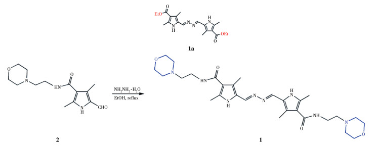
Scheme 1.
Synthesis route of probe 1

Pyrrole-based hydrazone for fluorescent imaging of Hg2+ in lysosomes
Zhen WANG , Si-Yuan LI , Yuan WANG , Wei-Na WU , Lei ZHANG , Zhong CHEN , Ling-Ling YAN


Fluorescent probes can be used as an efficient tool for the detection and imaging of biological trace species, pesticide residues and heavy metal cations[1]. Among various heavy metal cations, Hg2+ is extremely toxic, and it becomes methylmercury which accumulates in the body through the food chain, leading to a series of mental illnesses and the well-known Minamata disease[2-4]. Consequently, selective and sensitive fluorescent probes for Hg2+ detection are highly desirable.
Generally, most of papers published about Hg2+ probes have investigated the high thiophilicity of Hg2+ [5-8]. Furthermore, rhodamine[6-7, 9, 12], coumarin[10], and naphthalimide[7] have been frequently employed as signal fluorophores for the probe design. Some of them have been utilized for fluorescence imaging of Hg2+ in living cells (Table S1, Supporting information) [5-14]. It is worth noting that lysosomes digest unwanted substances and cell fragments and contain a variety of hydrolases, so they are considered"enzyme banks"or"cell cleaners"in cells[15]. In fact, Hg2+ has been found to accumulate in lysosomes, causing abnormal lysosomal pH fluctuation, which is closely associated with lysosome dysfunctions[16]. In this regard, lysosomes are one of the important organelle targets for Hg2+ toxicity[17]. However, the probes that can be used in lysosomes for Hg2+ detection are barely reported[5-6, 11]. It is well known that the pH of lysosomes in normal cells ranges from 4.5 to 6.0, while that of cancer cells is even lower[18]. Nevertheless, some of the reported probes, especially those based on rhodamine backbone, could not perform normally under such extremely acidic conditions[12]. In this regard, it is still a great challenge to the development of lysosome-targeting probes toward Hg2+.
Recently, our group reported a pyrrole-containingbis-hydrazone (1a, Scheme 1) capable of detecting Hg2+ in real water samples[14]. Herein, a lysosome - targeting group of 4-(2-aminoethyl)morpholine was introduced to 1a to fabricate a simple and effective fluorescent turn-on probe 1 for Hg2+ detection in lysosomes of HeLa cells. It is worth noting that the primary starting materi-al, 5 - formyl - 2, 4 - dimethyl - 1H - pyrrole - 3 - carboxylic acid, is a commercial intermediate of Sunitinib (an inhibitor used in cancer therapies) favoring the real -world application of the as-synthesized probe.
Solvents and starting materials for syntheses were purchased commercially and used as received. Elemental analyses were carried out on an Elemental Vario ELanalyzer. 1H NMR spectra were recorded on a Bruker AV400 NMR spectrometer in DMSO - d6 solution. The UV spectra were recorded on a Purkinje General TU-1800 spectrophotometer. Fluorescence spectra were determined on a Varian CARY Eclipse spectrophotometer, in the measurements of emission and excitation spectra the pass width was 5 nm. Time-resolved photoluminescence spectra were determined on an Edinburgh FLS980 spectrophotometer. ESI-MS spectra were obtained on a Bruker Daltonics Esquire 6000 mass spectrometer. The cytotoxic effect exerted by 1 on cultured HeLa cells was ascertained by a standard MTT assay according to the literature method[19]. Fluorescent images were taken on Zeiss Leica inverted epifluorescence/reflectance laser scanning confocal microscope.
5-Formyl-2, 4-dimethyl-1H-pyrrole-3-carboxylic acid (2-morpholin-4-yl-ethyl)-amide (2) was prepared from 5-formyl-2, 4-dimethyl-1H-pyrrole-3-carboxylic acid according to literature method[20]. Hydrazinium hydroxide (100 mg, 85%, 1.7 mmol) and 2 (279 mg, 1mmol) were added to an EtOH solution (10 mL). The mixture was refluxed for 3 h with two drops of acetic acid. After cooling to room temperature, the separated solid was filtered, washed with EtOH, and then dried in air. Yield: 58%. Anal. Calcd. for C28H42N8O4(%): C, 60.63; H, 7.63; N, 20.20. Found(%): C, 60.48; H, 7.78;N, 20.33. 1H NMR (400 MHz, DMSO - d6): δ 11.35 (s, 1H, NH), 8.40 (s, 1H, CH), 7.24 (s, 1H, NH), 3.57 (t, 4H, 2CH2), 3.30 (2H, merged by peak of H2O), 2.41 -2.44 (m, 8H, 4CH2), 2.35 (s, 3H, CH3), 2.25 (s, 3H, CH3). 13C NMR was not recorded due to the poor solubility of 1. ESI-MS: m/z=555.321 8 for [M+H] + (Calcd.555.330 0), 278.179 8 for [M+2H]2+ (Calcd. 278.1750), 185.787 0 for [M+3H]3+ (Calcd. 185.786 6).
The spectral analyses were accomplished in EtOH/H2O (7∶3, V/V) solution at room temperature. The concentration of probe 1 for UV-Vis and fluorescence measurement was 10 µmol·L-1. Solutions of metal ions were prepared with nitrate or chloride salts in H2O. UV-Vis and fluorescence spectrophotometric titration were conducted directly in a 2 mL cuvette by successive addition of corresponding chemical reagent using a microliter syringe. Upon addition of every aliquot, the solution was well mixed then the spectrumwas measured.
Probe 1 was produced by the condensation of 2 and hydrazinium hydroxide in a moderate yield, and was further characterized by elemental analyses, 1H NMR, and ESI-MS methods. It should be noted that the peak of one CH2 group was covered by the H2O peak in the 1H NMR spectrum of 1 (Fig. S1), which is assumed according to the spectrum of 2 shown in Fig.S2.
The absorption spectrum of probe 1 (10 µmol·L-1) in EtOH/H2O (7∶3, V/V) solution displayed one band centered at 380 nm (Fig. 1a), attributed to the n - π*transition of imine units[14]. When probe 1 bound to Hg2+ (3 equiv.), the band had an obvious redshift to 413 nm, a hyperchromatic effect, along with the generation of a shoulder at 440 nm, assigned to the ligand to metal charge transfer (LMCT) [13]. This finding indicates that the N atom of the imine bond is involved in the coordination with Hg2+. The change of the color of the liquid in the cuvette from colorless to yellow leads to the conclusion that the response of probe 1 to Hg2+ is more pronounced and more selective than the other metal ions (3 equiv.) tested (Fig. 1a, Inset), including Ag+, Al3+, Ca2+, Cd2+, Co2+, Cr3+, Cu2+, Fe3+, K+, Mg2+, Mn2+, Na+, Ni2+, Pb2+, and Zn2+, which enables the potential application of detection of Hg2+ in water samples by naked eyes. The absorbance ratio A440/A380 linearly increased by increasing the Hg2+ concentration from 4.5 to 16.5 µmol·L-1 (Fig. 1b, Inset). The two isosbestic points at 308 and 403 nm reveal the existence of only one intermediate complex (Fig. 1b). The Job′s plot obtained by varying the concentration ratio of Hg2+ and 1 gained the 1∶1 combined stoichiometric ratio of 1 and Hg2+ (Fig.S3), with an association constant (Ka) of 3.48×10⁴ L·mol-1 according to the Benesi - Hildebrand expression (Fig. S4) [21]. The limit of detection (LOD) of 1 to Hg2+ was 59.6 nmol·L-1 based on 3σ/k (σ: standard deviation, k: slope)[20], which was lower than the LOD of 1a.
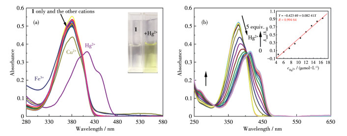
Inset in a: the color change of 1 solution in the presence of Hg2+; Inset in b: the absorbance ratio of A440/A380 as a function of Hg2+ concentration (4.5-16.5 µmol·L-1)
Fluorescence emission spectral changes of 1, caused by the presence of several metal ions in the EtOH/H2O (7: 3, V/V) solution, are demonstrated in Fig. 2. When excited at 410 nm, the initial solution of 1 displayed a weak emission at 475 nm (quantum yield Φ =0.02, determined with quinine sulfate, and Φc=0.546 in 0.05 mol·L-1 H2SO4)[22]. Significant changes in the emission spectrum were observed in the presence of Hg2+ (3 equiv.), except in the case of other metal cations (3 equiv.). When 1 was combined with Hg2+ (3equiv.), the fluorescence intensity was significantly enhanced as the color changed from colorless to cyan (Fig. 2a, Inset) under a 365 nm UV lamp (Φ =0.21).Furthermore, it can be seen from Fig.S5 that the average decay constant (τ) value of 1 obtained by the singleexponential decay fitting method was 1.56 ns, which was far less than that of 1-Hg2+ (6.12 ns), indicating thecoordination between 1 and Hg2+.
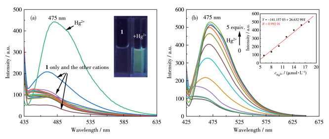
Inset in a: the color change of 1 solution in the presence of Hg2+ under 365 nm UV lamp; Inset in b: the fluorescence intensity at 475 nm as a function of Hg2+ concentration (6.0-18.0 µ mol·L-1); λex=410 nm
Similarly, the linear response concentration range of Hg2+ was 6.0-18.0 µmol·L-1 according to the fluorescent titration results (Fig. 2b, Inset). The Ka and LOD were 2.61×10⁴ L·mol-1 (Fig. S6) and 45.8 nmol·L-1, respectively, which are in the same order of magnitude when compared with the data from UV spectra. All these facts show that the response of probe 1 to Hg2+ is characterized by efficient selectivity and sensitivity in colorimetric and fluorescent detection, so it can be used to research Hg2+ quantitatively in water or biological samples.
The specificity of Hg2+ to probe 1 was determined by testing the interference of other metal cations (Fig.S7 and S8). No distinct changes in the UV and fluorescence spectra of 1+Hg2+ were observed in the presence of other metal cations, in addition to trivalent cations (Al3+, Cr3+, and Fe3+) in which their strong Lewis acidity results in imine - bond cleavage[23], thereby indicating that the ability of probe 1 to detect Hg2+ is less affected by other metal cations and has high selectivity.
It was concluded that 1 can detect Hg2+ in a pH range of 4.0 to 10.0 by studying the fluorescence intensity changes of probe 1 and probe 1+Hg2+ at different pH values (Fig.S9). Due to this unique property, probe 1 can be used for practical studies of Hg2+ in lysosomes. Compared with the probe 1a described in our previous work[14], the introduction of 4 - (2 - aminoethyl) morpholine not only improves the selectivity of the probe (interference from Cu2+ in the case of 1a) but also enhances the tolerance to acidic environments.
A fast time response is an important characteristic of chemosensors. The changes in the fluorescence of 1 (10 mol·L-1) in EtOH/H2O (7∶3, V/V) solution with 3 equiv. of Hg2+ was detected in less than 2 min, which is a faster analysis time for the Hg2+ detection than that of 1a (ca. 3 min) (Fig.S10).
The reversibility of the probe toward Hg2+ was investigated by adding EDTA to the 1+Hg2+ system. The results showed that the emission of 1+Hg2+ wastotally quenched upon the addition of EDTA (3 equiv.), and it was recovered by further addition of Hg2+. The on-off phenomenon was reversible even after four cycles (Fig.S11), indicating that the probe can be easily regenerated for repeated use.
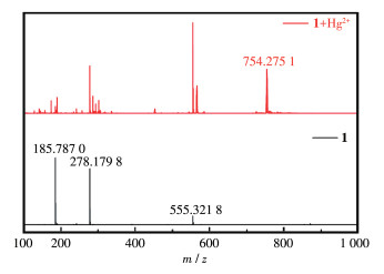
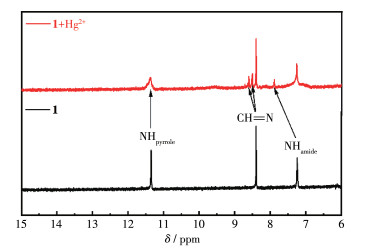
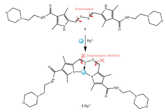
To analyze the coordination mechanism of 1 with Hg2+, ESI-MS measurements were performed in EtOH solution (Fig. 3). The spectrum of 1+Hg2+ displayed a peak at m/z 754.275 1 corresponding to the protonated cation of [Hg(1-2H)] (1 - Hg2+, C28H40N8O4Hg, Calcd.753.29). Furthermore, in the 1H NMR spectrum of 1+Hg2+ (Fig. 4), the signal of the CH=N groups of 1 at δ 8.40 shifted to the lower field and split into two peaks at δ 8.50 and 8.60, indicating the coordination of one imine N atom and the change in the environment of the other imine N atom. The amide NH signal of 1 was greatly broadened at δ 7.24 and significantly shifted to δ 7.89 in the presence of Hg2+, it may be because of the effect of the two pyrrole N atoms to protons and coordination effect. Therefore, a donor set of two pyrrole and one imine N atoms involving the binding of Hg2+ is suggested, as illustrated in Scheme 2.
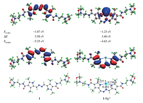
To gain more insights into the sensing mechanism, 1 and 1-Hg2+ were examined by density functional theory (DFT) calculation[24]. The results show that both HOMO and LUMO are dispersed on pyrrole and imine moieties in probe 1 (Fig. 5), while the C=N isomerization leads to weak fluorescence. In contrast, the C=N isomerization is not possible in the 1 - Hg2+ complex. Meanwhile, the HOMO of 1-Hg2+ is localized on the pyrrole and imine moieties, while the LUMO is almost centered on the Hg2+ ion, clearly indicating that the emission of 1-Hg2+ is mainly from the LMCT. The corresponding energy difference (ΔE) of 1 - Hg2+ (3.40eV) is lower than that of 1 (3.58 eV), reflecting the longer maximum absorbance wavelength of 1-Hg2+ compared to that of 1. The Hg—Npyrrole bond lengths in complex 1-Hg2+ were 0.236 0 and 0.237 5 nm, respectively, which are shorter than that of Hg—Nimine (0.253 2 nm). Nevertheless, these bond lengths are comparable with those in the calculated 1a-Hg2+ complex, thus suggesting stable emission of complex 1-Hg2+ in an aqueous solution[14].
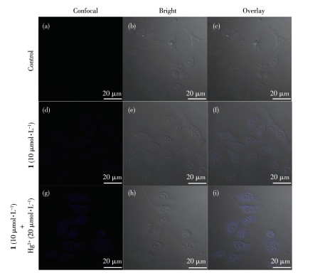
The MTT assay was used to detect the effect of probe 1 on cell viability as shown in Fig.S12, the cell viability was still 82.6% even while the probe concentration reached 40 µmol·L-1, which indicates that probe 1 had low cytotoxicity. The biological suitability of probe 1 was assessed by detecting its ability to examine Hg2+ in HeLa cells. HeLa cells without the treatment of probe 1 exhibited no fluorescence signal(Fig. 6a), while after incubation with 1 (10 µmol·L-1) at 37 ℃ for 30 min, a faint blue fluorescence was observed in the cells (Fig. 6d), which should be attributed to the emission of probe 1. Next, the brighter cellular blue fluorescence was shown after the cells were treated with 20 µmol·L-1 Hg2+ (Fig. 6g). This finding indicates the capability of 1 in imaging Hg2+ in living cells.
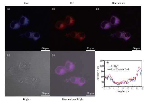
To determine the capability of 1 to detect Hg2+ in the lysosomes of HeLa cells, colocalization images were performed with LysoTracker Red (Fig. 7). Whenthe image was merged, it could be observed that the blue fluorescence channel of 1+Hg2+ has a coincidence rate of 0.91 with the red channel of the lysosome tracker. The above results indicate that the probe can detect Hg2+ in the lysosomes of living cells.
In summary, a pyrrole-based bis-hydrazone appended with 4-(2-aminoethyl)morpholine moiety has been prepared, which acts as a fluorescent off-on sensor toward Hg2+. The mechanism was completely investigated by UV - Vis, fluorescence spectroscopy, time -resolved fluorescence spectroscopy, ESI-MS, 1H NMR, and DFT calculations. probe 1 can be used as an Hg2+ sensor. In addition, the costained experiment reveals that the probe was able to image Hg2+ in lysosomes.
Supporting information is available at
成昭, 梁静, 任熙玲, 崔誉文, 杨莉宁. 有机小分子Zn2+荧光探针与生命体中Zn2+示踪的研究进展[J]. 化学世界, 2021,62,(12): 707-716. CHENG Z, LIANG J, REN X L, CUI Y W, YANG L N.. Research progress in organic small-molecule fluorescent Zn2+ probes and fluo- rescent imaging analysis of biological Zn2+.[J]. Chemical World, 2021, 62(12): 707-716.
Marumoto M, Sakamoto M, Marumoto K, Tsuruta S, Komohara Y. Mer- cury and selenium localization in the cerebrum, cerebellum, liver, and kidney of a Minamata disease case[J]. Acta Histochem.Cytochem., 2020, 53(6): 147-155. doi: 10.1267/ahc.20-00009
Kim K H, Kabir E, Jahan S A. A review on the distribution of Hg in the environment and its human health impacts[J]. J. Hazard. Mater., 2016, 306: 376-385. doi: 10.1016/j.jhazmat.2015.11.031
Erdemir S, Kocyigit O, Malkondu S. Detection of Hg2+ ion in aqueous media by new fluorometric and colorimetric sensor based on triazole- rhodamine[J]. J. Photochem. Photobiol. A, 2015, 309: 15-21. doi: 10.1016/j.jphotochem.2015.04.017
Sarkar A, Chakraborty S, Lohar S, Ahmmed E, Saha N C, Mandal S K, Dhara K. Chattopadhyay P. A lysosome- targetable fluorescence sen- sor for ultrasensitive detection of Hg2+ in living cells and real sam- ples[J]. Chem. Res. Toxicol., 2019, 32(6): 1144-1150. doi: 10.1021/acs.chemrestox.9b00005
Yuan X, Leng T H, Guo Z Q, Wang C Y, Li J Z, Yang W W, Zhu W H. A FRET - based dual - channel turn - on fluorescence probe for the detection of Hg2+ in living cells[J]. Dyes Pigm., 2018, 161: 403-410.
Wang Y S, Ding H C, Wang S, Fan C B, Tu Y Y, Liu G, Pu S Z. A rati- ometric and colorimetric probe for detecting Hg2+ based on naph- thalimide rhodamine and its staining function in cell imaging[J]. RSC Adv., 2019, 9(21): 11664-11669. doi: 10.1039/C9RA01459D
Lan L X, Niu Q F, Li T D.. A highly selective colorimetric and ratio- metric fluorescent probe for instantaneous sensing of Hg2+ in water, soil and seafood and its application on test strips[J]. Anal. Chim. Acta, 2018, 1023: 105-114. doi: 10.1016/j.aca.2018.03.023
侯淑华, 李仕琦, 汤立军. 新型水溶性罗丹明类荧光探针的合成及其对游离3价金属离子的识别[J]. 应用化学, 2022,39,(2): 241-246. HOU S H, LI S Q, TANG L J.. Synthesis of a novel water-soluble rho- damine-based fluorescent probe and its selective detection of free tri- valent ions.[J]. Chinese Journal of Applied Chemistry, 2022, 39(2): 241-246.
Wu C J, Wang J B, Shen J J, Bi C, Zhou H W. Coumarin-based Hg2+ fluorescent probe: Synthesis and turn - on fluorescence detection in neat aqueous solution[J]. Sens. Actuator B-Chem., 2017, 243: 678-683. doi: 10.1016/j.snb.2016.12.046
Zhang Y F, Chen H Y, Chen D, Wu D, Chen Z, Zhang J, Chen X Q, Liu S H, Yin J. A colorimetric and ratiometric fluorescent probe for mercury in lysosome[J]. Sens. Actuator B-Chem.., 2016, 224: 907-914. doi: 10.1016/j.snb.2015.11.018
孙京府, 钱鹰. 一种萘酰亚胺-罗丹明衍生物的合成、聚集诱导荧光增强及其双通道荧光探针性能[J]. 有机化学, 2016,36,(1): 151-157. SUN J F, QIAN Y.. A novel naphthalimide- rhodamine fluorescence sensor: synthesis, aggregation - induced emission enhancement and its dual-channel detection property[J]. Chin. J. Org. Chem., 2016, 36(1): 151-157.
Vinoth Kumar G G, Kesavan M P, Tamilselvi A, Rajagopal G, Raja J D, Sakthipandi K, Rajesh J, Sivaraman G. A reversible fluorescent chemosensor for the rapid detection of Hg2+ in an aqueous solution: Its logic gates behavior[J]. Sens. Actuator B-Chem., 2018, 273: 305-315. doi: 10.1016/j.snb.2018.06.067
Wang Y, Mao P D, Wu W N, Mao X J, Fan Y C, Zhao X L, Xu Z Q, Xu Z H. New pyrrole-based single-molecule multianalyte sensor for Cu2+, Zn2+, and Hg2+ and its AIE Activity[J]. Sens. Actuator B -Chem., 2018, 255: 3085-3092. doi: 10.1016/j.snb.2017.09.133
Zhu J L, Xu Z, Yang Y, Xu L. Small-molecule fluorescent probes for specific detection and imaging of chemical species inside lysosomes[J]. Chem. Commun., 2019, 55(47): 6629-6671. doi: 10.1039/C9CC03299A
Yuan L L, Shi X J, Tang B Z, Wang W X. Real-time in vitro monitor- ing of the subcellular toxicity of inorganic Hg and methylmercury in zebrafish cells[J]. Aquat. Toxicol., 2021, 236: 105859. doi: 10.1016/j.aquatox.2021.105859
Lauwerys R, Buchet J P. Study on the mechanism of lysosome labili- zation by inorganic mercury in vitro[J]. Eur. J. Biochem., 1972, 26(4): 535-542. doi: 10.1111/j.1432-1033.1972.tb01796.x
He D D, Liu W, Sun R, Fan C, Xu Y J, Ge J F. N-pyridineium-2 -yl Darrow Red analogue: Unique near-infrared lysosome-biomarker for the detection of cancer cells[J]. Anal. Chem., 2015, 87(3): 1499-1502. doi: 10.1021/ac5045912
普特, 杨云海, 王凯博, 叶敏, 范黎明, 付立新, 苏发武, 查友贵. MTT比色法测定烟酰胺类化合物对植物病原细菌的抑菌活性[J]. 云南农业大学学报(自然科学), 2019,34,(1): 15-21. PU T, YANG Y H, WANG K B, YE M, FAN L M, FU L X, SU F W, ZHA Y G. Determination of the nicotinamide compounds by using MTT colorimetric assay against pathogenic bacteria[J]. J. Yunnan Agric. Univ. (Natural Science), 2019, 34(1): 15-21.
Wu W N, Wu H, Zhong R B, Wang Y, Xu Z H, Zhao X L, Xu Z Q, Fan Y C. Ratiometric fluorescent probe based on pyrrole - modified rhodamine 6G hydrazone for the imaging of Cu2+ in lysosomes[J]. Spectroc. Acta Pt. A-Molec. Biomolec. Spectr., 2018, 212: 121-127.
Wang Y, Chang H Q, Wu W N, Zhao X L, Yang Y, Xu Z Q, Xu Z H, Jia L. Rhodamine-2 -thioxoquinazolin -4-one conjugate: A highly sen- sitive and selective chemosensor for Fe3+ ions and crystal structures of its Ag and Hg complexes[J]. Sens. Actuator B-Chem., 2017, 239: 60-68. doi: 10.1016/j.snb.2016.07.170
Wu W N, Mao P D, Wang Y, Zhao X L, Xu Z Q, Xu Z H, Xue Y. Quinoline containing acetyl hydrazone: An easily accessible switch- on optical chemosensor for Zn2+[J]. Spectroc. Acta Pt. A-Molec. Biomolec. Spectr., 2018, 188: 324-331. doi: 10.1016/j.saa.2017.07.020
Qin J C, Fan L, Yang Z Y. A small - molecule and resumable two- photon fluorescent probe for Zn2+ based on a coumarin Schiff - base[J]. Sens. Actuator B-Chem., 2016, 228: 156-161.
Jia F Y, Luo J W, Zhang B. Mechanistic insight into palladium- catalyzed enantioselective remote meta-C-H arylation and alkylation by using density functional theory (DFT) calculations[J]. Adv. Synth. Catal., 2020, 362(8): 1686-1695.

Figure 1 (a) UV-Vis spectra of 10 µmol·L-1 probe 1 in EtOH/H2O (7∶3, V/V) solution with 3 equiv. of metal ions: Ag+, Al3+, Ca2+, Cd2+, Co2+, Cr3+, Cu2+, Fe3+, Hg2+, K+, Mg2+, Mn2+, Na+, Ni2+, Pb2+, and Zn2+ ions and blank; (b) UV-Vis spectra of 10 µmol·L-1 probe 1 upon the addition of Hg2+ (0-5 equiv.) in EtOH/H2O (7∶3, V/V) solution
Inset in a: the color change of 1 solution in the presence of Hg2+; Inset in b: the absorbance ratio of A440/A380 as a function of Hg2+ concentration (4.5-16.5 µmol·L-1)

Figure 2 (a) Fluorescence emission spectra of 10 µmol·L-1 probe 1 in EtOH/H2O (7∶3, V/V) solution with 3 equiv. of metal ions: Ag+, Al3+, Ca2+, Cd2+, Co2+, Cr3+, Cu2+, Fe3+, Hg2+, K+, Mg2+, Mn2+, Na+, Ni2+, Pb2+, and Zn2+ ions and blank. The inset shows. (b) Fluorescence emission spectra of 10 µmol·L-1 probe 1 upon the addition of Hg2+ (0-5 equiv.) in EtOH/H2O (7∶3, V/V) solution
Inset in a: the color change of 1 solution in the presence of Hg2+ under 365 nm UV lamp; Inset in b: the fluorescence intensity at 475 nm as a function of Hg2+ concentration (6.0-18.0 µ mol·L-1); λex=410 nm

Figure 6 Confocal fluorescence images of Hela cells: confocal fluorescence (a), brightfield (b), and overlay (c) images of HeLa cells incubated for 30 min at 37 ℃; confocal fluorescence (d), brightfield (e), and overlay (f) images of HeLa cells incubated with 10 µmol·L-1 of 1 for 30 min at 37 ℃; confocal fluorescence (g), brightfield (h), and overlay (i) images of HeLa cells incubated with 10 µmol·L-1 of 1 for 30 min at 37 ℃ and then incubated with 20 µmol·L-1 Hg2+ for another 30 min at 37 ℃

Figure 7 Bright field and fluorescence images of HeLa cells stained with 10 µmol·L-1 of probe 1+Hg2+ (20 µmol·L-1) and LysoTracker Red: (a) from the blue channel, (b) from the red channel (lysosomes staining), (c) an overlay blue and red channels, (d) bright field image, (e) an overlay of bright field, blue, and red channels; (f) Intensity profile of the linear region of interest across the HeLa cell co-stained with LysoTracker Red and the blue channel of 1+Hg2+

 扫一扫看文章


扫一扫关注我们
 下载:
下载:

 下载:
下载:


