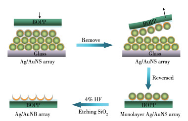
Scheme 1.
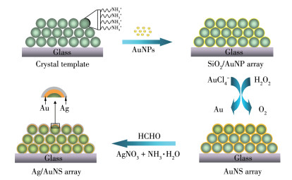
Schematic illustration of the Ag/AuNS array forming process
Facile fabrication and surface-enhanced Raman scattering properties of ordered Au/Ag nanobowl array
Yan-Ying RAO , Bing-Gui WANG , Zhang-Liang LI , Xue-Wen CHEN , Li-Rong QIU , Hong XU , Jian-Hui HUANG

Recently, the incorporation of metal nanoparticles, especially porous metallic materials into ordered structures, has been extensively investigated since such materials may find exciting applications in catalysis[1-2], photonic crystals[3], as substrates for surface-enhanced Raman scattering (SERS) [4-6], as chemical and biological sensors[7-8], etc. The success of these applications strongly relies on the structure of porous metallic materials with specific pore sizes. In the quest to fabricate porous metallic materials with desired pore sizes and structures, attempts have been made to use templates in a replication process[9]. Self-assembled colloidal microspheres or nanospheres are effective templates for the fabrication of ordered nanostructures. The colloidal template with microscale feature, which is afforded by SiO2 or copolymer sphere, is an excellent platform for other materials to be deposited on and can be used as masks or templates for creating ordered porous metal materials[10].
Since it was discovered in the late 1970s, SERS has been widely used in many fields, such as trace detection[11], clinical diagnosis[12-13], chemical and biochemical monitoring[14-16]. However, all the applications of SERS are based on the substrate of SERS-active structures. Physical parameters such as shapes, roughness, sizes, and separation distances of Ag or Au nanoparticles were found to be the key factors that influence the enhancement of SERS[8]. Ag nanoparticles and their arrays are among the best materials for both SERS and enhancing fluorescence, due to the intense surface plasmon resonance in the visible-wavelength range[17-18]. Unfortunately, the biotoxicity and easy oxidation of silver nanoparticles confined its applications in biomedical and biosensors fields. Gold, due to its strong bio-compatibility, stability, non-biotoxicity, and super optical properties show superiority in biosensor applications[19-20]. The use of Au/Ag bimetallic nanoparticles which are coated by a thin gold film on the surface of silver nanoparticles is one of the most popular materials for SERS active substrates because of their high-intensity enhancement in previous reports[21-22]. Unfortunately, the disadvantages of such nanoparticles include repeatability and reproducibility limited their applications in sensors and chemical assays. The development of an efficient method for fabricating ordered Au/Ag nanohole array with controlled shape, roughness, size, and separation distances has become a hot issue in the field of nano-science.
In the previous research, we reported a novel method of fabricating large-area ordered Au nanoshell and then nanobowl arrays by wet chemosynthesis[23]. This article is a further study of our previous work to fabricate an ordered Au/Ag nanobowl (Au/AgNB) array which showed higher SERS activity. Firstly, a thin layer of Au nanoshell (AuNS) was generated by the in-situ growth method on the close-packed 3D SiO2 colloidal crystals surface. Secondly, a thin layer of AuNS was deposited on the surface of the AuNS array to form an ordered Ag/Au double nanoshell (Ag/AuNS) array. A monolayer-ordered reversed the Ag/AuNS array was conveniently obtained by an acrylic estermodified biaxial-oriented polypropylene (BOPP), also called an acrylic tap. The ordered Au/AgNB array attached to the acrylic ester-modified BOPP could be obtained when the SiO2 core was etched by HF solution. The method presented in this article is cost-effective and convenient. The ordered Au-up and Ag-down nanobowl array showed higher SERS enhancement compared with the AuNB array. Furthermore, the produced Au/AgNB array with Au-up had prominent stability and bio-compatibility due to the nature of gold, showed high SERS-enhancing activity, and can be used as universal SERS substrates for biosensors and biomedical applications.
SiO2 colloidal spheres (ca. 100, 180, and 330 nm in diameter) and 3-(aminopropyl)-triethoxysilane (APTES, 98%) were obtained from Nissan Chemical Ind., Ltd., Japan, and Sigma, respectively. Hydrogen peroxide (H2O2, 30%), sodium borohydride (NaBH4), potassium carbonate (K2CO3), chloroauric acid tetrahydrate (HAuCl4·4H2O), silver nitrate (AgNO3, 99.8%), ammonia (NH3·H2O, 30%), formaldehyde solution (HCHO, 40%), and anhydrous ethanol were bought from Nanjing Sunshine Biotechnology Ltd., China. Nile blue A sulfate (NBA) was purchased from Sigma-Aldrich. All of the chemical reagents were used without further purification. And all of them were analytical grades. Acrylic ester-modified BOPP (pressure-sensitive adhesive transparent tap) was bought from Deli Ltd., China. K2CO3/HAuCl4 solution (growth solution) was prepared as follows[24] : 400 mL of aqueous K2CO3 solution (0.25 g·L-1) was mixed with 6 mL of HAuCl4 (1%) stock solution under continuous stirring for 20 min and aging in the dark at 4 ℃ for 4 d. The aqueous solution used in the experiments was prepared with Milli-Q water from the Milli-Q system (resistivity > 18 MΩ).
Firstly, SiO2 colloidal sphere templates were purified by centrifugation and redispersion in ethanol at least five times. Secondly, 1.783 mL APTES was added to 180 mL SiO2 colloids sphere (100 nm) suspension (85 g·L-1 in ethanol), 0.5 mL APTES was added to 140 mL the SiO2 colloidal sphere (180 nm) suspension (50 g·L-1 in ethanol), 0.3 mL APTES was added to 125 mL SiO2 colloidal sphere (330 nm) suspension (56 g·L-1 in ethanol). Then, the mixture was vigorously stirred to react at 40 ℃ overnight. Eventually, APTES groups covalently bonded to the surfaces of SiO2 spheres, extending their amine groups outward as a new termination of the SiO2 surface[25]. To remove excess reactants from the reaction mixture, the APTES-functionalized SiO2 spheres had to be purified by centrifugation and redispersion in ethanol at least five times. Then the amino-functionalized SiO2 spheres were packed onto the templates with 3D ordered arrays on glass substrates with the vertical deposition method[25]. The SiO2 crystal templates were dried at ambient temperature before use.
AuNP (3-5 nm) was prepared by the reduction of HAuCl4 with NaBH4 according to our previous method[25]: 3 mL of HAuCl4 (1%) was mixed with 200 mL of H2O under vigorous stirring for about 5 min, followed by the addition of 1 mL of K2CO3 (0.2 mol·L-1). Then 9 mL of freshly prepared NaBH4 (0.5 g·L-1) was quickly added to the mixture. The mixture will turn into a wine red color rapidly, which indicated the generation of AuNP. The obtained solution was stored at 4 ℃ until use. The SiO2 sphere templates (2.5 cm×1.5 cm) were fixed in a beaker containing 40 mL as-prepared AuNP solution (gold seeds) and vigorously stirred for about 6 h[24]. Then the SiO2/AuNP array was rinsed 3 times with Milli-Q water to remove the unattached AuNP and dried in air.
To initiate the growth of the attached AuNP, four of the SiO2/AuNP arrays (2.5 cm×1.5 cm) were exposed to 80 mL K2CO3/HAuCl4 solution and the reducing agent 200 μL H2O2 (100 μmol·L-1) under vigorous stirring at room temperature for 20 min. Then the AuNS array was rinsed three times with Milli-Q water to remove the growth solution. After that the AuNS array was exposed to 30 mL, 0.06 mmol·L-1 AgNO3 solution, 0.6 mL NH3·H2O (30%), and the reducing agent 0.6 mL HCHO under vigorous stirring at room temperature for 5 min. Then the Ag/AuNS array was rinsed three times with Milli-Q water to remove the growth solution and dried in air.
A single layer of close-packed reversed Ag/AuNS array was lifted by an acrylic ester-modified BOPP (acrylic tape) from the ordered Ag/AuNS array. Then the SiO2 sphere template was removed by treatment with 4% hydrofluoric (HF) acid, producing a monolayer-ordered Au/AgNB array (with Au-up and Ag-down) attached to the acrylic ester-modified BOPP. The morphology of the AuNS array, Ag/AuNS array, and Au/AgNB array produced under different conditions were characterized by Zeiss ULTRA-plus scanning electron microscope (SEM) at 15 kV.
To determine the SERS-activity of the ordered AuNS array, Ag/AuNS array, Au/AgNB array, and AuNB array, all the nanomaterials were soaked in NBA solution for 2 h. Raman spectra were collected with a Renishaw Invia Reflex system equipped with Peltier-cooled charge-coupled device (CCD) detectors and a Leica microscope. Samples were excited with a 785 nm diode laser under line focus mode and the laser power was adjusted to 0.000 1% which was about 0.12 μW. The corresponding laser was focused onto the sample surface using a 50× long working distance objective. Spectra were collected in a continuous mode with 10 s exposure time and a grating of 1 200 mm-1 was used. Every SERS spectrum was averaged from five measurements. All the experiments were performed in triplicate and the values were averaged. The microscope should be refocused after every measurement and all Raman experiments were carried out at room temperature (ca. 20 ℃).
Scheme 1 is the schematic illustration of the growth of the Ag/AuNS array. Firstly, the APTES-functionalized SiO2 spheres were packed as 3D-ordered hexagonal close-packed arrays on glass substrates by the vertical deposition method[25]. Here, SiO2 spheres with different diameters of 100, 180, and 330 nm were arranged uniformly over areas on a scale of several square centimeters for further study. Then the SiO2 templates were immersed into a beaker of aqueous AuNP (3-5 nm) under vigorous stirring. During this process, the negatively charged AuNP would attach to the amino groups′ modified SiO2 sphere surfaces by electrostatic interactions to form the SiO2/AuNP array[24]. Afterward, we used the AuNP attached to the surface of the SiO2/AuNP array as the nucleation sites to guide the growth of a thin gold shell layer (5-10 nm) by H2O2, which was an active reducing agent for the reduction of AuCl4- to Au0, and then the AuNS array formed. Finally, the AuNS array was used as a template for the growth of a silver layer by HCHO reduced Ag+ to Ag0 which deposited on its surface, and then the Ag/AuNS array formed.
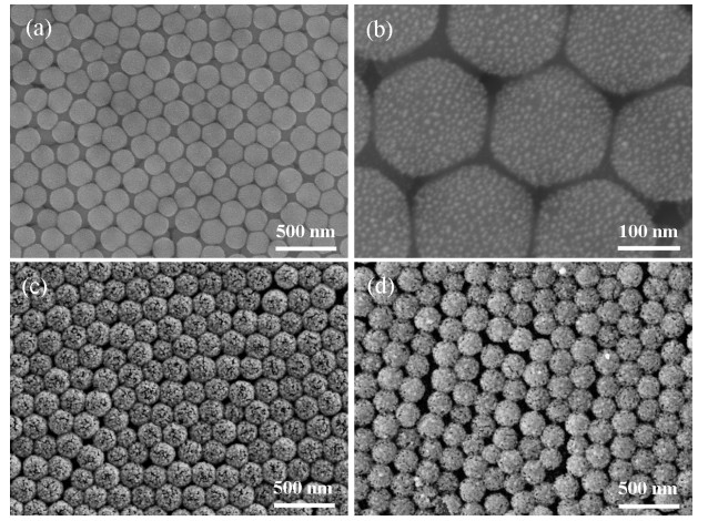
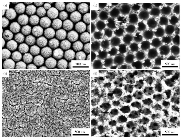
Fig. 1a shows a typical top-view SEM image of the ordered SiO2/AuNP array templates. The SEM image reveals that the SiO2 spheres with a diameter of 180 nm were organized in a close-packed arrangement with long-range hexagonal order. These high-quality colloidal crystals can be used as templates for the fabrication of ordered metallic shell array structures. Using the attached AuNP on the surface of the SiO2/AuNP array (Fig. 1b) as the nucleation sites to guide the growth of the AuNS array (Fig. 1c). Fig. 1c shows the top view SEM image of the ordered AuNS array, there were many nanogaps on the AuNS array surface, which was proved to greatly enhance the SERS-activity from the point of electromagnetic (EM) theory[26]. The morphology and thickness of the AuNS array could be well controlled by the concentration of H2O2, the amount of K2CO3/HAuCl4 growth solution, and the reaction time. Furthermore, the growth program is simple and convenient, a batch of the SiO2/AuNP array can be reacted in one beaker. In this work, we used four SiO2/AuNP arrays to react in 60 mL K2CO3/HAuCl4 solution and 200 μL H2O2 (100 μmol·L-1) under vigorous stirring at room temperature for 20 min. Fig. 1d shows the SEM image of the Ag/AuNS array produced from the AuNS array as shown in Fig. 1c. As can be seen, the surface of the AuNS array was covered with reduced Ag, and then the Ag/AuNS array formed.
Scheme 2 is the schematic illustration of the ordered Au/Ag bimetallic nanobowl array forming process, which is a very flexible fabrication strategy. A monolayer-ordered Ag/AuNS array can be easily reversed by an acrylic ester-modified BOPP if the thickness of the Ag/AuNS is well controlled, which can be replaced by any tube or needle if only the surface was functionalized by an appropriate viscous molecule. A monolayer-ordered Au/AgNB array attached to the acrylic ester-modified BOPP could be obtained when the SiO2 core was etched by HF solution. The method exhibited versatility in the fabrication of the ordered Au/AgNB array with different core sizes, and the core size of the nanobowl can be well controlled by the SiO2 core size. In this article, the ordered Au/AgNB arrays with a core size of 100, 180, and 330 nm were fabricated respectively. The ordered Au/AgNB array fabricated by this approach allows better control of the nanostructures, resulting in uniform roughness and a core size of the metal film and turn providing better reproducibility and SERS enhancement for the substrate.
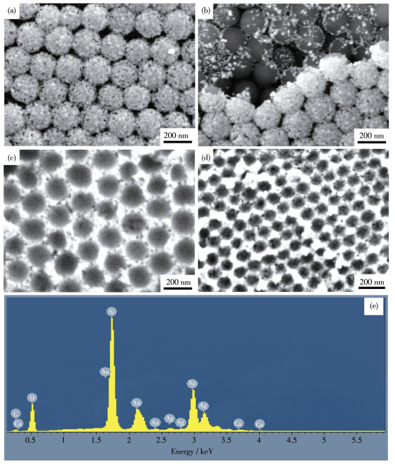
Fig. 2 shows the SEM images of the Ag/AuNS array and the BOPP tape reverse-ordered Au/AgNB array with diameters of 180 and 100 nm. The Ag/AuNS array was homogeneously and integrally arranged on the glass substrates as shown in Fig. 2a, it can be seen that the surface structure of Ag/AuNS was rough, there was a certain gap between the nanoparticles, and the size of AgNP was 20-30 nm. It shows that the nanoshell array being taken off by the BOPP tape was monolayer from the SEM image of the remaining Ag/AuNS array boundary (Fig. 2b). It can be seen that the top of the first layer of SiO2 core was covered with an Ag/Au bimetallic thickness of about 30 nm, the atomic ratio of Ag and Au was about 4∶6, and the energy dispersive spectroscopy (EDS) analysis was shown in Fig. 2e. The second layer was covered by a thimbleful of Au/Ag bimetallic. This is because the upside of the first layer SiO2 or nanoshell array was exposed to the reaction solution and it is easy to deposit reduced Au or Ag; While the downside of the first layer connected with the second layer, and the second layer is not as exposed to the reaction solution as the top side of the first layer so that there is little metal deposition on them. The force between the Ag/AuNS array on the first layer and the second layer is faint which leads to the nanoshell array being taken off to be monolayer by the BOPP tape. Because the reversed ordered Ag/AuNS array was only half shell, the Au/AgNB array but not the nanocage array would be formed after the SiO2 cores were etched by HF solution. As shown in Fig. 2c and 2d, reversed the ordered Au/AgNB array attached to the BOPP with core sizes of 180 and 100 nm, respectively, were gotten after being etched the SiO2 core off.
Compared with those arrays with the core size of 100 and 180 nm, the ordered Au/AgNB array with the core size of 330 nm was controlled by much more parameters due to its bigger interspace between every two or three SiO2 spheres. In the process, besides the diameter of the SiO2 core, a uniform and appropriate thickness of the AuNS array was firstly pivotal, which can be well controlled by the concentration of H2O2 and the amount of K2CO3/HAuCl4 solution. Likewise, the thickness of AuNS can be well controlled by the concentration and the amount of AgNO3 solution, NH3·H2 O (30%), and HCHO, which is crucial for the growth of the Ag/AuNS array. And the appropriate thickness of the Ag/AuNS array is essential to the reversed monolayer Ag/AuNS array and the Au/AgNB array. Fig. 3a and 3c show SEM images of the Ag/AuNS array with a core size of 330 nm growing in the different concentrations and amounts of AgNO3 solution, NH3·H2O (30%), and HCHO. When the AuNS array was exposed to 30 mL, 0.06 mmol·L-1 AgNO3 solution, 0.6 mL NH3·H2O (30%), and with 0.6 mL HCHO as reducing agent, under vigorous stirring at room temperature for 5 min, the thickness of the Ag/AuNS array (Fig. 3a) would be optimal for the fabrication of the ordered Au/AgNB array (Fig. 3b). But if the AuNS array was reacted into the mixed solution of 60 mL AgNO3 solution (0.12 mmol·L-1), 2.4 mL NH3·H2O (30%), and 2.4 mL HCHO, more Ag0 will be reduced and deposited onto the surface of the AuNS array and fill the interspaces between two layers of the AuNS array ultimately, as shown in Fig. 3c. Because the diameter of SiO2 is big (ca. 330 nm), there will be more interspaces between SiO2 cores, and the overdosed reduced Ag0 will be easy to fill the interspaces between two layers of the AuNS array. The overly deposited metal will result in a double layer or multilayer of the reversed Ag/AuNS array, which produced the corresponding ordered porous Au/Ag bimetallic film once the SiO2 sphere template was removed by HF solution (Fig. 3d). In virtue of the smaller interspace between every two or three SiO2 spheres (5-10 nm), it is harder for the overdosed reduced Au0 or Ag0 to fill the interspaces between two layers of the AuNS array. What′s more, the production of the Ag/AuNS array with core sizes of 100 and 180 nm has much more excellent repeatability and uniformity.
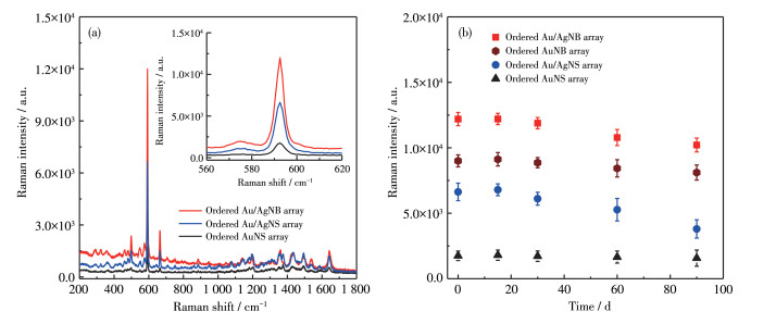
Considering the excellent SERS performance of metal Ag and its strong bio-compatibility, stability, and exceptional optical properties of metal Au as well as the nanobowl array structures that may provide"hot spots "to enhance the Raman signals[24, 27], it was reasonably expected that the obtained Au/AgNB array could act as effective SERS substrate to check the SERS activity of the produced Au/AgNB array, we chose NBA as a Raman report molecule. The spectra which had high specificity presented the characteristic Raman shifts of NBA at 592 and 1 638 cm-1, whose modes were formed by the positively charged nitrogen[28]. In this work, we chose the core diameter of 180 nm as an example, and we measured the SERS signals of NBA molecules adsorbed on the surface of the ordered AuNS array, 3D ordered Ag/AuNS array, and ordered Au/AgNB array, which are presented in Fig. 4a. It can be seen that the SERS spectral intensity of the ordered Au/AgNB array was about 7 times that of the ordered AuNS array, and two times that of the ordered Ag/AuNB array, and it is clear that the ordered Au/AgNB array exhibit the highest SERS signal intensity among those three SERS substrates.
Inset: the corresponding enlarged spectra
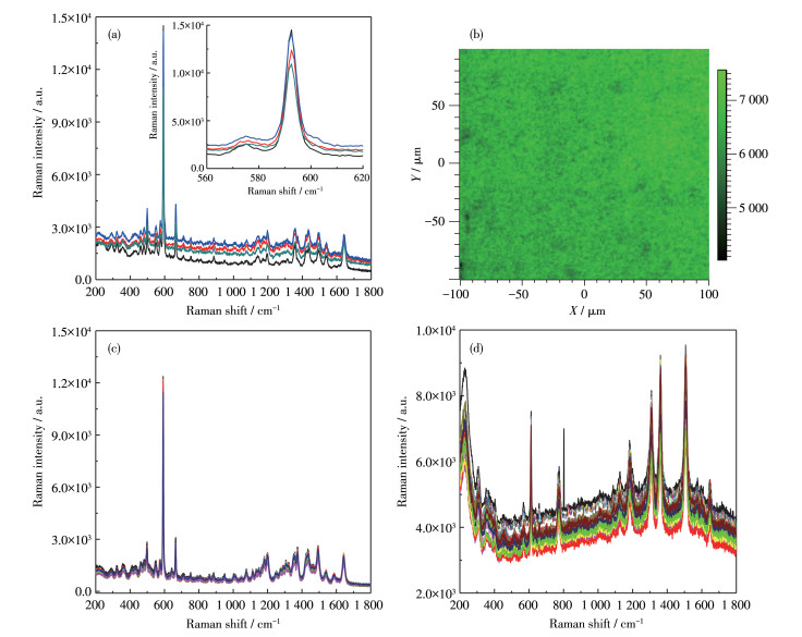
The ideal SERS substrates should have good stability, high reproducibility, and repeatability, together with high SERS activity. Reproducibility of SERS substrate refers to the possibility to produce an enhanced signal at various parts of the substrate with minimum intensity variations. Repeatability refers to the minimum batch-to-batch variation of the substrate. Repro-ducibility and repeatability of the substrates are highly critical for SERS-based sensing. The ordered Au/AgNB array with uniform roughness and core size provides good reproducibility, repeatability, stability, and SERS enhancement. Fig. 5a shows the SERS spectra of 10-6 mol·L-1 NBA on four Au/AgNB arrays which were produced from four batches, whose SERS intensity of NBA in 592 cm-1 were 10 948, 12 387, 14 169, and 14 497 counts, respectively. Fig. 5b shows the corresponding SERS mapping image of the Au/AgNB arrays, the image was measured at 592 cm-1 band of NBA and it could be seen that excepting some inevitable defect spots, most of them were between 6 500 and 7 200 counts. Fig. 5c shows the enhanced Raman signal at 10 random spots of the same substrate, and Fig. 5d shows the enhanced Raman signal at 35 random spots of one Au/AgNB array by using R6G (rhodamine 6G) as a Raman probe molecule. As can be seen, the difference between the 10 and 35 random spots was small, which indicates the Au/AgNB arrays showed excellent reproducibility. In addition to the strong SERS enhancement effect, the Au/AgNB arrays were also stable and could be produced with high repeatability. The stability is a significant parameter for a good SERS substrate. Fig. 4b shows the intensity of the prominent peak at 592 cm-1 of NBA using different SERS substrates from 0-100 d, and further studies were carried out to compare the SERS activities and stability of different nanostructure arrays together with batch-to-batch variation as shown in it. Compared with the ordered AuNS array, AuNB array[24], and Ag/AuNS array, the ordered Au/AgNB array exhibited the highest SERS signal intensity. Fig. 4a shows the SERS spectra of 10-6 mol·L-1 NBA on the ordered AuNS array, Ag/AuNS array, and Au/AgNB array, whose SERS intensity of NBA in 592 cm-1 were 1 750, 6 629, and 12 194 counts, respectively. It can be seen that compared with the SERS spectrum of the ordered AuNS array, about four times stronger the relative intensity of bands can be observed in the SERS spectrum of the ordered Ag/AuNS array, and the SERS spectrum of the ordered Au/AgNB array showed the highest SERS activity. This indicated that the SERS enhancement effect of Au/Ag bivalve nanostructures is mainly derived from the combined effect of Au, Ag shells, and their porous ordered structures.
Inset: the corresponding SERS spectra of the 592 cm-1 band of NBA
The SERS enhancement factor (EF) was calculated by using the analytical chemistry point of view through the analytical EF (AEF) defined as following Eq.1[29]:
|
|
(1) |
Where ISERS corresponds to the Raman intensity obtained for the SERS substrate under a certain concentration of cSERS and IRS corresponds to the Raman intensity obtained under non-SERS conditions at an analyte concentration of cRS. The experimental conditions, such as the laser wavelength, laser power, microscope objective or lenses, spectrometer, and measuring conditions on the substrate, are taken into account and are identical in all cases.
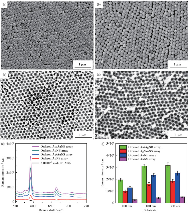
According to the established definition, the AEF value of the substrates was estimated by considering the 592 cm-1 Raman band of NBA, when using cSERS= 1.0×10⁻⁷ mol·L-1 and cRS=5.0×10⁻² mol·L-1 at the same experimental conditions (as shown detail in SERS measurements of experimental section, but changed the laser power from 0.000 1% to 0.1% which was better for detecting the Raman spectra of NBA with non-SERS substrate). Fig. 6a-6d show the SEM images of the ordered AuNS array, Ag/AuNS array, AuNB array, and Au/AgNB array with a core diameter of 180 nm, respectively. Fig. 6e shows the Raman spectra of 5.0×10⁻² mol·L-1 NBA on the ordered AuNS array, Ag/AuNS array, AuNB array and Au/AgNB array with a core diameter of 180 nm, respectively. Fig. 6f shows SERS intensities of 1.0×10⁻⁷ mol·L-1 NBA on 592 cm-1 band by the four different substrates with a core diameter of 100, 180, and 330 nm, respectively. And the AEF values calculated for the ordered Au/AgNB array, AuNB array[24], Au/AgNSs array, and AuNS array with different core diameters were shown in Table 1, the ordered Au/AgNB array shows the highest AEF value in all three core size (1.37×10⁷, 2.19×10⁷, and 2.23×10⁷, respectively).
 下载:
导出CSV
下载:
导出CSV
| SERS substrate | ISERS / a.u. | AEF | |||||
| 100 nm | 180 nm | 330 nm | 100 nm | 180 nm | 330 nm | ||
| AuNS array | 27 198 | 42 422 | 51 109 | 1.93×10⁶ | 3.01×10⁶ | 3.62×10⁶ | |
| AuNB array | 126 254 | 232 861 | 250 642 | 8.96×10⁶ | 1.65×10⁷ | 1.78×10⁷ | |
| Au/AgNS array | 98 342 | 158 933 | 182 432 | 6.98×10⁶ | 1.13×10⁷ | 1.27×10⁷ | |
| Au/AgNB array | 192 875 | 308 838 | 314 563 | 1.37×10⁷ | 2.19×10⁷ | 2.23×10⁷ | |
In summary, a novel in-situ growth method of the Ag/AuNS array based on 3D SiO2 colloidal crystal for fabricating large-area ordered Au/AgNB arrays with adjustable periodic holes, shapes, and sizes were reported in this paper. The method presented here is cost-effective, convenient, and productive, and also has the potential for being a practical way to produce porous-film-related nanostructure devices shortly. Furthermore, the Au/AgNB array with controlled pore size, good stability, and reproducible preparation shows high-intensity enhancement, its SERS analytical enhancement factor (AEF) could reach 2.23×10⁷, which has been well recognized in its practical applications as a universal excellent SERS substrate.
Zhai Q G, Bu X H, Mao C Y, Zhao X, Daemen L, Cheng Y Q, Ramirez-Cuesta A J, Feng P Y. An ultra-tunable platform for molecular engineering of high-performance crystalline porous materials[J]. Nat. Commun., 2016, 7: 13645. doi: 10.1038/ncomms13645
Feng J R, Lv F, Zhang W Y, Li P H, Wang K, Yang C, Wang B, Yang Y, Zhou J H, Lin F, Wang G C, Guo S J. Iridium-Based multimetallic porous hollow nanocrystals for efficient overall-water-splitting catalysis[J]. Adv. Mater., 2017, 29(47): 1703798. doi: 10.1002/adma.201703798
Seo J, Lee J, Kim Y. Ultrasensitive plasmon-free surface-enhanced Raman spectroscopy with femtomolar detection limit from 2D van der Waals heterostructure[J]. Nano Lett., 2020, 20(3): 1620-1630. doi: 10.1021/acs.nanolett.9b04645
Zheng Y H, Soeriyadi A H, Rosa L, Ng S H, Bach U, Gooding J J. Reversible gating of smart plasmonic molecular traps using thermoresponsive polymers for single-molecule detection[J]. Nat. Commun., 2015, 6(1): 8797-8803. doi: 10.1038/ncomms9797
Kong X M, Xi Y T, Le Duff P, Chong X Y, Li E, Ren F H, Rorrer G L, Wang A X. Detecting explosive molecules from nanoliter solution: A new paradigm of SERS sensing on hydrophilic photonic crystal biosilica[J]. Biosens. Bioelectron., 2017, 88: 63-70. doi: 10.1016/j.bios.2016.07.062
Zou X, Silva R, Huang X, Alsharab J F, Asefa T. A self-cleaning porous TiO2-Ag core-shell nanocomposite material for surface-enhanced Raman scattering[J]. Chem. Commun., 2013, 49(4): 382-384. doi: 10.1039/C2CC35917K
Zhong L B, Yin J, Zheng Y M, Liu Q, Cheng X X, Luo F H. Self-assembly of Au nanoparticles on PMMA template as flexible, transparent, and highly active SERS substrates[J]. Anal. Chem., 2014, 86(13): 6262-6267. doi: 10.1021/ac404224f
Gahlaut S K, Savargaonkar D, Sharan C, Yadav S, Singh J P. SERS platform for dengue diagnosis from clinical samples employing a hand held Raman spectrometer[J]. Anal. Chem., 2020, 92(3): 2527-2534. doi: 10.1021/acs.analchem.9b04129
Imai R, Tanaka M, Hashimoto H, Asoh H. Facile synthesis of size-and shape-controlled freestanding Au nanohole arrays by sputter deposition using anodic porous alumina templates[J]. Nanotechnology, 2020, 31(41): 415303. doi: 10.1088/1361-6528/ab9f76
Hong G S, Li C, Qi L M. Facile fabrication of two-dimensionally ordered macroporous silver thin films and their application in molecular sensing[J]. Adv. Funct. Mater., 2010, 20(21): 3774-3783. doi: 10.1002/adfm.201001177
Dong Y H, Laaksonen A, Huo F, Gao Q W, Ji X Y. Hydrated ionic liquids boost the trace detection capacity of proteins on TiO2 support[J]. Langmuir, 2021, 37(16): 5012-5021. doi: 10.1021/acs.langmuir.1c00525
Reza K K, Wang J, Vaidyanathan R, Dey S, Wang Y L, Trau M. Electrohydrodynamic-induced SERS immunoassay for extensive multiplexed biomarker sensing[J]. Small, 2017, 13(9): 1602902. doi: 10.1002/smll.201602902
Wang Z Y, Zong S F, Li W, Wang C L, Xu S H, Chen H, Cui Y P. SERS-fluorescence joint spectral encoding using organic-metal-QD hybrid nanoparticles with a huge encoding capacity for high-throughput biodetection: Putting theory into practice[J]. J. Am. Chem. Soc., 2012, 134(6): 2993-3000. doi: 10.1021/ja208154m
Fang S, Hung H C, Sinclair A, Peng Z, Tao B, Galvan D D, Jain P, Li B W, Jiang S Y, Yu Q M. Hierarchical zwitterionic modification of a SERS substrate enables real-time drug monitoring in blood plasma[J]. Nat. Commun., 2016, 7(7): 13437.
Sandeep S P, Gonzalo R G, Siraj S, Tazara L L, Claramaria R G, Inocencio H C, Pedro C S, Tanya C V, Elder D R. Ultra-sensitive SERS substrate for label-free therapeutic drug monitoring of paclitaxel and cyclophosphamide in blood serum[J]. Anal. Chem., 2019, 91(3): 2100-2111. doi: 10.1021/acs.analchem.8b04523
Panikar S S, Banu N, Escobar E R, García G R, Cervantes-Martínez J, Villegas T C, Salas P, Rosa E D. Stealth modified bottom up SERS substrates for label-free therapeutic drug monitoring of doxorubicin in blood serum[J]. Talanta, 2020, 218: 121138. doi: 10.1016/j.talanta.2020.121138
Tang H B, Meng G W, Huang Q, Zhang Z, Huang Z L, Zhu C H. Arrays of cone-shaped ZnO nanorods decorated with Ag nanoparticles as 3D surface-enhanced Raman scattering substrates for rapid detection of trace polychlorinated biphenyls[J]. Adv. Funct. Mater., 2012, 22: 218-224. doi: 10.1002/adfm.201102274
Sánchez-Iglesias A, Aldeanueva-Potel P, Ni W, Pérez-Juste J, Pastoriza-Santos I, Alvarez-Puebla R A, Mbenkum B N, Liz-Marzán L M. Chemical seeded growth of Ag nanoparticle arrays and their application as reproducible SERS substrates[J]. Nano Today, 2010, 5(1): 21-27. doi: 10.1016/j.nantod.2010.01.002
Ding C Q, Tian Y. Gold nanocluster-based fluorescence biosensor for targeted imaging in cancer cells and ratiometric determination of intracellular pH[J]. Biosens. Bioelectron., 2015, 65: 183-190. doi: 10.1016/j.bios.2014.10.034
Li Y, Zhang Y L, Zhao M, Zhou Q Q, Wang L L, Wang H Z, Wang X H, Zhan L S. A simple aptamer-functionalized gold nanorods based biosensor for the sensitive detection of MCF-7 breast cancer cells[J]. Chem. Commun., 2016, 52: 3959-3961. doi: 10.1039/C6CC01014H
Liao S, Luo Z, Metternich J B, Zenobi R, Stellacci F. Quantification of surface composition and segregation on AuAg bimetallic nanoparticles by MALDI MS[J]. Nanoscale, 2020, 12(44): 22639-22644. doi: 10.1039/D0NR05061J
Liao W L, Liu K, Chen Y J, Hu J P, Gan Y. Au-Ag bimetallic nanoparticles decorated silicon nanowires with fixed and dynamic hot spots for ultrasensitive 3D SERS sensing[J]. J. Alloy. Compd., 2021, 868: 159136. doi: 10.1016/j.jallcom.2021.159136
Rao Y Y, Zhao X X, Li Z L, Huang J H. Phenolic acids induced growth of 3D ordered gold nanoshell composite array as sensitive SERS nanosensor for antioxidant capacity assay[J]. Talanta, 2018, 190: 174-181. doi: 10.1016/j.talanta.2018.07.069
Rao Y Y, Tao Q, An M, Rong C H, Dong J, Dai Y R, Qian W P. Novel and simple route to fabricate 2D ordered gold nanobowl arrays based on 3D colloidal crystals[J]. Langmuir, 2011, 27(21): 13308-13313. doi: 10.1021/la203158q
饶艳英, 李章良, 黄建辉, 姜宇杭, 赵晓旭. 三维有序金纳米壳结构的可控制备及其SERS性能[J]. 无机化学学报, 2018,34,(7): 1231-1239. RAO Y Y, LI Z L, HUANG J H, JIANG Y H, ZHAO X X. Preparation and SERS properties of 3D ordered gold nanoshells arrays[J]. Chinese J. Inorg. Chem., 2018, 34(7): 1231-1239.
Jiang J, Bosnick K, Maillard M, Brus L. Single molecule Raman spectroscopy at the junctions of large Ag nanocrystals[J]. J. Phys. Chem. B, 2003, 107(37): 9964-9972. doi: 10.1021/jp034632u
Kim K, Choi J Y, Shin K S. Raman scattering characterization of 1,4-phenylenediisocyanide in Au-Au and Ag-Au Nanogaps[J]. Spectroc. Acta Pt. A-Molec. Biomolec. Spectr., 2013, 100: 3-9. doi: 10.1016/j.saa.2012.01.045
Do Nascimento G M, Temperini M. Studies on the resonance Raman spectra of polyaniline obtained with near-IR excitation[J]. J. Raman Spectrosc., 2008, 39(7): 772-778. doi: 10.1002/jrs.1841
Su Q Q, Ma X Y, Dong J, Jiang C Y, Qian W P. A reproducible SERS substrate based on electrostatically assisted APTES-functionalized surface-assembly of gold nanostars[J]. ACS Appl. Mater. Interfaces, 2011, 3(6): 1873-1879. doi: 10.1021/am200057f

Figure 1 SEM images of (a, b) the SiO2/AuNP array, (c) the AuNS array, and (d) the Ag/AuNS array (with a core diameter of 180 nm)

Figure 2 SEM images of (a) the ordered Ag/AuNS array; Boundary views of (b) the ordered Ag/AuNS array after being taken off by acrylic ester modified BOPP partly, (c) the corresponding ordered Au/AgNB array (with a core diameter of 180 nm), and (d) the ordered Au/AgNB array (with a core diameter of 100 nm); (e) EDS spectrum of the ordered Ag/AuNS array in (a)

Figure 3 SEM images of (a) the ordered Ag/AuNS array produced by 30 mL, 0.06 mmol·L-1 AgNO3 solution, (b) the corresponding ordered Au/AgNB array, (c) the thinker Ag/AuNS array produced by 60 mL, 0.12 mmol·L-1 AgNO3 solution, and (d) the corresponding ordered porous Au/Ag bimetallic film (with a core diameter of 330 nm)

Figure 4 (a) SERS spectra of 10-6 mol·L-1 NBA on the ordered AuNS array, Ag/AuNS array, and ordered Au/AgNB array (with a core diameter of 180 nm) substrates, respectively; (b) Time stability of four different substrates
Inset: the corresponding enlarged spectra

Figure 5 (a) SERS spectra of 10-6 mol·L-1 NBA on four Au/AgNB arrays from four batches; (b) Raman image of the 592 cm-1 band of NBA by using the Au/AgNB array as substrate (the exciting laser for streamlined images were in a 785 diode laser with pinhole and the exposure time was 2 s); SERS spectra of (c) 10-6 mol·L-1 NBA taken from 10 random spots and (d) 10-6 mol·L-1 R6G taken from 35 random spots on the Au/AgNB array
Inset: the corresponding SERS spectra of the 592 cm-1 band of NBA

Figure 6 SEM images of (a) the ordered AuNS array, (b) the ordered Ag/AuNS array, (c) the ordered AuNB array, and (d) the ordered Au/AgNB array with a core diameter of 180 nm, and (e) corresponding Raman spectra of 5.0×10⁻² mol·L-1 NBA; (f) SERS intensity of 1.0×10⁻⁷ mol·L-1 NBA on 592 cm-1 band by four different substrates (core diameter was 100, 180 and 330 nm, respectively)
Table 1. AEF value calculated for different SERS substrates
| SERS substrate | ISERS / a.u. | AEF | |||||
| 100 nm | 180 nm | 330 nm | 100 nm | 180 nm | 330 nm | ||
| AuNS array | 27 198 | 42 422 | 51 109 | 1.93×10⁶ | 3.01×10⁶ | 3.62×10⁶ | |
| AuNB array | 126 254 | 232 861 | 250 642 | 8.96×10⁶ | 1.65×10⁷ | 1.78×10⁷ | |
| Au/AgNS array | 98 342 | 158 933 | 182 432 | 6.98×10⁶ | 1.13×10⁷ | 1.27×10⁷ | |
| Au/AgNB array | 192 875 | 308 838 | 314 563 | 1.37×10⁷ | 2.19×10⁷ | 2.23×10⁷ | |
 下载: 导出CSV

 扫一扫看文章

扫一扫关注我们