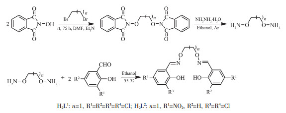
Scheme 1.
Syntheses routes to H2L1 and H2L2

Multihalogen-Substituted Salamo-Type Mn(Ⅱ) Complexes: Syntheses, Crystal Structures, Hirshfeld Analyses and Fluorescence Properties
Lan WANG , Zhi-Li WEI , Meng YU , Ying-Qi PAN , Yang ZHANG , Wen-Kui DONG


As is generally known, salen-type ligands (R1-CH=N-(CH2)2-N=CH-R2) and their derivatives are a class of important N2O2 chelating ligands[1-3], their metal complexes have been extensively investigated in modern coordination chemistry for the past several decades[4-7], because they not only show a variety of properties in catalytic and optical properties[8-13], but also have some advantages in the fields of antibacterial activities[14-21], electrochemical conducts[22-24], supramole-cular buildings[25-32], ions recognitions[33-38] and so on. Recently, new researches have been expanded in the use of salamo-type ligands (R1-CH=N-O-(CH2)n-O-N=CH-R2)[39-43], and their complexes have been applied in a very wide and diversified subjects such as supra-molecular architectures[44-45] and luminescence properties[46-49]. As derivatives of salen-type compounds, salamo -type compounds are more significant, because the electronic and steric effects of the ligands on salamo-metal-assisted catalysis may be controlled by the introduction of proper substituents into the benzene rings. Furthermore, it is important to introduce suitable functional groups into the organic moieties of the ligands in order to improve the properties of these metal complexes[50-51]. Now, the synthesis of structurally-specific, superior properties of salamo-type metal complexes, and study of their applications have become an important chemical field and potential areas.
Based on previous works, we have designed and synthesized two multihalogen-substituted salamo-type manganese(Ⅱ) complexes by the coordination reactions of salamo-type bisoxime ligands H2L1, H2L2 with manganese(Ⅱ) acetate tetrahydrate, respectively. To the best of our knowledge, the aforementioned manganese(Ⅱ) salamo-type complexes haven′t been reported[52-54]. Here, elemental analyses, FT-IR, UV-visible absorption spectra, single-crystal X-ray diffractions, fluorescence spectra and Hirshfeld surfaces of the two manganese(Ⅱ) complexes were investigated in detail to study their structural features and spectral characteristics.
2-Hydroxy-3, 5-dichlorobenzaldehyde and 2-hydroxy-5-nitrobenzaldehyde (99%) were purchased from Alfa Aesar and used directly without purification. All other solvents and chemicals were analytical grade and obtained from Tianjin Chemical Reagent Factory. Elemental analysis data of C, H and N were performed on a GmbH VarioEL V3.00 automatic elemental analysis instrument. Elemental analysis for manganese(Ⅱ) was detected with an IRIS ER/S-WP-1 ICP atomic emission spectrometer. Melting points were measured with a microscopic melting point apparatus made by Beijing Taike Instrument Limited Company and were uncorrected. Fourier transform IR spectra were recorded with a VERTEX70 FT-IR spectro-photometer, with samples prepared as KBr (500~4 000 cm-1) and CsI (100~500 cm-1) pellets. UV-visible absorption spectra were collected on a Shimadzu UV-3900 spectrophotometer. Fluorescent spectra were recorded on F-7000 spectrophotometer. Single crystal X-ray structure determinations were made by a SuperNova and Bruker D8 Venture diffractometers, respectively. Hirshfeld surfaces analyses and two-dimensional fingerprint plots of complexes 1 and 2 were calculated using Crystal Explorer program.
The major reaction steps, containing the synthesis of 4, 4′, 6, 6′-tetrachloro-2, 2′ -(ethylenediyldioxybis(nitrilomethylidyne))diphenol (H2L1) and 5-nitro-4′, 6′-dichloro-2, 2′-(ethylenedioxybis(azomethine))diphenol (H2L2) are given in Scheme 1.
1, 2-Bis(aminooxy)ethane was prepared in accor-dance with the previously literatures[55-56]. Yield: 56.8%. Anal. Calcd. for C2H8N2O2(%): C, 26.08; H, 8.76; N, 30.42. Found(%): C, 25.97; H, 8.70; N, 30.49.
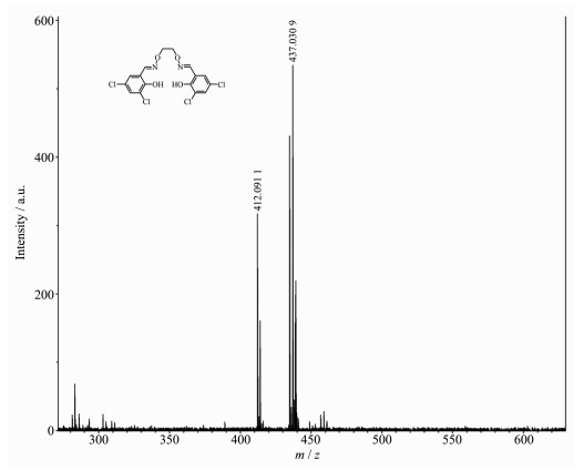
The salamo-type bisoxime ligand (H2L1) was synthesized according to our previous work[54, 57]. Fig. 1 shows the high resolution mass spectrum of ligand H2L1. In the high resolution mass spectrum, a strong peak appeared at m/z=437.030 9, which is consistent with the relative molecular mass determined by the elemental analysis. 1H NMR (500 MHz, DMSO-d6): δ 4.48 (s, 4H, -CH2), 7.53 (t, J=2.6 Hz, 2H, -ArH), 7.61 (t, J=2.6 Hz, 2H, -ArH), 8.47 (d, J=2.0 Hz, 2H, -N=CH), 10.38 (s, 2H, -OH). Yield: 72.8%. m.p. 204~205 ℃. Anal. Calcd. for C16H12Cl4N2O4(%): C, 43.87; H, 2.76; N, 6.39. Found(%): C, 43.95; H, 2.68; N, 6.28.
A solution of 2-hydroxy-3, 5-dichlorobenzaldehyde (382.02 mg, 2.0 mmol) in ethanol (100 mL) was added dropwise to a solution of 1, 2-bis(aminooxy)ethane (368.4 mg, 4.0 mmol) in ethanol (40 mL), then the mixture was subjected to heating at 55 ℃ for 6 h. The solution was concentrated in vacuum and the residue was purified by column chromatography (SiO2, chloro-form/ethyl acetate, 25:1, V/V) to afford 2-(O-(1-ethylo-xyamide))oxime-4, 6-dichlorophenol. Yield: 65.6%, m.p. 92~93 ℃. Anal. Calcd. for C9H10Cl2N2O3(%): C, 40.78; H, 3.80; N, 10.57. Found(%): C, 40.92; H, 3.69; N, 10.63.
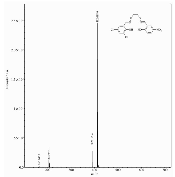
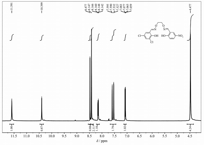
2-Hydroxy-5-nitrobenzaldehyde (334.24 mg, 2.0 mmol) was dissolved in the ethanol solution (15 mL) and added to a stirred colorless ethanol solution (20 mL) of the above-obtained 2-(O-(1-ethyloxyamide))oxime-4, 6-dichlorophenol (530.18 mg, 2 mmol). The mixture was heated and stirred at 55 ℃ about 6 h before being allowed to cool to room temperature. Then the suspension solution was filtered and washed with ethanol/hexane (1:4, V/V). After dried under vacuum, the resulting white solid of H2L2 was collected. The high resolution mass spectrum and nuclear magnetic resonance spectrum of the ligand H2L2 are shown in Fig. 2 and 3, respectively. A strong peak appeared at m/z=412.096 0 in the high resolution mass spectrum, which is consistent with the relative molecular mass determined by the elemental analysis. 1H NMR (500 MHz, DMSO-d6): δ 4.48 (s, 4H, -CH2), 7.07 (dd, J=9.0, 3.0 Hz, 1H, -ArH), 7.53 (d, J=2.5 Hz, 1H, -ArH), 7.59 (d, J=2.5 Hz, 1H, -ArH), 8.15 (dd, J=9.0, 3.0 Hz, 1H, -ArH), 8.42 (s, 2H, -N=CH), 8.48 (s, 1H, -ArH), 10.40 (s, 1H, -OH), 11.59 (s, 1H, -OH). Yield: 68%. m.p. 174 ~175 ℃. Anal. Calcd. for C16H13Cl2N3O6(%): C 46.40; H 3.16; N 10.14. Found(%): C 46.59; H 3.05; N 10.03.
A solution of manganese(Ⅱ) acetate tetrahydrate (2.45 mg, 0.014 mmol) in mixed solvent of methanol (2.0 mL) and ethanol (3.0 mL) was added dropwise to a chloroform solution (3.0 mL) of H2L1 (2.19 mg, 0.005 mmol) at room temperature. The color of the mixing solution turned brown immediately. After the mixture was stirred for 1 h, the mixture was filtered. The resulting filtrate was left undisturbed for about several days to form clear light brown block-like crystals suitable for X-ray crystallographic analysis. Yield: 45.7%. Anal. Calcd. for C16H14Cl4MnN2O6(%): C, 36.46; H, 2.68; N, 5.32; Mn, 10.42. Found(%): C, 36.74; H, 2.52; N, 5.19; Mn, 10.59.
A mixed solution of methanol (2.0 mL) and ethanol (4.0 mL) of manganese(Ⅱ) acetate tetrahydrate (3.72 mg, 0.015 mmol) was added to a chloroform solution (3.0 mL) of H2L2 (2.07 mg, 0.005 mmol) at room temperature. After the mixture was stirred for 2 h, the mixture was fltered off. The resulting fltrate was left undisturbed for about one week to form block-like crystals suitable for X-ray crystallographic analysis. Yield: 40.5%. Anal. Calcd. for C16H15.73Cl2MnN3O8.37(%): C, 37.70; H, 3.11; N, 8.24; Mn, 10.78. Found(%): C, 37.75; H, 3.22; N, 8.06; Mn, 10.64.
The single crystal of complex 1 with approxi-mated dimensions of 0.21 mm×0.15 mm×0.12 mm was placed on a SuperNova diffractometer. The diffraction data were collected using a graphite monochromated Mo Kα radiation source (λ=0.071 073 nm) at 293(2) K. While the X-ray diffraction data of complex 2 with approximated dimensions of 0.26 mm×0.22 mm×0.20 mm was placed on a Bruker D8 Venture diffractometer. The diffraction data were collected using a graphite monochromated Mo Kα radiation source (λ=0.071 073 nm) at 291(2) K. In addition, multi-scan absorption corrections were applied. The crystal structures were solved using direct methods and refined anisotropically by the full-matrix least-squares techniques based on c with SHELXL-2014/7[58] and SHELXL-2018 prog-rams[58], respectively. The positions for hydrogen atoms were fixed on geometrically idealized positions and refined via a riding model. The non-hydrogen atoms were refined anisotropically with displacement para-meters. Selected data of collection parameters and structure refinement of complexes 1 and 2 are summ-arized in Table 1.

 下载:
导出CSV
下载:
导出CSV
| Complex | 1 | 2 |
| Formula | C16H14Cl4MnN2O6 | C16H15.73Cl2MnN3O8.37 |
| Formula weight | 527.03 | 509.81 |
| Crystal system | Monoclinic | Triclinic |
| Space group | C2/c | P1 |
| a/nm | 1.728 35(17) | 0.764 9(2) |
| b/nm | 1.515 65(17) | 1.159 4(2) |
| c/nm | 0.776 92(6) | 1.427 0(2) |
| α/(°) | 110.121(6) | |
| β/(°) | 97.161(9) | 100.257(9) |
| γ/(°) | 102.123 4(19) | |
| V/nm3 | 2.019 3(3) | 1.117 7(4) |
| Z | 4 | 2 |
| Dc/(g·cm-3) | 1.734 | 1.515 |
| μ/mm-1 | 1.220 | 0.877 |
| F(000) | 1 060 | 517 |
| θ range/(°) | 3.314~26.993 | 3.329~25.996 |
| Index ranges | -21 ≤ h ≤ 21, -19 ≤ k ≤ 18, -9 ≤ l ≤ 5 | -7 ≤ h ≤ 9, -14 ≤ k ≤ 14, -17 ≤ l ≤ 17 |
| Reflection collected | 4 100 | 8 032 |
| Independent reflection | 2 172 | 4 234 |
| Rint | 0.016 2 | 0.028 6 |
| Data, restraint, parameter | 2 172, 1, 127 | 4 234, 3, 291 |
| GOF | 1.099 | 0.995 |
| Final R1, wR2 indices [I>2σ(I)] | 0.043 8, 0.125 4 | 0.054 9, 0.134 1 |
| R1, wR2 indices (all data) | 0.057 6, 0.136 8 | 0.075 4, 0.140 5 |
| Largest differences peak and hole/(e·nm-3) | 990, -488 | 425, -330 |
CCDC: 1813346, 1; 1816807, 2.
Infrared spectroscopy of complexes 1 and 2 are measured at various bands within the region of 4 000~100 cm-1 and major infrared bands are delineated in Table 2.
下载:
导出CSV
| cm-1 | |||||
| Compound | Ar-O | C=N | M-N | M-O | C=C |
| H2L1 | 1 217 | 1 609 | — | — | 1 462, 1 575, 1 595 |
| 1 | 1 209 | 1 607 | 463 | 511 | 1 486, 1 579, 1 597 |
| H2L2 | 1 315 | 1 614 | 1 475, 1 572, 1 594 | ||
| 2 | 1 285 | 1 602 | 507 | 474 | 1 492, 1 578, 1 598 |
It can be seen from the IR spectra that the C=N stretching vibration band of H2L1 exhibited a characteristic absorption at 1 609 cm-1, and that of complex 1 appeared at 1 607 cm-1. A typical Ar=O vibration band in H2L1 emerged at 1 217 cm-1, and that of complex 1 was at approximately 1 209 cm-1. The changes in the spectrum of complex 1 show that M-N/O interactions were formed in the IR spectrum upon complexation. In addition, the appearance of new medium intensity bands for complex 1 at approximately 463 and 511 cm-1 were attributed to M-N and M-O stretching vibration bands. The infrared spectrum of complex 1 showed the expected absorption bands at ca. 3 395, 1 637 and 557 cm-1 which could be assigned to the coordinated water molecules, indicating the presence of coordinated water molecules[59]. The facts mentioned above are in accordance with the results of crystal X-ray diffractions.
A typical C=N stretching band of the free ligand H2L2 appeared at 1 614 cm-1, and that of complex 2 appeared at 1 602 cm-1. The Ar-O vibration band in H2L2 emerged at 1 315 cm-1, and that of complex 2 was at approximately 1 285 cm-1. The shift of Ar-O stretching frequencies could be evidence of the Mn-O bond formation between the Mn(Ⅱ) ion and oxygen atoms of phenolic groups. In the far-IR spectrum of complex 2, the M-O and M-N stretching frequencies were also obtained[60]. The bands at ca. 507 and 474 cm-1 could be attributed to M-N and M-O vibration bands. The changes in the spectra of complex 2 show that H2L2 coordinates with Mn(Ⅱ) ions[61].
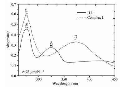
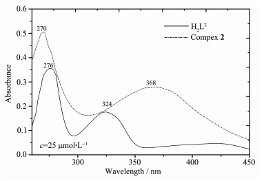
UV-Vis spectra of the free ligands in 25 μmol·L-1 ethanol solution and their corresponding complexes 1 and 2 in 1.0 mmol·L-1 ethanol solution at 298 K were measured within a range of 220~550 nm (Fig. 4 and 5).
Obviously, the absorption peaks of ligand H2L1 are different from those of complex 1. The UV-Vis spectrum of H2L1 had two relatively intense absorption peaks at ca. 276 and 324 nm. The absorption band at 276 nm could be assigned to the π-π* transition for the benzene rings; and the second peak at 324 nm could be assigned to the n-π* transition for the C=N group. In contrast to H2L1, the absorption peaks were bathochromically shifted[62] (Fig. 4). These phenomena are owing to the reaction of H2L1 with the Mn(Ⅱ) ions.
The absorption spectrum of ligand H2L2 shown in Fig. 5 included two relatively intense peaks centered at ca. 276 and 324 nm, which could be assigned to the π-π* transition for the benzene rings and the n-π* transition for the C=N group. But in complex 2, the absorption peak at 276 nm showed a hypsochromic shift to 270 nm, and the disappearance of the absorption peak of the ligand at 324 nm might be caused by coordination of the two N atoms of C=N groups with the Mn(Ⅱ) ion. For complex 2, a new absorption peak appeared at 368 nm due to the transfer of electrons from the ligand to the Mn(Ⅱ) ion.
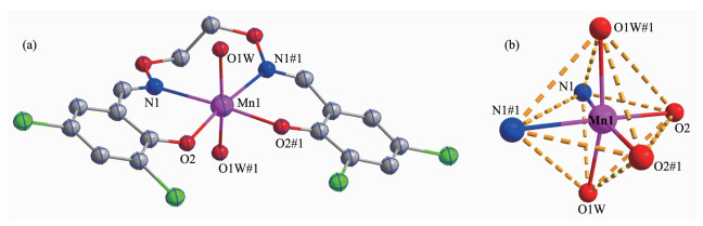
X-ray diffraction analysis demonstrated that complex 1 possesses a symmetric mononuclear structure and crystallizes in the monoclinic system with C2/c space group. Complex 1 is built up by one C16H14Cl4MnN2O6 molecule, which contains one Mn(Ⅱ) ion, one completely deprotonated ligand (L1)2- and two coordinated water molecules. The molecular structure with atomic labeling of complex 1 is depicted in Fig. 6. Selected bond lengths and angles of complex 1 are listed in Table 3.
Symmetry codes: #1: 2-x, y, 1/2-z; Hydrogen atoms are omitted for clarity
下载:
导出CSV
| 1 | |||||
| Mn1-O2 | 0.211 97(19) | Mn1-O1W | 0.216 3(2) | Mn1-N1 | 0.229 3(3) |
| Mn1-O2#1 | 0.211 98(19) | ||||
| O2-Mn1-O2 | 93.69(11) | O2-Mn1-O1W | 94.44(8) | O1W-Mn1-O1W | 167.23(12) |
| O2-Mn1-N1 | 80.00(9) | O2-Mn1-O1W#1 | 94.28(8) | O1W-Mn1-N1 | 84.88(9) |
| O2-Mn1-N1#1 | 173.57(9) | N1-Mn1-N1 | 106.33(13) | O1W-Mn1-N1#1 | 87.47(9) |
| 2 | |||||
| Mn1-O4 | 0.205 6(3) | Mn1-O1 | 0.207 7(2) | Mn1-O1W | 0.219 9(2) |
| Mn1-N1 | 0.221 9(3) | Mn1-N2 | 0.229 7(3) | Mn1-O2W | 0.220 1(2) |
| O4-Mn1-O1 | 91.83(10) | O4-Mn1-O1W | 83.72(11) | O1-Mn1-O1W | 98.09(9) |
| O4-Mn1-O2W | 101.12(11) | O1-Mn1-O2W | 85.57(9) | O1W-Mn1-O2W | 173.88(9) |
| O4-Mn1-N1 | 171.40(11) | O1-Mn1-N1 | 82.98(9) | O1W-Mn1-N1 | 90.20(9) |
| O2W-Mn1-N1 | 85.37(10) | O4-Mn1-N2 | 82.46(10) | O1-Mn1-N2 | 173.75(9) |
| O1W-Mn1-N2 | 83.85(9) | O2W-Mn1-N2 | 93.02(10) | N1-Mn1-N2 | 102.99(9) |
| Symmetry codes: #1: 2-x, y, 1/2-z for 1. | |||||
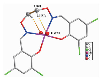
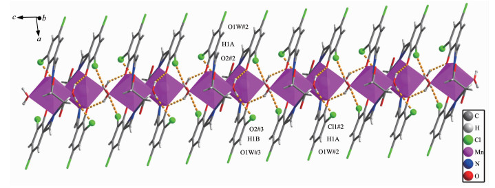
The coordination sphere of the hexa-coordinated Mn(Ⅱ) ion is completed by two phenolic oxygen atoms (Mn1-O2 0.211 97(19) nm and Mn1-O2#1 0.211 98(19) nm) and two nitrogen atoms (Mn1-N1 0.229 3(3) nm and Mn1-N1#1 0.229 3(3) nm) coming from the completely deprotonated unit (L1)2- and two oxygen atoms (Mn1-O1W, 0.216 3(2) nm and Mn1-O1W#1, 0.216 3(2) nm) of the coordinated water molecules. Therefore, the Mn(Ⅱ) ion forms a slightly distorted octahedral geometry. The distances of the Mn(Ⅱ) ion and the phenolic oxygen atoms (O2 and O2#1) of the fully deprotonated (L1)2- unit are clearly shorter than those of the nitrogen atoms (N1 and N1#1) from the (L1)2- unit (Fig. 6). There are one intramolecular hydrogen bond (C8#1-H8B…O1W#1), three intermolecular hydrogen bonds (O1W#2-H1A…O2#2, O1W#2-H1A…Cl1#2, O1W#3-H1B…O2#3) in complex 1, finally forming a 1D supramolecular structure (Fig. 7 and 8).
Symmetry codes: #1: 2-x, y, 1/2-z
Symmetry codes: #2: 2-x, 1-y, -z; #3: x, 1-y, -1/2+z
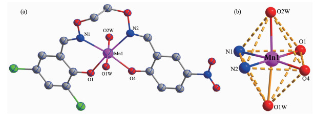
X-ray crystallographic analysis reveals that complex 2 belongs to triclinic crystal system with space group P1, and has a symmetric mononuclear structure. Fig. 9(a) and (b) showed the molecular structure and the octahedral unit of complex 2. Selected bond lengths and angles are provided in Table 4.
Hydrogen atoms are omitted for clarity
下载:
导出CSV
| D-H…A | d(D-H)/nm | d(H…A)/nm | d(D…A)/nm | ∠DHA/(°) |
| C8#1-H8B…O1W#1 | 0.097 | 0.252 | 0.337 9(4) | 148 |
| O1W#2-H1A…O2#2 | 0.089 | 0.210 | 0.290 9(3) | 151.7 |
| O1W#2-H1A…Cl1#2 | 0.089 | 0.278 | 0.338 4(2) | 127 |
| O1W#3-H1B…O2#3 | 0.088 | 0.192 | 0.272 1(3) | 149.5 |
| O1W-H1WA…N2 | 0.089 | 0.251 | 0.300 5(4) | 116 |
| O1W-H1WB…O4 | 0.089 | 0.240 | 0.284 1(4) | 111 |
| O2W-H2WA…N1 | 0.089 | 0.251 | 0.299 6(4) | 115 |
| O2W-H2WB…O1 | 0.089 | 0.246 | 0.290 7(4) | 111 |
| C8-H8A…N2 | 0.097 | 0.249 | 0.287 9(5) | 104 |
| C9-H9A…N1 | 0.097 | 0.245 | 0.286 7(5) | 106 |
| O1W#1-H1WB…O1#1 | 0.089 | 0.213 | 0.283 1(3) | 136 |
| O2W#1-H2WB…O4#1 | 0.089 | 0.220 | 0.286 4(4) | 131 |
| C3-H3#2…O3#2 | 0.093 | 0.258 | 0.346 0(5) | 158 |
| C14#3-H14…O6#3 | 0.093 | 0.252 | 0.321 1(5) | 131 |
| Symmetry codes: #1: 2-x, y, 1/2-z; #2: 2-x, 1-y, -z; #3: x, 1-y, -1/2+z for 1; #1: 1-x, 2-y, 1-z; #2: x, 1+y, z; #3: 1-x, 2-y, 2-z for 2. | ||||
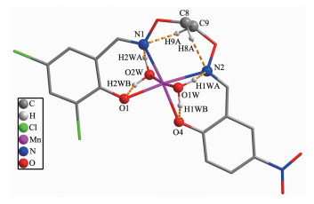
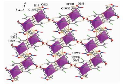
Single crystal X-ray diffraction analysis showed that the crystal structure of complex 2 was similar to that of complex 1, forming a 1:1 ((L2)2- and Mn(Ⅱ)) mononuclear structure. Complex 2 contained one Mn(Ⅱ) ion, one full deprotonated (L2)2- unit and two coor-dinated water molecules. The hexa-coordinated Mn(Ⅱ) ion (Mn1) is located in the cis-N2O2 coordination cavity (Mn1-O1, 0.207 7(2) nm, Mn1-O4, 0.205 6(3) nm, Mn1-N1 0.221 9(3) nm and Mn1-N2 0.229 7(3) nm) of the completely deprotonated (L2)2- unit, and two oxygen atoms (Mn1-O1W, 0.219 9(2) nm and Mn1-O2W 0.220 1(2) nm) of the coordinated H2O molecules occupied together the axial positions. All of the six oxygen atoms coordinate to the Mn(Ⅱ) ion (Mn1) constituting an octahedral geometry, as shown in Fig. 9(b). Six intramolecular hydrogen bonds (O1W-H1WA…N2, O1W-H1WB…O4, O2W-H2WA…N1, O2W-H2WB…O1, C8-H8A…N2 and C9-H9A…N1) and four intermolecular hydrogen bonding interactions(O1W#1-H1WB…O1#1, O2W#1-H2WB…O4#1, C3-H3#2…O3#2 and C14#3-H14…O6#3) are observed and shown in Fig. 10. With the help of hydrogen bond interactions in complex 2, the adjacent complex 2 molecule units are linked together forming an infinite 2D supramolecular network, which is shown in Fig. 11.
Symmetry codes: #1: 1-x, 2-y, 1-z; #2: x, 1+y, z; #3: 1-x, 2-y, 2-z
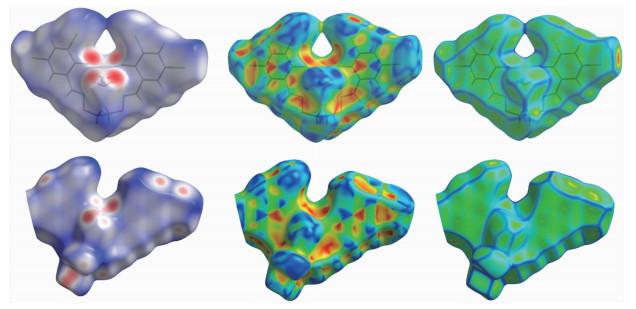
The Hirshfeld surfaces of the free ligands and their complexes 1 and 2 were analyzed using Crystal Explorer program. The surface analyses of H2L1, H2L2 and complexes 1 and 2 have been mapped with dnorm (standard high resolution), curvedness and shape index(Fig. 12)[63]. When mapped with the function of dnorm, the surface of H2L produces several spherical red depressions, indicating the main presence of type C-H…O interactions, and other visible depressions correspond to H…H and C…H interactions. For complexes 1 and 2, more intense red depressions can be seen on the Hirshfeld surfaces which marks the O…H/H…O interactions persisting. The 2D fingerprint plots are used to explain the atom pair contacts of the crystal, which can quantify the intermolecular interactions. Meantime, it could be decomposed to highlight contributions from different interactions. The 2D fingerprint plot of H…H, C…H/H…C and O…H/H…O interactions in the complexes are depicted in Fig. 13.
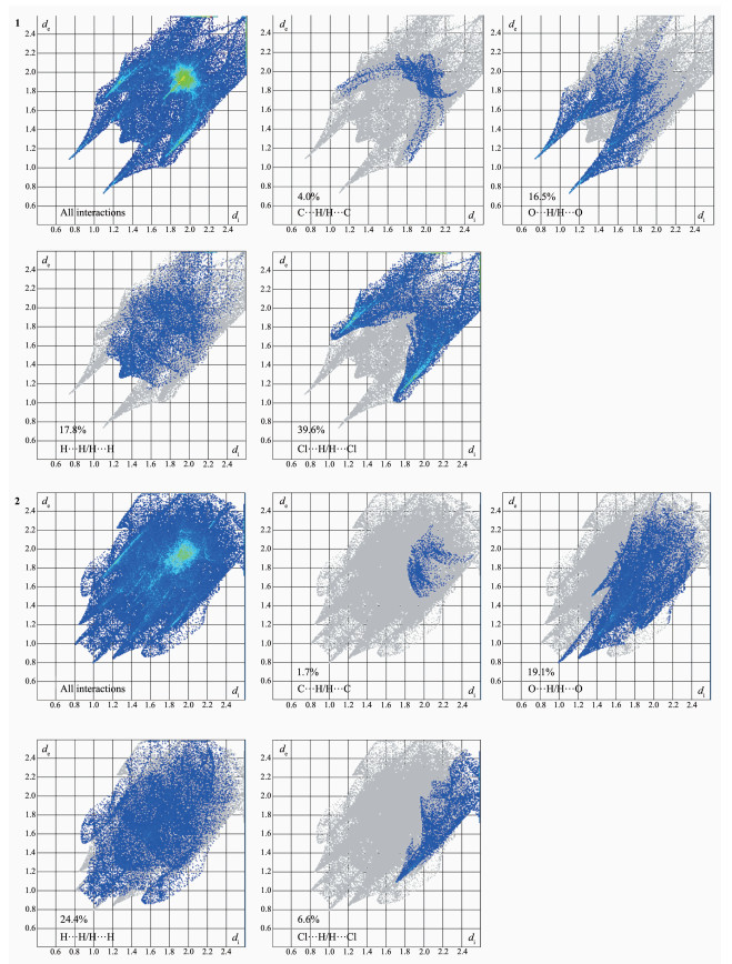
In complex 1, the proportions of C…H/H…C, O…H/H…O, H…H and Cl…H interactions cover 4.1%, 16.5%, 17.8% and 39.7% of the Hirshfield surfaces, respectively, while the proportions of C…H/H…C, O…H/H…O, H…H and Cl…H interactions in complex 2 cover 1.7%, 19.1%, 24.4% and 6.6%, respectively. Because of the existence of hydrogen bonds, complexes 1 and 2 can be more stable[64].
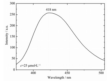
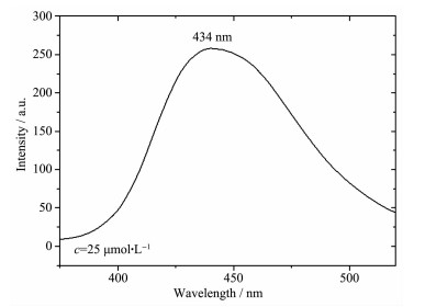
The fluorescence behaviors of H2L1, H2L2 and their corresponding complexes 1 and 2 were measured in ethanol solution (25 μmol·L-1) at room temperature within wavelength range of 350~600 nm and displayed in Fig. 14~15.
The spectrum of H2L1 is non-fluorescent upon excitation at 370 nm in ethanol solution. However, complex 1 exhibited an intense fluorescence intensity (Fig. 14) at 418 nm upon excitation at 370 nm, because of the photoinduced electron transfer mechanism. In complex 1, the Mn(Ⅱ) ion is coordinated with the lone pair electrons of C=N and phenol oxygen atoms of benzene rings, which enhances the planar steel of the molecules, resulting in enhancement of fluorescence[65].
The spectrum of H2L2 is also non-fluorescent upon excitation at 375 nm. But complex 2 exhibited an intense emission peak at ca. 434 nm upon excitation at 375 nm (Fig. 15), which can be attributed to the photoinduced electron transfer mechanism. As same as complex 1, the Mn(Ⅱ) ion in complex 2 is coordinated with the N atoms of the C=N group and phenol oxygen atoms of benzene rings, leading to the increased coplanarity of molecule and enhanced fluorescence.
In conclusion, we have designed and synthesized two Mn(Ⅱ) complexes with two salamo-type ligands H2L1 and H2L2, and characterized them by physico-chemical methods and single-crystal X-ray diffractions. X-ray crystal structures revealed that the structural features of complexes 1 and 2 are very similar, and the water molecules participate in the coordination. In the IR spectra of complexes 1 and 2, the M-O and M-N vibrational absorption bands were observed. The UV-Vis spectra clearly indicate that the ligand (L1)2- or (L2)2- units of complexes 1 or 2 are coordinated with the Mn(Ⅱ) ions. The fluorescent results of complexes 1 and 2 showed relatively strong emission peaks compared to the corresponding free ligands. The Hirshfeld surfaces and 2D fingerprint plots could explain the atom pair contacts of the crystal, which could quantify the intermolecular interactions.
Liu P P, Wang C Y, Zhang M, et al. Polyhedron, 2017, 129:133-140 doi: 10.1016/j.poly.2017.03.019
Song X Q, Liu P P, Liu Y A, et al. Dalton Trans., 2016, 45:8154-8163 doi: 10.1039/C6DT00212A
Subramaniam P, Anbarasan S, Devi S S, et al. Polyhedron, 2016, 119:14-22
Akine S, Miyashita M, Piaob S, et al. Inorg. Chem. Front., 2014, 1:53-57 doi: 10.1039/C3QI00067B
Dong X Y, Zhao Q, Kang Q P, et al. Crystals, 2018, 8:230 doi: 10.3390/cryst8050230
Zhang L W, Li X Y, Kang Q P, et al. Crystals, 2018, 8:173 doi: 10.3390/cryst8040173
Li X Y, Kang Q P, Liu L Z. Crystals, 2018, 8:43 doi: 10.3390/cryst8010043
Peng Y D, Li X Y, Kang Q P, et al. Crystals, 2018, 8:1079
Peng Y D, Wang F, Gao L, et al. J. Chin. Chem. Soc., 2018, 65:893-899 doi: 10.1002/jccs.201800038
Li X Y, Chen L, Gao L, et al. RSC Adv., 2017, 7:35905-35916 doi: 10.1039/C7RA06796H
Xu L, Li N, Zhao L. Z. Kristallogr.:New Cryst. Struct., 2016, 231:967-968
Li L H, Dong W K, Zhang Y, et al. Appl. Organomet. Chem., 2017, e3818
Chin T K, Endud S, Jamil S, et al. Catal. Lett., 2013, 143:282-288 doi: 10.1007/s10562-012-0959-1
Gao L, Liu C, Wang F, et al. Crystals, 2018, 8:77 doi: 10.3390/cryst8020077
Wu H L, Bai Y H, Zhang Y H, et al. Z. Anorg. Allg. Chem., 2014, 640:2062-2071 doi: 10.1002/zaac.201400109
Wu H L, Pan G L, Wang H, et al. J. Photochem. Photobiol. B, 2014, 135:33-43 doi: 10.1016/j.jphotobiol.2014.04.005
Wu H L, Pan G L, Bai Y C, et al. J. Chem. Res., 2014, 38:211-217 doi: 10.3184/174751914X13933417974082
Wu H L, Bai Y C, Zhang Y H, et al. J. Coord. Chem., 2014, 67:3054-3066 doi: 10.1080/00958972.2014.959507
Wang F, Xu Y L, Aderinto S O, et al. J. Photochem. Photobiol. A, 2017, 332:273-282 doi: 10.1016/j.jphotochem.2016.09.004
Wu H L, Wang H, Wang X L, et al. New J. Chem., 2014, 38:1052-1061 doi: 10.1039/c3nj01145c
Wu H L, Pan G L, Bai Y C, et al. Res. Chem. Intermed., 2015, 41:3375-3388 doi: 10.1007/s11164-013-1440-5
Hao J, Li X Y, Wang L, et al. Spectrochim. Acta Part A, 2018, 204:388-402 doi: 10.1016/j.saa.2018.06.074
Wang L, Hao J, Zhai L X, et al. Crystals, 2017, 7:277 doi: 10.3390/cryst7090277
Ömer S, Ümmühan Ö Ö, Nurgul S, et al. Tetrahedron, 2016, 72:5843-5852 doi: 10.1016/j.tet.2016.08.004
Li J, Zhang H J, Chang J, et al. Crystals, 2018, 8:176 doi: 10.3390/cryst8040176
Chai L Q, Zhang K Y, Tang L J, et al. Polyhedron, 2017, 130:100-107 doi: 10.1016/j.poly.2017.04.010
Dong X Y, Li X Y, Liu L Z, et al. RSC Adv., 2017, 7:48394-48403 doi: 10.1039/C7RA07826A
Jia H R, Chang J, Zhang H J, et al. Crystals, 2018, 8:272 doi: 10.3390/cryst8070272
Chen L, Dong W K, Zhang H, et al. Cryst. Growth Des., 2017, 17:3636-3648 doi: 10.1021/acs.cgd.6b01860
Zhang L W, Liu L Z, Wang F, et al. Molecules, 2018, 23:1141 doi: 10.3390/molecules23051141
Dong X Y, Zhao Q, Wei Z L, et al. Molecules, 2018, 23:1006 doi: 10.3390/molecules23051006
Chai L Q, Tang L J, Chen L C, et al. Polyhedron, 2017, 122:228-240 doi: 10.1016/j.poly.2016.11.032
Dong W K, Li X L, Wang L, et al. Sens. Actuators B, 2016, 229:370-378 doi: 10.1016/j.snb.2016.01.139
Wang B J, Dong W K, Zhang Y, et al. Sens. Actuators B, 2017, 247:254-264 doi: 10.1016/j.snb.2017.02.154
Wang F, Gao L, Zhao Q, et al. Spectrochim. Acta Part A, 2018, 190:111-115 doi: 10.1016/j.saa.2017.09.027
Dong Y J, Li X L, Zhang Y, et al. Supramol. Chem., 2017, 29:518-527 doi: 10.1080/10610278.2017.1285031
Hao J, Li X Y, Zhang Y, et al. Materials, 2018, 11:523 doi: 10.3390/ma11040523
Dong W K, Akogun S F, Zhang Y, et al. Sens. Actuators B, 2017, 238:723-734 doi: 10.1016/j.snb.2016.07.047
Dong W K, Ma J C, Zhu L C, et al. Cryst. Growth Des., 2016, 16:6903-6914 doi: 10.1021/acs.cgd.6b01067
Wang L, Li X Y, Zhao Q, et al. RSC Adv., 2017, 7:48730-48737 doi: 10.1039/C7RA08789F
Dong W K, Li G, Wang Z K, et al. Spectrochim. Acta Part A, 2014, 133:340-347 doi: 10.1016/j.saa.2014.04.190
Hao J, Li L H, Zhang J T, et al. Polyhedron, 2017, 134:1-10 doi: 10.1016/j.poly.2017.05.060
Zhang H, Dong W K, Zhang Y, et al. Polyhedron, 2017, 133:279-293 doi: 10.1016/j.poly.2017.05.051
Ma J C, Dong X Y, Dong W K, et al. J. Coord. Chem., 2016, 69:149-159 doi: 10.1080/00958972.2015.1108410
Hao J, Liu L Z, Dong W K, et al. J. Coord. Chem., 2017, 70:1936-1952 doi: 10.1080/00958972.2017.1324152
杨玉华, 郝静, 董银娟, 等.无机化学学报, 2017, 33:1280-1292 doi: 10.11862/CJIC.2017.150YANG Yu-Hua, HAO Jing, DONG Yin-Juan, et al. Chinese. J. Inorg. Chem., 2017, 33:1280-1292 doi: 10.11862/CJIC.2017.150
Akine S, Taniguchi T, Saiki T, et al. J. Am. Chem. Soc., 2005, 127:540-541 doi: 10.1021/ja046790k
Zhao Q, Wei Z L, Kang Q P, et al. Spectrochim. Acta Part A, 2018, 203:472 doi: 10.1016/j.saa.2018.06.007
Dong W K, Zheng S S, Zhang J T, et al. Spectrochim. Acta Part A, 2017, 184:141-150 doi: 10.1016/j.saa.2017.04.061
张宏佳, 常健, 贾浩然, 等.无机化学学报, 2018, 34:2261-2270 doi: 10.11862/CJIC.2018.261ZHANG Hong-Jia, CHANG Jian, JIA Hao-Ran, et al. Chinese J. Inorg. Chem., 2018, 34:2261-2270 doi: 10.11862/CJIC.2018.261
常健, 张宏佳, 贾浩然, 等.无机化学学报, 2018, 34:2097-2107 doi: 10.11862/CJIC.2018.256CHANG Jian, ZHANG Hong-Jia, JIA Hao-Ran, et al. Chinese J. Inorg. Chem., 2018, 34:2097-2107 doi: 10.11862/CJIC.2018.256
Dong W K, Sun Y X, Zhao C Y, et al. Polyhedron, 2010, 29:2087-2097 doi: 10.1016/j.poly.2010.04.006
Dong X Y, Wang L, Yang Y H, et al. Asian J. Chem., 2013, 8:4289-4292
Dong W K, Zhao C Y, Sun Y X, et al. Inorg. Chem. Commu., 2009, 12:234-236 doi: 10.1016/j.inoche.2008.12.019
Zheng S S, Dong W K, Zhang Y, et al. New J. Chem., 2017, 41:4966-4973 doi: 10.1039/C6NJ04090J
Dong W K, Lan P F, Zhou W M, et al. J. Coord. Chem., 2016, 69:1272-1283 doi: 10.1080/00958972.2016.1168520
Wang L, Dong X Y, Zhao L, et al. Asian J. Chem., 2013, 6:3440-3442
Sheldrick G M. Acta Crystallogr. Sect. A:Found. Crystallogr., 2008, A64:112-122
王莉, 康全鹏, 郝静, 等.无机化学学报, 2018, 34:525-533 doi: 10.11862/CJIC.2018.035WANG Li, KANG Quan-Peng, HAO Jing, et al. Chinese J. Inorg. Chem., 2018, 34:525-533 doi: 10.11862/CJIC.2018.035
Dong X Y, Gao L, Wang F, et al. Crystals, 2017, 7:267 doi: 10.3390/cryst7090267
Dong W K, Ma J C, Dong Y J, et al. Polyhedron, 2016, 115:228-235 doi: 10.1016/j.poly.2016.05.017
Gao L, Wang F, Zhao Q, et al. Polyhedron, 2018, 139:7-16 doi: 10.1016/j.poly.2017.10.004
Ghosh K, Harms K, Chattopadhyay S. Polyhedron, 2017, 123:162-175 doi: 10.1016/j.poly.2016.10.027
Wang F, Liu L Z, Gao L, et al. Spectrochim. Acta Part A, 2018, 203:56-64 doi: 10.1016/j.saa.2018.05.088
Kang Q P, Li X Y, Zhao Q, et al. Appl. Organomet. Chem., 2018, 32:e4379 doi: 10.1002/aoc.4379

Figure 6 (a) Molecular structure of complex 1; (b) Coordination polyhedron for Mn(Ⅱ) ion
Symmetry codes: #1: 2-x, y, 1/2-z; Hydrogen atoms are omitted for clarity

Figure 7 View of intramolecular hydrogen bonding interactions of complex 1
Symmetry codes: #1: 2-x, y, 1/2-z

Figure 8 View of 1D supramolecular architecture of complex 1 formed by hydrogen bonding interactions
Symmetry codes: #2: 2-x, 1-y, -z; #3: x, 1-y, -1/2+z

Figure 9 (a) Molecular structure of complex 2; (b) Coordination polyhedron for Mn(Ⅱ) ion
Hydrogen atoms are omitted for clarity

Figure 11 View of 2D supramolecular architecture of complex 2 formed by hydrogen bonding interactions
Symmetry codes: #1: 1-x, 2-y, 1-z; #2: x, 1+y, z; #3: 1-x, 2-y, 2-z

Figure 12 Hirshfeld surfaces of complexes 1 and 2 mapped with dnorm, shape index and curvedness

Figure 13 Two dimensional fingerprint plots of complexes 1 and 2 with major decomposition plots
Table 1. Crystal data and refinement parameter for complexes 1 and 2
| Complex | 1 | 2 |
| Formula | C16H14Cl4MnN2O6 | C16H15.73Cl2MnN3O8.37 |
| Formula weight | 527.03 | 509.81 |
| Crystal system | Monoclinic | Triclinic |
| Space group | C2/c | P1 |
| a/nm | 1.728 35(17) | 0.764 9(2) |
| b/nm | 1.515 65(17) | 1.159 4(2) |
| c/nm | 0.776 92(6) | 1.427 0(2) |
| α/(°) | 110.121(6) | |
| β/(°) | 97.161(9) | 100.257(9) |
| γ/(°) | 102.123 4(19) | |
| V/nm3 | 2.019 3(3) | 1.117 7(4) |
| Z | 4 | 2 |
| Dc/(g·cm-3) | 1.734 | 1.515 |
| μ/mm-1 | 1.220 | 0.877 |
| F(000) | 1 060 | 517 |
| θ range/(°) | 3.314~26.993 | 3.329~25.996 |
| Index ranges | -21 ≤ h ≤ 21, -19 ≤ k ≤ 18, -9 ≤ l ≤ 5 | -7 ≤ h ≤ 9, -14 ≤ k ≤ 14, -17 ≤ l ≤ 17 |
| Reflection collected | 4 100 | 8 032 |
| Independent reflection | 2 172 | 4 234 |
| Rint | 0.016 2 | 0.028 6 |
| Data, restraint, parameter | 2 172, 1, 127 | 4 234, 3, 291 |
| GOF | 1.099 | 0.995 |
| Final R1, wR2 indices [I>2σ(I)] | 0.043 8, 0.125 4 | 0.054 9, 0.134 1 |
| R1, wR2 indices (all data) | 0.057 6, 0.136 8 | 0.075 4, 0.140 5 |
| Largest differences peak and hole/(e·nm-3) | 990, -488 | 425, -330 |
 下载: 导出CSV
Table 2. Main infrared data of H2L1, H2L2 and their complexes 1 and 2
| cm-1 | |||||
| Compound | Ar-O | C=N | M-N | M-O | C=C |
| H2L1 | 1 217 | 1 609 | — | — | 1 462, 1 575, 1 595 |
| 1 | 1 209 | 1 607 | 463 | 511 | 1 486, 1 579, 1 597 |
| H2L2 | 1 315 | 1 614 | 1 475, 1 572, 1 594 | ||
| 2 | 1 285 | 1 602 | 507 | 474 | 1 492, 1 578, 1 598 |
下载: 导出CSV
Table 3. Selected bond lengths (nm) and angles (°) for complexes 1 and 2
| 1 | |||||
| Mn1-O2 | 0.211 97(19) | Mn1-O1W | 0.216 3(2) | Mn1-N1 | 0.229 3(3) |
| Mn1-O2#1 | 0.211 98(19) | ||||
| O2-Mn1-O2 | 93.69(11) | O2-Mn1-O1W | 94.44(8) | O1W-Mn1-O1W | 167.23(12) |
| O2-Mn1-N1 | 80.00(9) | O2-Mn1-O1W#1 | 94.28(8) | O1W-Mn1-N1 | 84.88(9) |
| O2-Mn1-N1#1 | 173.57(9) | N1-Mn1-N1 | 106.33(13) | O1W-Mn1-N1#1 | 87.47(9) |
| 2 | |||||
| Mn1-O4 | 0.205 6(3) | Mn1-O1 | 0.207 7(2) | Mn1-O1W | 0.219 9(2) |
| Mn1-N1 | 0.221 9(3) | Mn1-N2 | 0.229 7(3) | Mn1-O2W | 0.220 1(2) |
| O4-Mn1-O1 | 91.83(10) | O4-Mn1-O1W | 83.72(11) | O1-Mn1-O1W | 98.09(9) |
| O4-Mn1-O2W | 101.12(11) | O1-Mn1-O2W | 85.57(9) | O1W-Mn1-O2W | 173.88(9) |
| O4-Mn1-N1 | 171.40(11) | O1-Mn1-N1 | 82.98(9) | O1W-Mn1-N1 | 90.20(9) |
| O2W-Mn1-N1 | 85.37(10) | O4-Mn1-N2 | 82.46(10) | O1-Mn1-N2 | 173.75(9) |
| O1W-Mn1-N2 | 83.85(9) | O2W-Mn1-N2 | 93.02(10) | N1-Mn1-N2 | 102.99(9) |
| Symmetry codes: #1: 2-x, y, 1/2-z for 1. | |||||
下载: 导出CSV
Table 4. Hydrogen bonding parameters of complexes 1 and 2
| D-H…A | d(D-H)/nm | d(H…A)/nm | d(D…A)/nm | ∠DHA/(°) |
| C8#1-H8B…O1W#1 | 0.097 | 0.252 | 0.337 9(4) | 148 |
| O1W#2-H1A…O2#2 | 0.089 | 0.210 | 0.290 9(3) | 151.7 |
| O1W#2-H1A…Cl1#2 | 0.089 | 0.278 | 0.338 4(2) | 127 |
| O1W#3-H1B…O2#3 | 0.088 | 0.192 | 0.272 1(3) | 149.5 |
| O1W-H1WA…N2 | 0.089 | 0.251 | 0.300 5(4) | 116 |
| O1W-H1WB…O4 | 0.089 | 0.240 | 0.284 1(4) | 111 |
| O2W-H2WA…N1 | 0.089 | 0.251 | 0.299 6(4) | 115 |
| O2W-H2WB…O1 | 0.089 | 0.246 | 0.290 7(4) | 111 |
| C8-H8A…N2 | 0.097 | 0.249 | 0.287 9(5) | 104 |
| C9-H9A…N1 | 0.097 | 0.245 | 0.286 7(5) | 106 |
| O1W#1-H1WB…O1#1 | 0.089 | 0.213 | 0.283 1(3) | 136 |
| O2W#1-H2WB…O4#1 | 0.089 | 0.220 | 0.286 4(4) | 131 |
| C3-H3#2…O3#2 | 0.093 | 0.258 | 0.346 0(5) | 158 |
| C14#3-H14…O6#3 | 0.093 | 0.252 | 0.321 1(5) | 131 |
| Symmetry codes: #1: 2-x, y, 1/2-z; #2: 2-x, 1-y, -z; #3: x, 1-y, -1/2+z for 1; #1: 1-x, 2-y, 1-z; #2: x, 1+y, z; #3: 1-x, 2-y, 2-z for 2. | ||||
下载: 导出CSV

 扫一扫看文章


扫一扫关注我们