Table 1.
Crystallographic data of 1
Citation:
LI Ji, HAO Yuan-Yuan, QIAN Yong, XUE Xu-Lin, SU Zhi, LIU Hong-Ke. DNA Targeting Rigid Dinuclear Ruthenium-Arene Complexes[J]. Chinese Journal of Inorganic Chemistry,
2019, 35(8): 1500-1508.
doi:
10.11862/CJIC.2019.184

靶向DNA的刚性配体的双核芳基钌配合物
摘要:
以刚性配体1,3-bib(1,3-二(1H-咪唑-1-基)苯)与[Ru(η6-p-bip)Cl2]2(p-bip,联苯基团)为原料,合成了3种双核芳基钌配合物[Ru2(η6-p-bip)2(1,3-bib)2XY]X2(X=Y=Cl-(1),X=Y=Br-(2),X=I-和Y=Cl-(3),并用核磁和质谱等对配合物进行了表征。配合物1的单晶衍射结果表明其具有一种刚性双核M2L2碗状结构,空腔中心有一个阴离子Cl-。配合物3对A549细胞有较高的抗癌活性(IC50=13.9 μmol·L-1),与顺铂细胞毒性(IC50=15.2 μmol·L-1)相当。紫外吸收光谱、圆二色谱、凝胶电泳法研究表明配合物1~3与DNA发生强烈的相互作用并且诱发DNA发生解旋。
English
DNA Targeting Rigid Dinuclear Ruthenium-Arene Complexes
Abstract:
Three dinuclear Ru(Ⅱ)-arene complexes[Ru2(η6-p-bip)2(1, 3-bib)2XY]X2 (X=Y=Cl- (1), X=Y=Br- (2), X=I- and Y=Cl- (3); 1, 3-bib=1, 3-di(1H-imidazol-1-yl)benzene, p-bip=biphenyl) were synthesized and fully characterized by 1H NMR and ESI-MS. Single crystal X-ray diffractions studies showed that complex 1 owns a rigid M2L2 bowl-like structure, where one Cl- is trapped inside the cavity to balance the charge. Complex 3 showed the best anticancer activities among complexes 1~3. The IC50 value of 3 towards to human lung cancer cells (A549) reached to 13.9 μmol·L-1, which is comparable to that of cisplatin (IC50=15.2 μmol·L-1). Complexes 1~3 have shown strong interactions with DNA and could induce the unwinding of the DNA superhelix structure.
-
Key words:
- Ru-arene complexes
- / crystal structure
- / anticancer activity
- / interaction with DNA
-
0. Introduction
Cisplatin, as the most widely used anticancer drug, has attracted more and more concerns due to the serious side-effects and resistance[1-3]. Piano-stool-like metal-arene complexes, especially the Ru(Ⅱ)-arene complexes, have been considered as the most potent substitutes for the cisplatin, because of their unique and versatile biochemical properties[4-5]. In addition, the multinuclear metal-arene complexes have indicated the enhanced anticancer activities than the correspon-ding mononuclear complexes[6-7]. In our previous study, dinuclear Ru(Ⅱ)-arene complexes based on the rigid imidazole-containing ligand 1, 3-di(1H-imidazol-1-yl)benzene (1, 3-bib) and caped p-cymene (p-cym) with various counter anions, have shown strong DNA interactions and distinct anticancer behaviors[8]. As is known, the arene group, as the core component in the metal-arene complex, has important impact on the anticancer activities, which could facilitate the entry of the substrate into cells[4].
In order to study the effect of arene ligands on the anticancer activities, we changed the arene group from the p-cym group to the biphenyl group (p-bip), since the p-bip has higher hydrophobicity than p-cym, which could increase cellular accumulation and the anticancer activity[9-10]. Three new dinuclear Ru(Ⅱ)-arene complexes have been synthesized and studied. These complexes were listed as [Ru2(η6-p-bip)2(1, 3-bib)2XY]X2 (X=Y=Cl- (1), X=Y=Br- (2), X=I- and Y=Cl- (3)) and the single crystal structure of 1 has been resolved. Bowl-like structure of 1 has been observed and one counter anion is included in the center of the structure. Among complexes 1~3, complex 3 indicated the best anticancer activity with IC50 values of 13.9 and 15.2 μmol·L-1 towards HeLa and A549 cancer cells, respectively, due to the existence of the I- anions. Strong interactions between complexes 1~3 to DNA have been studied and confirmed by UV-Vis spectra, CD spectra, and gel electrophoresis experiments.
1. Experimental
1.1 Materials and methods
Reactions were carried out under argon by using standard Schlenk techniques. All solvents were of analytical grade. Chemicals were obtained from commercial sources and used without further purification. The ligand 1, 3-bib and dimer [RuCl2(η6-p-bip)]2 were synthesized according to the literature methods[11-12]. Deuterated solvent for NMR purposes were obtained from Merck and Cambridge Isotopes. 1H NMR spectra were recorded on a Bruker AVANCE 400 spectrometer at ambient temperature. Electrospray ionization mass spectra (ESI-MS) were obtained using an LCQ spectrometer (Thermo Scientific). A Varian Cary 50 Probe UV-Vis spectrophotometer was used for UV scanning. The circular dichroic spectra of DNA in the region between 200 and 350 nm were obtained by using an Applied-Photophysics Chriascan spectro-photometer operating at 25 ℃.
1.2 Syntheses of complexes
1.2.1 [Ru2(η6-p-bip)2(1, 3-bib)2Cl2]Cl2 (1)
Ligand 1, 3-bib (42.0 mg, 0.2 mmol) and Ru2(η6-p-bip)2Cl4 (65.2 mg, 0.1 mmol) were stirred in 20 mL CH3OH solution at 65 ℃. After refluxing for 12 h under the N2 protection, the solvent was removed under vacuum and the crude product was further purified by silica gel chromatography using CH2Cl2/CH3OH as the elute. Pale yellow product was collected with a yield of 85% based on the consumed Ru(Ⅱ) dimer. Elemental analysis Calcd. for Ru2C48H44Cl4N8O2(%): C 51.99, H 4.00, N 10.10; Found(%): C 51.48, H 3.96, N 10.19. 1H NMR (DMSO-d6, 400 MHz): δ 10.31 (s, 4H, 1, 3-bib), 8.95 (s, 2H, 1, 3-bib), 7.98 (t, 4H, J=1.7 Hz, 1, 3-bib), 7.85~7.80 (m, 4H, 1, 3-bib), 7.70 (d, 6H, J=1.5 Hz, 1, 3-bib), 7.44~7.36 (m, 10H, p-bip), 6.71 (d, 4H, J=6.0 Hz, p-bip), 6.50 (t, 4H, J=6.3, 5.5 Hz, p-bip), 6.21 (t, 2H, J=5.6 Hz, p-bip). ESI-MS(+): Calcd. for [Ru2(η6-p-bip)2(1, 3-bib)2Cl3]+ ([Ru2C48H40N8Cl3]+) m/z: 1 037.38, Found: 1 039.08; Calcd. for [Ru2(η6-p-bip)2(1, 3-bib)2Cl2]2+ ([Ru2C48H40N8Cl2]2+) m/z: 500.96, Found: 501.25.
1.2.2 [Ru2(η6-p-bip)2(1, 3-bib)2Br2]Br2 (2)
To a CH3OH solution (5 mL) of complex 1 (16.09 mg, 0.015 mmol), 5 mL CH3OH containing AgCF3SO3 (15.42 mg, 0.06 mmol) was added. After stirred for 12 h at room temperature, the AgCl precipitate was removed by filtration, and the CH3OH solution (15 mL) contain-ing KBr (89.25 mg, 0.75 mmol) was added dropwise to the resulting solution. After 12 h, the solvent was removed under vacuum and the crude product was further purified by silica gel chromatography using CH2Cl2/CH3OH as the eluent. Pale yellow product was collected with a yield of 79% based on the consumed complex 1. Elemental analysis Calcd. for Ru2C48H42Br4N8O(%): C 45.44, H 3.34, N 8.83; Found(%): C 44.99, H 3.31, N 8.90. 1H NMR (DMSO-d6, 400 MHz): δ 9.87 (s, 4H, 1, 3-bib), 8.78 (s, 2H, 1, 3-bib), 7.94 (s, 4H, 1, 3-bib), 7.86~7.61 (m, 10H, p-bip), 7.53 (s, 4H, p-bip), 7.48~7.30 (m, 6H, p-bip), 6.78 (d, J=5.9 Hz, 4H, p-bip), 6.52 (t, J=5.7 Hz, 4H, p-bip), 6.13 (t, J=5.5 Hz, 2H, p-bip). ESI-MS(+): Calcd. for [Ru2(η6-p-bip)2(1, 3-bib)2Br3]+ ([Ru2C48H40N8Br3]+) m/z: 1 170.74, Found: 1 170.83.
1.2.3 [Ru2(η6-p-bip)2(1, 3-bib)2ClI]I2 (3)
To a CH3OH solution (5 mL) of complex 1 (16.09 mg, 0.015 mmol), 5 mL CH3OH containing AgCF3SO3 (15.42 mg, 0.06 mmol) was added. After stirred for 12 h at room temperature, the AgCl precipitate was removed by filtration, and the CH3OH solution (15 mL) containing KI (124.50 mg, 0.75 mmol) was added dropwise to the resulting solution. After 12 h, the solvent was removed under vacuum and the crude product was further purified by silica gel chromato-graphy using CH2Cl2/CH3OH as the eluent. Pale red product was collected with a yield of 80% based on the consumed complex 1. Elemental analysis Calcd. for Ru2C48H42ClI3N8O(%): C 42.23, H 3.10, N 8.21; Found(%): C 41.81, H 3.06, N 8.29. 1H NMR (DMSO-d6, 400 MHz): δ 10.15 (s, 4H, 1, 3-bib), 8.81 (s, 2H, 1, 3-bib), 7.93 (d, J=16.6 Hz, 4H, 1, 3-bib), 7.78 (d, J=6.9 Hz, 4H, 1, 3-bib), 7.75~7.62 (m, 6H, 1, 3-bib), 7.44~7.35 (m, 10H, p-bip), 6.70 (d, J=5.9 Hz, 4H, p-bip), 6.47 (t, J=5.7 Hz, 4H, p-bip), 6.19 (t, J=5.5 Hz, 2H, p-bip). ESI-MS(+): Calcd. for [Ru2(η6-p-bip)2(1, 3-bib)2ClI2]+ ([Ru2C48H40N8ClI2]+) m/z: 1 220.29, Found: 1 220.83.
1.3 X-ray crystallographic study
Diffraction data were collected using a Bruker Apex 2 CCD with Kα radiation (λ=0.071 073 nm) at 296(2) K for complex 1 using the ω-2θ scan method. Absorption corrections were applied using a multi-scan technique[13]. An empirical absorption correction was applied. All the structures were solved by direct methods using SHELXS-2014 and refined by the full-matrix least-squares techniques using the SHELXL-2014[14] program within WINGX. All non-hydrogen atoms were refined anisotropically and the hydrogen atoms of the organic molecule were refined at calculated positions, assigned isotropic thermal para-meters, and allowed to ride their parent atoms. The crystallographic data and detailed bond distances and angles were listed in Table 1 and 2.
Table 1

 下载:
导出CSV
下载:
导出CSV
Formula Ru2C48H44Cl4N8O2 Formula weight 1 108.85 Crystal system Monoclinic Space group P21/c a / nm 1.383 13(19) b / nm 0.925 39(13) c / nm 3.478 1(5) β/(°) 94.371(2) Z 4 Dc/ (g·cm-3) 1.659 F(000) 2 240 θ range / (°) 1.2-28.1 Limiting indices -17≤h≤17, -12≤k≤12, -45≤l≤45 Reflection collected, unique 35 046, 10 018 Rint 0.061 Completeness to θ=25.00° / % 92.70 Data, restraint, parameter 10 018, 0, 583 Goodness-of-fit on F2 1.043 Final R indices [I>2σ(I)]* R1=0.048 0, 6wR2=0.102 6 *R1=∑||Fo|-|Fc||/∑|Fo|; wR2=|∑w(|Fo|2-|Fc|2)/∑|w(Fo)2|1/2, where w=1/[σ2(Fo2)+(aP)2+bP], P=(Fo2+2Fc2)/3. Table 2
Table 2. Selected bond lengths (nm) and angles (°) for 1下载:
导出CSV
Ru1-N1 0.210 3(3) Ru1-Cl1 0.240 9(1) Ru2-N5 0.209 5(3) Ru1 -N8 0.210 1(3) Ru2-N4 0.211 1(4) Ru2-Cl2 0.240 0(1) N1-Ru1-Cl1 86.47 (8) N1 -Ru1 -N8 89.44 (12) N5-Ru2-Cl2 86.95 (9) N8-Ru1-Cl1 85.06 (10) N4-Ru2-Cl2 88.17 (9) N4-Ru2-N5 87.28(13) CCDC: 1875666, 1.
1.4 UV studies
Complexes 1~3 were characterized by UV-Vis spectroscopy. All the complexes were dissolved in DMSO/H2O (1:9, V/V), and the final concentration is 100 μmol·L-1. The absorbance was recorded at time intervals of 1 h at 298 K for 17 h.
1.5 Cell viability assay
HeLa, A549 and LO2 cells were obtained from the Experimental Animal Centre of Sun Yat-Sen University (Guangzhou, China) and cultured in DMEM (Dulbecco′s modified Eagle′s medium, Gibco BRL) containing 10% FBS (fetal bovine serum, Gibco BRL), 100 μg·mL-1 streptomycin and 100 U·mL-1 penicillin (Gibco BRL). Cells were grown at 37 ℃ in a 5%(V/V) CO2 humidified incubator. The cytotoxicity of the tested complexes towards different cell lines was determined by MTT assay. Cells were cultured in 96-well plates for 24 h. Cells were incubated with a series of concentrations of complexes for another 48 h. 20 μL MTT solution (5 mg·mL-1) was added into each well and incubated for another 4 h. The culture media was removed and 150 μL DMSO was added into each well. The plate was shaken for 10 min. The absorbance at 492 nm was measured by a microplate reader[15-16].
1.6 UV-Vis spectroscopy studies of DNA binding property
Calf thymus DNA (CT-DNA) was purchased from Sigma. Phosphate buffer saline (PBS, 5 mmol·L-1, pH =7.4) was used for the spectral studies on DNA binding properties of the complexes. The PBS was prepared by ultra-pure water at room temperature. The stock CT-DNA solution was stored at 4 ℃. The concentration of CT-DNA was determined by UV-Vis absorption measurements at 260 nm after proper dilution with PBS buffer, taking 6 600 L·mol-1·cm-1 as the molar absorption coefficient. The ratio of UV absorbance (A260/A280) was ca. 1.9, indicating that the CT-DNA solution was sufficiently free of protein. Titration experiments were performed at room temperature. The working solution of complexes (100 μmol·L-1) were first placed in a 1 cm path quartz cuvette and aliquots of a CT-DNA stock solution was added, with mixing for 8 min to allow equilibrium to be reached. The absorption studies were conducted with a fixed complex concentration and alternating the CT-DNA concentra-tion. We observed a subtractive role from each reading on the UV-Vis when the complex and CT-DNA was incubated. With the increasing concentration of CT-DNA, the absorbance change at the metal-to-ligand charge transfer (MLCT) region. The binding constants (Kb) were determined from the spectroscopic titration data with the following equation[8, 17-18]:
$ {c_{{\rm{DNA}}}}/({\varepsilon _{\rm{a}}} - {\varepsilon _{\rm{f}}}) = {c_{{\rm{DNA}}}}/({\varepsilon _{\rm{b}}} - {\varepsilon _{\rm{f}}}) + 1/[{{\rm{K}}_{\rm{b}}}({\varepsilon _{\rm{b}}} - {\varepsilon _{\rm{f}}})] $
where cDNA is the concentration of the DNA; εa (εa=Aobsd/ccomplex) is the apparent absorption coefficient; εf is the extinction coefficient for the free complex; εb stands for the absorption coefficient of the complex when fully bounded to DNA. Linear fitting of cDNA/(εa-εf) with cDNA from the above equation allowed us to calculate Kb for the complex binding to CT-DNA.
1.7 Circular dichroism spectroscopy studies of DNA binding property
The interaction of complexes with CT-DNA was studied by CD spectroscopy in PBS buffer (50 mmol·L-1, pH=7.4). A series of samples containing different concentration ratios (ccomplex/cDNA) were prepared and incubated at 25 ℃ for 24 h. The concentration of CT-DNA was set at 100 μmol·L-1 and the ratio increased from 0.02 to 0.12. CD spectra were recorded in the cuvette of 1 cm path length, with the range of 200~350 nm, the scan rate of 100 nm·min-1 and the slit width of 1 nm.
1.8 Gel electrophoresis experiments
Closed circular supercoiled pBR322 plasmid DNA was used for the gel electrophoresis experiments, which was purchased from Sangon Biotech (Shanghai, China). DNA (1 μL, 200 μmol·L-1) was treated with ultrapure water (4 μL), PBS (5 μL, 100 mmol·L-1), and different concentrations of complexes (10 μL, ccomplex/cDNA=0.25, 0.5, 0.75, 1.0, 1.5 and 2.0, respectively). The mixture was incubated at 37 ℃ for 12 h in the dark, then loading buffer (4 μL, 0.05% bromophenol blue, 30 mmol·L-1 EDTA, 36% glycerol, 0.05% xylene cyanol FF) was added. Electrophoresis was carried out in a native agarose gel (0.8%) in 1×TAE (tris base, acetic acid, and EDTA) buffer for 2 h at 70 mA. The gel was subsequently stained with 1 μg·mL-1 ethidium bromide and visualized under a UV trans-illuminator and photographed by using a digital camera[19].
2. Results and discussion
2.1 Design and syntheses of macrocyclic di-ruthenium complexes 1~3
Macrocyclic di-ruthenium complex 1 was obtained from the direct reaction of the ligand (1, 3-bib) with the dimer [Ru(η6-p-bip)Cl2]2, while complexes 2 and 3 were prepared by reaction of complex 1 with appropriate silver salts (Scheme 1)[11]. All complexes were obtained in good yields and characterized by 1H NMR and ESI-MS (Fig.S1~S4, Supporting Information). In the complex 2, the coordinated and the counter anions Cl- were fully replaced by anion Br-, which were confirmed by the ESI-MS results. However, only one coordinated Cl- in complex 3 was replaced by a I- and the formula of [Ru2(η6-p-bip)2(1, 3-bib)2ClI]I2 was confirmed by the ESI-MS results.
Scheme1
2.2 X-ray crystallography
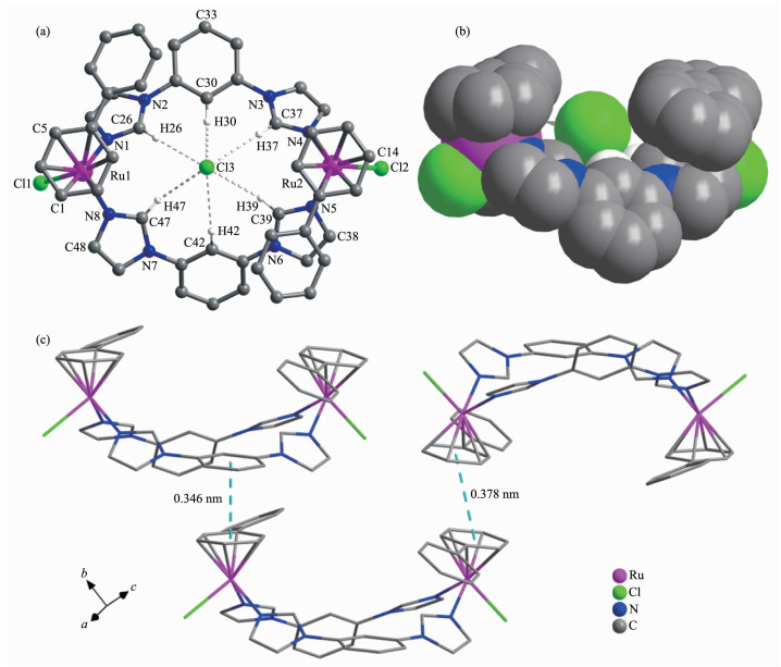
Complex 1 crystallizes in P21/c space group, and shows a classic "bowl-like" macrocyclic structure with Cl- as the counter anions (Fig. 1). Each Ru(Ⅱ) is coor-dinated by six carbon atoms from one phenyl ring, two nitrogen atoms from two 1, 3-bib ligands and one Cl- anion. The bond lengths of Ru-N and Ru-Cl are in a range of 0.208 6(8)~0.211 2(8) nm and 0.239 8(3)~0.240 5(3) nm, respectively, and the bond angles of N-Ru-Cl and N-Ru-N are in a range of 85.3°~88.4° and 87.1(3)°~89.3(3)°, respectively. The bond lengths and angles are similar to the previously reported Ru(Ⅱ)-arene complexes[8, 20]. One Cl- is captured into the bowl-like structure through the hydrogen bonds (Table S1), which is similar to the previously reported complex [Ru2(η6-p-cymene)2(1, 3-bib)2Cl2](NO3)2[8]. The trapped Cl- could be ensured from the results of ESI-MS. For example, for complex 1, the peak at 1 039.08 was clearly observed for [Ru2(η6-p-bip)2(1, 3-bib)2Cl3]+ (Calcd. m/z: 1 037.38) (Fig.S2)[8]. The upper and bottom planes of the bowl-like complex 1 are defined by C1, C5, C14 from the arene group and C48, C38 and C33 from the 1, 3-bib ligands, respectively (Fig. 1a). The diameter of the upper and bottom bases of the "bowl" were 1.25 or 0.94 nm, with a distance of 0.43 nm between the two bases. Comparing to [Ru2(η6-p-cymene)2(1, 3-bib)2Cl2](NO3)2, complex 1 owned a larger open window(0.94 nm vs 0.91 nm) and longer distance between two bases(0.43 nm vs 0.4 nm). The Ru1…Ru2 distance is 0.97 nm in complex 1. The dinuclear units are further connected by the π-π interactions between two paralleled phenyl rings from two adjacent units with the "center-to-center" distances of 0.346 and 0.378 nm, and the dihedral angle of 0° and 4.8°, respectively.
Figure 1
 Figure 1. (a) X-ray structure of complex 1, where one Cl- anion was enveloped as a guest molecular, and the other Cl-, partial hydrogen atoms, and the free water molecules are omitted for clarity; (b) Side-viewing of bowl-like complex 1 in space filling mode; (c) π-π stacking interactions between two parallel phenyl rings of two adjacent units, with a "center-to-center" distance of 0.346 and 0.378 nm, respectively
2.3 Cytotoxicity of complexes 1~3 in vitro
The cytotoxicity was evaluated in A549 (human lung cancer), HeLa (human cervical cancer), and LO2 (human normal liver cell line) by the colorimetric MTT assay for 48 h. Cisplatin was also tested as a positive control. The results are listed in Table 3 and show that complexes 1~3 own less toxicity to normal human cells (LO2) with an IC50 value of 28.5, 13.6 and 13.0 μmol·L-1, respectively, comparing to the cisplatin with the IC50 value of 1.5 μmol·L-1. Among 1~3, complex 3 shows the best anticancer activity to the cancer cells HeLa and A549 with IC50 value of 15.2 and 13.9 μmol·L-1, respectively, which is comparable to that of cisplatin. In general, the anti-proliferative efficacy of dinuclear Ru(Ⅱ) complexes are in the following order: 3 > 2 > 1, which indicates the important role of the counter anions, since the halide exchange could modify the cellular uptake and accumulation pathways[9]. Comparing to the other dinuclear Ru(Ⅱ)-arene with I- as the counter anions, [Ru2(η6-p-cymene)2(m-bib)2I2]I2 (m-bib=1, 3-bis(1H-imidazol-1-yl)methyl)benzene) and [Ru2(η6-p-cymene)2(1, 3-bib)2Cl2]I2, com-plex 3 showed a significant enhancement in the anti-cancer activity to A549 cancer cells with IC50 value of 13.9 μmol·L-1 vs 150.1 μmol·L-1 and 30.9 μmol·L-1, respectively[8, 19]. The results here indicate that both the ligand and the arene group play important roles for the enhancement of the anticancer activity of these dinuclear Ru-arene complexes[9]. In addition, complex 3 exhibits much higher anticancer activity comparing to several reported mononuclear complexes towards A549 cell lines[21].
Table 3
Table 3. IC50 values of complexes 1~3 as obtained from MTT assay下载:
导出CSV
μmol·L-1 Compound HeLa A549 LO2 1 25.6±1.7 45.4±2.3 28.5±0.8 2 21.9±2.1 28.9±1.4 13.6±0.8 3 15.2±1.1 13.9±1.1 13.0±0.5 Cisplatin 7.9±0.4 15.2±0.5 1.5±0.1 2.4 UV studies
The UV studies of the complex in solution is important for biological application. As shown in Fig.S5, the characteristic absorption bands for the complexes 1 and 2 showed only minimum or no change, while the bands for complex 3 displayed slight hypochromicity, indicating that complexes 1~3 could maintain their structural integrity in aqueous solution at 298 K. Previously reported dinuclear Ru(Ⅱ)-arene complex with rigid ligand have also showed high stability under physiological conditions[22-23].
2.5 Interaction with CT DNA
In order to understand the interactions of Ru(Ⅱ)-arene complexes with potent targets in the cancer cells, especially DNA, the DNA titration experiment was studied by the UV-Vis, CD and DNA electrophoresis.
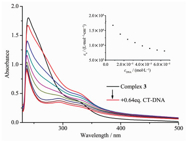
The UV-Vis spectra of the titration experiments of CT-DNA by complexes 1~3 at 25 ℃ are shown in Fig. 2 and S6. Complex 3 showed a maximum absorption wavelength at 240 nm and it slightly blue-shifted to 237 nm with the addition of 0.66 equal amounts of CT-DNA. The hypochromicity percentage of the MLCT band (metal to ligand charge transfer) was observed to be 50.3% at 241 nm, which suggests the strong stacking interaction between 1, 3-bib ligands and the base pairs of DNA[8]. Similar blue-shift bands were detected for complexes 1 or 2 from 241 to 238 nm or 244 to 242 nm, respectively. The hypochromicity percentage of complexes 1 and 2 were observed much less than that of complex 3, as 15.1% and 24.1% with the addition of 0.8 and 0.88 equal amount of CT-DNA, respectively, which suggests that complex 3 showed the strongest binding ability to DNA[24]. The intrinsic binding cons-tants (Kb) of complexes 1~3 to CT-DNA were 1.62×10⁴, 6.27×10³ and 2.07×10⁴ L·mol-1, respectively. Comparing to our previous reported dinuclear Ru(Ⅱ)-arene complexes with p-cymene as the arene group, the binding capacity to DNA has been dramatically decreased, which may result from the substitution of biphenyl group[25].
Figure 2
 Figure 2. Absorption spectra of complex 3 in PBS buffer at 25 ℃ on addition of CT-DNA
Arrow in the picture shows the absorbance change by the increasing of CT-DNA concentration; Inset: plot of εa versus cDNA for the titration of DNA with Ru(Ⅱ) complexes
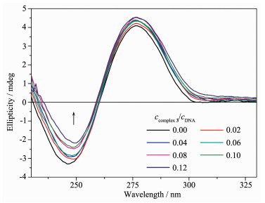
To further examine the binding mode between complexes 1~3 with CT-DNA, CD titration experiments were also performed. The positive band at about 278 nm is due to the base stacking and the negative band at about 248 nm is due to the right-handed helicity, which are both the characteristics of B-DNA. The increasing of the ratio of ccomplex/cDNA and dramatic decreasing of the ellipticity for negative bands suggest that 1~3 could unwind the DNA helix and lead to the loss of helicity through rotation of the bases (Fig. 3 and S7)[26].
Figure 3
 Figure 3. CD spectra of CT-DNA bound by 3 with ccomplex/cDNA ratio ranging from 0 to 0.12
Original DNA concentration: 0.1 mmol·L-1
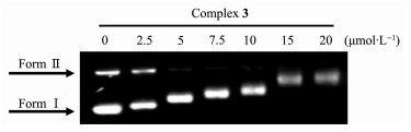
The effect of the binding of complex 3 on plasmid DNA was also investigated by DNA electrophoresis studies. Closed circular DNA can reduce its superh-elical density and decrease the DNA migration rate in agarose by binding of unwinding agents[27]. Positive charged metal complexes could bind to the negative charge part of DNA[27-28]. Complex 3 induced significant changes in the migration of DNA (Form Ⅰ) at 2.5 μmol ·L-1 (Fig. 4), suggesting that complex 3 could unwind the DNA superhelix.
Figure 4
 Figure 4. Gel electrophoresis assay of pBR322 DNA treated with complex 3 with variable concentrations
Comparing complexes 1~3 with other dinuclear [Ru2(η6-p-cymene)2(1, 3-bib)2Cl2]X2 complexes (X=Cl-, I-, NO3-, BF4-, PF6-, CF3SO3-)[8], even all complexes share the similar bowl-like structure, the best cytotoxi-city to A549 cancer cell among complexes 1~3 (13.9 μmol·L-1) is much better than that of Ru-η6-p-cymene complexes (30.9 μmol·L-1)[8]. In addition, even all complexes could interact with DNA, the binding capacity of complexes 1~3 (~104 L·mol-1) is much weaker than that of Ru-η6-p-cymene complexes (~105 L·mol-1). The differences may result from the effect of distinct arene groups (η6-p-cymene vs biphenyl).
3. Conclusions
In summary, three Ru(Ⅱ)-arene complexes contain-ing the rigid imidazole ligand with distinct counter anions were successfully synthesized and characterized. The single crystal diffraction displays that one counter anion Cl- could locate in the cavity of dinuclear structure of complex 1. The cytotoxicity assay indicates that complex 3 exhibits the best cytotoxicity toward to A549 cell line, which is comparable to that of cisplatin. All three complexes show strong interactions with CT-DNA and could unwind the DNA superhelix. The different anticancer behaviors of 1~3 suggest that the coordinative and counter anions and the arene groups in the metal-arene complexes might play very important roles to adjust their anticancer capacities. Further studies will be continued and focused on the mechanism of the anticancer activity of these Ru(Ⅱ)-arene complexes.
Supporting information is available at http://www.wjhxxb.cn
-
-
[1]
Wang X, Wang X, Jin S, et al. Chem. Rev., 2019, 119(2):1138-1192
-
[2]
Bergamo A, Dyson P J, Sava G. Coord. Chem. Rev., 2018, 360:17-33 doi: 10.1016/j.ccr.2018.01.009
-
[3]
Johnstone T C, Suntharalingam K, Lippard S J. Chem. Rev., 2016, 116(5):3436-3486 doi: 10.1021/acs.chemrev.5b00597
-
[4]
Zeng L, Gupta P, Chen Y, et al. Chem. Soc. Rev., 2017, 46(19):5771-5804 doi: 10.1039/C7CS00195A
-
[5]
Liu H K, Kostrhunova H, Habtemariam A, et al. Dalton Trans., 2016, 45(46):18676-18688 doi: 10.1039/C6DT03356C
-
[6]
Fu Y, Romero M J, Salassa L, et al. Angew. Chem. Int. Ed., 2016, 55(31):8909-8912 doi: 10.1002/anie.201602995
-
[7]
Davey G E, Adhireksan Z, Ma Z, et al. Nat. Commun., 2017, 8:1575 doi: 10.1038/s41467-017-01680-4
-
[8]
Wu Q, Liu L Y, Li S, et al. J. Inorg. Biochem., 2018, 189:30-39 doi: 10.1016/j.jinorgbio.2018.08.013
-
[9]
Coverdale J P C, Bridgewater H E, Song J I, et al. J. Med. Chem., 2018, 61(20):9246-9255 doi: 10.1021/acs.jmedchem.8b00958
-
[10]
Liu H K, Sadler P J. Acc. Chem. Res., 2011, 44(5):349-359 doi: 10.1021/ar100140e
-
[11]
Peacock A F A, Habtemariam A, Fernandez R, et al. J. Am. Chem. Soc., 2006, 128(5):1739-1748 doi: 10.1021/ja055886r
-
[12]
Tronnier A, Strassner T. Dalton Trans., 2013, 42(27):9847-9851 doi: 10.1039/c3dt50841b
-
[13]
张复兴, 何唐锋, 姚淑芬, 等.无机化学学报, 2019, 35(4):598-604ZHANG Fu-Xing, HE Tang-Feng, YAO Shu-Fen, et al. Chinese J. Inorg. Chem., 2019, 35(4):598-604
-
[14]
Sheldrick G M. Acta Crystallogr. Sect. A:Found. Crystallogr., 2015, A71:3-8 https://www.ncbi.nlm.nih.gov/pubmed/25537383
-
[15]
Guan R, Chen Y, Zeng L, et al. Chem. Sci., 2018, 9(23):5183-5190 doi: 10.1039/C8SC01142G
-
[16]
高安丽, 熊庆丰, 姜婧, 等.无机化学学报, 2018, 34(9):1649-1654 http://www.cnki.com.cn/Article/CJFDTotal-WJHX201809008.htmGAO Aan-Li, XIONG Qing-Feng, JIANG Jing, et al. Chinese J. Inorg. Chem., 2018, 34(9):1649-1654 http://www.cnki.com.cn/Article/CJFDTotal-WJHX201809008.htm
-
[17]
Kumar C V, Barton J K, Turro N J. J. Am. Chem. Soc., 1985, 107(19):5518-5523 doi: 10.1021/ja00305a032
-
[18]
莫慧雯, 刘雅娴, 蔡戴宏, 等.无机化学学报, 2019, 35(3):477-484 http://www.cnki.com.cn/Article/CJFDTotal-WJHX201903013.htmMO Hui-Wen, LIU Ya-Xian, CAI Dai-Hong, et al. Chinese J. Inorg. Chem., 2019, 35(3):477-484 http://www.cnki.com.cn/Article/CJFDTotal-WJHX201903013.htm
-
[19]
Wang H Y, Qian Y, Wang F X, et al. Eur. J. Inorg. Chem., 2017(12):1792-1799
-
[20]
Li J, Zhang P, Xu Y, et al. Dalton Trans., 2017, 46(46):16205-16215 doi: 10.1039/C7DT03374E
-
[21]
Clavel C M, Paunescu E, Nowak-Sliwinska P, et al. Chem. Sci., 2014, 5(3):1097-1101 doi: 10.1039/c3sc53185f
-
[22]
Murray B S, Menin L, Scopelliti R, et al. Chem. Sci., 2014, 5(6):2536-2545 doi: 10.1039/c4sc00116h
-
[23]
Jeyalakshmi K, Haribabu J, Balachandran C, et al. Organo-metallics, 2019, 38(4):753-770 doi: 10.1021/acs.organomet.8b00702
-
[24]
Zhou W, Wang X, Hu M, et al. Chem. Sci., 2014, 5(7):2761-2770 doi: 10.1039/C4SC00384E
-
[25]
Li J, Tian M, Tian Z, et al. Inorg. Chem., 2018, 57(4):1705-1716 doi: 10.1021/acs.inorgchem.7b01959
-
[26]
Karidi K, Garoufis A, Hadjiliadis N, et al. Dalton Trans., 2005(4):728-734 doi: 10.1039/b410402a
-
[27]
Zhu Z, Wang X, Li T, et al. Angew. Chem. Int. Ed., 2014, 53(48):13225-13228 doi: 10.1002/anie.201407406
-
[28]
Keck M V, Lippard S J. Tetrahedron Lett., 1993, 34(9):1415-1416 doi: 10.1016/S0040-4039(00)60306-4
-
[1]
-
Figure 1 (a) X-ray structure of complex 1, where one Cl- anion was enveloped as a guest molecular, and the other Cl-, partial hydrogen atoms, and the free water molecules are omitted for clarity; (b) Side-viewing of bowl-like complex 1 in space filling mode; (c) π-π stacking interactions between two parallel phenyl rings of two adjacent units, with a "center-to-center" distance of 0.346 and 0.378 nm, respectively
Figure 2 Absorption spectra of complex 3 in PBS buffer at 25 ℃ on addition of CT-DNA
Arrow in the picture shows the absorbance change by the increasing of CT-DNA concentration; Inset: plot of εa versus cDNA for the titration of DNA with Ru(Ⅱ) complexes
Figure 3 CD spectra of CT-DNA bound by 3 with ccomplex/cDNA ratio ranging from 0 to 0.12
Original DNA concentration: 0.1 mmol·L-1
Figure 4 Gel electrophoresis assay of pBR322 DNA treated with complex 3 with variable concentrations
Table 1. Crystallographic data of 1
Formula Ru2C48H44Cl4N8O2 Formula weight 1 108.85 Crystal system Monoclinic Space group P21/c a / nm 1.383 13(19) b / nm 0.925 39(13) c / nm 3.478 1(5) β/(°) 94.371(2) Z 4 Dc/ (g·cm-3) 1.659 F(000) 2 240 θ range / (°) 1.2-28.1 Limiting indices -17≤h≤17, -12≤k≤12, -45≤l≤45 Reflection collected, unique 35 046, 10 018 Rint 0.061 Completeness to θ=25.00° / % 92.70 Data, restraint, parameter 10 018, 0, 583 Goodness-of-fit on F2 1.043 Final R indices [I>2σ(I)]* R1=0.048 0, 6wR2=0.102 6 *R1=∑||Fo|-|Fc||/∑|Fo|; wR2=|∑w(|Fo|2-|Fc|2)/∑|w(Fo)2|1/2, where w=1/[σ2(Fo2)+(aP)2+bP], P=(Fo2+2Fc2)/3.  下载: 导出CSV
Table 2. Selected bond lengths (nm) and angles (°) for 1
Ru1-N1 0.210 3(3) Ru1-Cl1 0.240 9(1) Ru2-N5 0.209 5(3) Ru1 -N8 0.210 1(3) Ru2-N4 0.211 1(4) Ru2-Cl2 0.240 0(1) N1-Ru1-Cl1 86.47 (8) N1 -Ru1 -N8 89.44 (12) N5-Ru2-Cl2 86.95 (9) N8-Ru1-Cl1 85.06 (10) N4-Ru2-Cl2 88.17 (9) N4-Ru2-N5 87.28(13)
下载: 导出CSV
Table 3. IC50 values of complexes 1~3 as obtained from MTT assay
μmol·L-1 Compound HeLa A549 LO2 1 25.6±1.7 45.4±2.3 28.5±0.8 2 21.9±2.1 28.9±1.4 13.6±0.8 3 15.2±1.1 13.9±1.1 13.0±0.5 Cisplatin 7.9±0.4 15.2±0.5 1.5±0.1
下载: 导出CSV
-


 扫一扫看文章
计量
- PDF下载量: 5
- 文章访问数: 819
- HTML全文浏览量: 44
