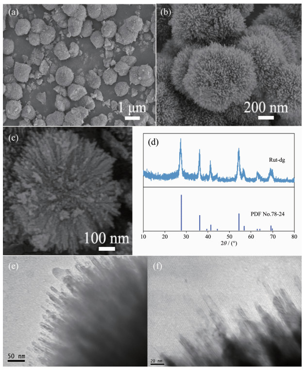
Figure 1.
(a~c) SEM images (d), XRD pattern and (e~f) TEM images of Rut-dg samples
Preparation of Coral-like Rutile Titania with Enhanced Photocatalytic Activity under UV and Visible Light
Jie ZHU , Feng-Juan GE , Yan CHEN , Yan XU , Xue-Yang ZHANG , Wei-Xin ZOU , Lin DONG

With the development of modern industry, the depletion of fossil fuels and the environmental pollu-tions have become serious problems. The development of renewable and clean energies become one of the most emergent research subjects[1-2]. Conversion of solar energy into hydrogen (H2) is considered as a perspective approach to solve these problems. Water splitting is an eco-friendly process that can be operated at ambient temperature and pressure[3-6]. Since 1972, Fujishima and Honda has reported photoassisted electrochemical water splitting on single-crystal titania and platinum electrodes[7], up to now, a large number of photocatalytic materials, with the enhanced light harvesting, photo-generated charge separation and transport, are designed for the photocatalytic hydrogen production, including binary metal oxides[8-14], complex metal oxides[15-18], metal sulfides[19-21] and metal-free materials[22-26].
TiO2 has been widely used as a good photocatalyst in H2 production from water splitting, due to its advantages of being cheap, stable, nontoxic and environmentally friendly. Generally, TiO2 has four polymorphs, including anatase, rutile, brookite and TiO2(B). These four polymorphs are used in different fields on the basis of their different physical and chemical properties, but the most frequently studied TiO2 based photocatalysts are anatase and rutile. Anatase, as it has higher surface area, more oxygen vacancies and lower conduction band (CB) potential, usually has better photocatalytic activity. However, anatase has poor visible light adsorption, which limits its application. It is reported that rutile has narrower band gap and faster charge carrier mobile, which make it a good candidate for photocatalytic reactions. There are still some shortcomings for rutile TiO2, such as lower surface area, less oxygen vacancies and faster e--h+ recombination[27]. Therefore, there are very few literatures that using pure rutile TiO2 as photocatalysts, especially in hydrogen production field. Even for those anatase-rutile combined catalysts, the catalysts with best activity usually compose only a small part of rutile[28-32]. Particularly, the commercial catalyst P25 (Degussa) is composed of about 71% anatase and 29% rutile. On the other hand, the morphology of TiO2 is an important factor for its photocatalytic activities. Some researchers prepared coral-like TiO2 with anatase phase or anatase-rutile mixed phases which have higher discoloration rate of organic dye solution[33-34]. However, the hydrogen generation by water splitting was seldom reported. Herein, we prepared coral-like rutile TiO2 in diethylene glycol solution by solvothermal method. The sample has high surface area and outstanding photocatalytic activities on H2 production from water splitting.
Titanium (Ⅳ) butoxide (TBOT, >99.0%), diethylene glycol (DG, >99.0%), chloroplatinic acid hexahydrate (H2PtCl6·6H2O, AR), triethanolamine (TEOA, 98%) and commercial rutile TiO2 (Rut-c for short, 99.8%) were purchased from Aladdin. Hydrochloric acid (HCl, 36%~38% (w/w)) was purchased from Shanghai Lingfeng. P25 was purchased from Degussa Corporation.
The coral-like rutile TiO2 was prepared by solvo-thermal method in reference[35] with some modification. In a typical synthesis, 2 mL TBOT was dissolved in 30 mL DG. After ultrasonication for 30 min, 10 mL HCl (5 mol·L-1) was added dropwise. The mixed solution was ultrasonicated for further 30 min before transferred into a 50 mL Teflon-lined stainless-steel autoclave. The autoclave was heated to 150 ℃ and maintained for 24 h. Then the autoclave was cooled down naturally to ambient temperature. The sample was centrifuged, washed several times by deionized water (DI) and ethanol and dried at 80 ℃ overnight. The sample was denoted as Rut-dg. As comparison, the DG solution was replaced by ethanol or ethylene glycol, and the resulted samples were denoted as Rut-et and Rut-eg.
Scanning electron microscopy (SEM) experiment was performed on a Philips XL30 electron microscope operated at beam energy of 10.0 kV. Transmission electron microscopy (TEM) images were taken on a JEM-2100 instrument at an acceleration voltage of 200 kV. The crystal structures were identified by X-ray diffraction (XRD) with Philips X′Pert Pro diffracto-meter by Ni-filtered Cu Kα radiation (λ=0.154 18 nm). The X-ray tube was operated at 40 kV and 40 mA. The scanning range was 10°~80°. Brunauer-Emmet-Teller (BET) surface area was measured by nitrogen adsorption-desorption at 77 K on Micrometrics ASAP-2020 adsorption apparatus. The FT-IR spectra were collected from 400 to 4 000 cm-1 at the spectral resolution of 4 cm-1 on Nicolet 5700 FT-IR spectro-meter. UV-Vis diffuse reflectance spectroscopy (UV-Vis DRS) were recorded in the range of 200~800 nm by a Shimadzu UV-2401 spectrophotometer with the reference of BaSO4.
The photocatalytic hydrogen evolution reaction was carried out in a top-irradiation vessel connected to a gas-closed glass system under an irradiation of 300 W Xe lamp with or without the 420 nm cut-off filter, to test the activity under visible or UV light. 50 mg catalyst was dispersed in 100 mL aqueous solution (90 mL DI water and 10 mL TEOA. H2PtCl6 aqueous solution was added which was in-situ reduced to Pt during the reaction (3%(w/w) Pt). The temperature of solution was kept around 6 ℃. The reaction system was sealed and evacuated for 30 min before irradiation. The amount of generated hydrogen was determined by a gas chromatograph.
The morphology of Rut-dg was investigated by SEM. Fig. 1(a, b) showed the spherical particles with uniform diameters at about 800~1 000 nm with coral-like surface. The intersecting surface of the broken spheres (Fig. 1c) exhibited the radical pore passages in it, and the branches could be estimated to have a length of ~200 nm and a diameter of ~10 nm.
The phase structure of the Rut-dg was chara-cterized by XRD (Fig. 1d). The sample showed typical diffraction peaks at about 27.40°, 35.84°, 41.22° and 54.28°. Compared with PDF No.78-2485, the peaks correspond with the (110), (101), (111) and (211) planes of rutile TiO2, respectively. Meanwhile, no peaks of anatase TiO2 was observed, indicating that pure rutile structure was prepared by this method. According to Scherrer equation D=Kγ/(Bcosθ), the mean particle size calculated though (110) plane diffraction peak was about 7.35 nm, where K is the Scherrer constant 0.89, B is the full width at half maximum (FWHM) at 2θ=27.4°, λ is the wavelength of X ray (0.154 18 nm). Combined with the SEM result, this size should be the diameter of the coral branches.
The detailed structure of the coral branches was examined by TEM in Fig. 1(e, f). It can be clearly seen that the diameters of the branches were about 7~10 nm, which is consistent with the SEM and XRD results.
As comparison, the XRD and SEM of Rut-et and Rut-eg were shown in Fig.S1. The samples were still rutile phase, while "coral branch" structures disapp-eared. The possible reason may be the higher viscosity of DG compared with ethanol and EG. The ion diffusion and nucleation process are hindered in DG solution, which constrains the isotropic growth of the crystal, leading to the branch structures.
The N2 adsorption/desorption isotherm of Rut-dg exhibited a type Ⅳ isotherm with a H2 hysteresis loop, demonstrating the mesoporous structures (Fig.S2). The average pore size calculated by BJH method was about 10 nm. The BET surface area of Rut-dg was 228 m2·g-1, which was more than 7 times of Rut-c (32 m2·g-1), indicating the branch structures enhanced the surface area dramatically.
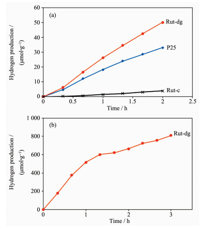
Fig. 2 shows the hydrogen production of Rut-dg, Rut-c and P25 samples under UV (Fig. 2a) or visible light irradiation (Fig. 2b), with an aqueous solution containing 10%(V/V) TEOA as a scavenger and 3%(w/w) Pt as a cocatalyst. It can be seen that under UV irradiation, Rut-dg had much higher hydrogen produc-tion (about 25 000 μmol·g-1·h-1 after 2 h irradiation), which was about 50% higher than commercial P25 and 13 times of Rut-c. Under visible light, only Rut-dg had an obvious photocatalytic activity about 270 μmol·g-1·h-1, while no obvious activities for Rut-c and P25 samples could be detected.
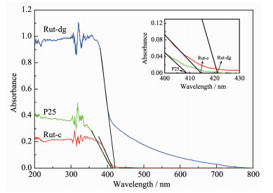
The optical absorption properties of different samples were investigated by UV-Vis DRS (Fig. 3). It can be seen that P25 and Rut-c had no obvious adsorption under visible light (>420 nm). For Rut-dg, the tangent line intersected the wavelength axis at about 421 nm, the values for Rut-c and P25 were 415 and 409 nm, respectively. The maximum absorption wavelength of Rut-dg had only slight redshift. However, it should be noted that the adsorption intensity of Rut-dg decreased slowly with the increase of wavelength and still maintained at about 20%~40% of the highest value under visible light range (420~500 nm). Thus, Rut-dg has considerable activity under visible irradiation.
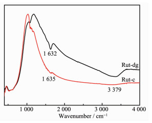
Organic compounds, such as alcohols (e.g., methanol, ethanol, isopropanol, etc.) and organic acids (formic acid, acetic acid, etc.), could be used as electron donors in photocatalytic process and promote the hydrogen evolution substantially. FT-IR spectra of Rut-dg and Rut-c were recorded to determine whether existed organics on the surface of the catalysts. As shown in Fig. 4, the peaks at ~1 632 cm-1 were commonly attributed to the stretching vibrations of C=C bonds or bending vibrations of H-O-H bonds. As no character-istic peaks of C-C, C-H or C-O bonds could be observed in the infrared spectrum, the peaks at 1 632 cm-1 should be attributed to absorbed water on the surface of samples. The peaks at 3 379 cm-1 should be O-H bending vibration of absorbed water. The FT-IR result showed that no detectable organics existed on the surface of the Rut-dg, which means that the better photocatalytic activity compared with Rut-c should be attributed to the special morphology of Rut-dg sample. It should be noted that the peaks intensity of Rut-dg was much higher than Rut-c. The possible reason should be Rut-dg has coral-like morphology and hydrophilic surface with a large number of hydroxyls on the surface, which leads to capillary condensation, thus more water molecules are absorbed on the surface of Rut-dg.
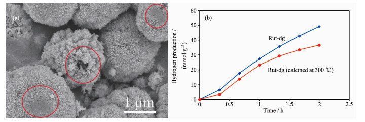
The Rut-dg was calcined at 300 ℃ for 1 h before photocatalytic test under UV irradiation. Obvious sintering of the branch structures could be observed in Fig. 5a, meanwhile, the BET surface area decreased sharply to 109 m2·g-1. From the N2 adsorption-desorption isotherm, it could be clearly seen that the isotherm exhibited a type Ⅱ isotherm, indicating the collapse of the mesoporous structures (Fig.S2). Accordingly the hydrogen production decreased by 15%~25% (Fig. 5b), further proving that the coral-like structure is the key factor for high activity.
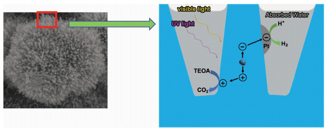
The coral-like structure can increase the hydrogen production owing to the following potential reasons (Fig. 6):
(1) As discussed in SEM and BET sections, Rut-dg has coral-like structure with high surface area. Compared with Rut-c, the high surface area equates to more surface-active sites in unit volume. The surface-active sites are required for the adsorption of H2O, which is a key step in photoreactivity between TiO2 and water[36]. Meanwhile, the coral structures also facilitate the adsorption of water, as discussed above.
(2) Rut-dg coral-like surface with branches diameter was less than 10 nm. According to the literature, when the grain size of titania decreases below 15 nm, the photocatalytic performance will increase abruptly because of quantum size effect and faster charge transfer[37].
(3) The higher surface area of Rut-dg makes more water adsorption and photogenerated carriers excited than Rut-c under same irradiation. Furthermore, the coral structures reduce the transfer distance of photon-generated carriers dramatically and so facilitate the oriented movement and efficient separation of electrons and holes, increase the probability of their reaching the surface without e--h+ recombination and enhance the activity remarkably[38].
In summary, the coral-like rutile TiO2 was synthesized by solvothermal method in DG solution. The coral branch structure increased the surface area of the sample substantially and facilitated the e--h+ separation, which lead to its superior performance on photocatalytic water splitting under both UV and visible light.
Acknowledgements: The financial support of the Key Research Project of Social Development of Xuzhou (Grant No.KC17154) is gratefully acknowledged.
Supporting information is available at http://www.wjhxxb.cn
Chu S, Majumdar A. Nature, 2012, 488:294-303 doi: 10.1038/nature11475
Xiang Q J, Yu J G, Jaroniec M. Chem. Soc. Rev., 2013, 41:782-796 https://www.ncbi.nlm.nih.gov/pubmed/21853184
Maeda K, Domen K. J. Phys. Chem. Lett., 2010, 1:2655-2661 doi: 10.1021/jz1007966
Hisatomi T, Kubota J, Domen K. Chem. Soc. Rev., 2014, 43:7520-7535 doi: 10.1039/C3CS60378D
Bowker M. Catal. Lett., 2012, 142:923-929 doi: 10.1007/s10562-012-0875-4
Chen X B, Shen S H, Guo L J, et al. Chem. Rev., 2010, 110:6503-6570 doi: 10.1021/cr1001645
Fujishima A, Honda K. Nature, 1972, 238:37-38 doi: 10.1038/238037a0
Guayaquil-Sosa J F, Serrano-Rosales B, Valades-Pelayo P J, et al. Appl. Catal. B, 2017, 211:337-348 doi: 10.1016/j.apcatb.2017.04.029
Wu M H, Zhang M, Lv T, et al. Appl. Catal. A, 2017, 547:96-104 doi: 10.1016/j.apcata.2017.08.027
Xiao Y, Yu X, Gao Y, et al. Catal. Commun., 2017, 102:1-4 doi: 10.1016/j.catcom.2017.07.017
Sui Y L, Liu S B, Li T F, et al. J. Catal., 2017, 353:250-255 doi: 10.1016/j.jcat.2017.07.024
Irfan R M, Jiang D C, Sun Z J, et al. J. Catal., 2017, 353:274-285 doi: 10.1016/j.jcat.2017.06.010
Imran M, Yousaf A B, Kasak P, et al. J. Catal., 2017, 353:81-88 doi: 10.1016/j.jcat.2017.06.019
Bharatvaj J, Preethi V, Kamnani S. Int. J. Hydrogen Energy, 2018, 43:3935-3945 doi: 10.1016/j.ijhydene.2017.12.069
Cui E T, Meng Q Q, Ge C Y, et al. Catal. Commun., 2018, 103:29-33 doi: 10.1016/j.catcom.2017.09.018
Chava R K, Do J Y, Kang M. J. Alloys Compd., 2017, 727:86-93 doi: 10.1016/j.jallcom.2017.08.108
Yu K, Zhang C X, Chang Y, et al. Appl. Catal. B, 2017, 200:514-520 doi: 10.1016/j.apcatb.2016.07.049
Qu Z P, Rong Y, Tang L, et al. Mol. Catal., 2017, 441:10-20 doi: 10.1016/j.mcat.2017.08.001
Sadovnikov S I, Kozlova E A, Gerasimov E Y, et al. Catal. Commun., 2017, 100:178-182 doi: 10.1016/j.catcom.2017.07.004
Lee W P C, Gui M M, Tan L L, et al. Catal. Commun., 2017, 98:66-70 doi: 10.1016/j.catcom.2017.05.004
Kumar D P, Park H, Kim E H, et al. Appl. Catal. B, 2018, 224:230-238 doi: 10.1016/j.apcatb.2017.10.051
Ding N, Zhang L S, Zhang H Y, et al. Catal. Commun., 2017, 100:173-177 doi: 10.1016/j.catcom.2017.06.050
Gao J T, Wang Y, Zhou S J, et al. ChemCatChem, 2017, 9:1708-1715 doi: 10.1002/cctc.201700492
Zhu Y P, Ren T Z, Yuan Z Y. ACS Appl. Mater. Interfaces, 2015, 7:16850-16856 doi: 10.1021/acsami.5b04947
Han Q, Wang B, Gao J, et al. ACS Nano, 2016, 10:2745-2751 doi: 10.1021/acsnano.5b07831
Zhang J Y, Wang Y H, Jin J, et al. ACS Appl. Mater. Interfaces, 2013, 5:10317-10324 doi: 10.1021/am403327g
Ma Y, Wang X L, Jia Y S, et al. Chem. Rev., 2014, 114:9987-10043 doi: 10.1021/cr500008u
Zhang J, Xu Q, Feng Z C, et al. Angew. Chem. Int. Ed., 2008, 47:1766-1769 doi: 10.1002/anie.200704788
Liu Y X, Wang Z L, Huang W X. Appl. Surf. Sci., 2016, 389:760-767 doi: 10.1016/j.apsusc.2016.07.173
Liu Y X, Wang Z L, Wang W D, et al. J. Catal., 2014, 310:16-23 doi: 10.1016/j.jcat.2013.03.024
Wahab A K, Ould-Chikh S, Meyer K, et al. J. Catal., 2017, 352:657-671 doi: 10.1016/j.jcat.2017.04.033
Wang P F, Zhou Q X, Xia Y G, et al. Appl. Catal. B, 2018, 225:433-444 doi: 10.1016/j.apcatb.2017.11.069
Shahi S K, Kaur N, Sandhu S, et al. J. Sci.:Adv. Mater. Devices, 2017, 2:347-353 doi: 10.1016/j.jsamd.2017.07.006
Hua X S, Zhang Y J, Ma N H, et al. J. Hazard. Mater., 2009, 172:256-261 doi: 10.1016/j.jhazmat.2009.06.166
Wang D D, Shan Z Q, Na R, et al. J. Power Sources, 2017, 337:11-17 doi: 10.1016/j.jpowsour.2016.10.115
Nowotny J, Bak T, Nowotny M, et al. J. Phys. Chem. B, 2006, 110:18492-18495 doi: 10.1021/jp063699p
Ren H J, Koshy P, Chen W F, et al. J. Hazard. Mater., 2017, 325:340-366 doi: 10.1016/j.jhazmat.2016.08.072
Chen X Y, Yu T, Fan X X, et al. Appl. Surf. Sci., 2007, 253:8500-8506 doi: 10.1016/j.apsusc.2007.04.035

Figure 2 H2 production of (a) Rut-dg, P25 and Rut-c samples under UV irradiation and (b) Rut-dg sample under visible light

Figure 5 (a) SEM image of Rut-dg calcined at 300 ℃ and (b) hydrogen production under UV irradiation of Rut-dg samples before and after calcination

 扫一扫看文章

扫一扫关注我们
 下载:
下载:

 下载:
下载:


