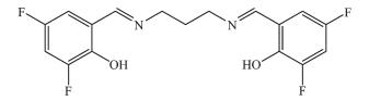
图 Scheme 1
Structure of the Schiff base H2L
Scheme 1.
Structure of the Schiff base H2L

Syntheses, Crystal Structures and Urease Inhibition of Two Manganese(Ⅲ) Complexes with Bis-Schiff Bases
Yun-Tong LI , Jing-Wen DONG , Yao LU , Yi-Tong GU , Chao-Nan SHANG , Fu-Yao LIU , Yu XIN , Chang-Ling JING , Zhong-Lu YOU


Urease (EC 3.5.1.5; urea amidohydrolase) is a binuclear nickel-dependent hydrolase enzyme, which can be synthesized by numerous organisms, including plants, bacteria, algae, fungi, and invertebrates, and occurs widely in animal and soil[1-2]. Urease enzyme catalyzes the decomposition of urea into ammonia and carbon dioxide in high efficiency[3], with the rate of catalyzed reaction 1014 times higher than the non-catalyzed reaction[4]. The enzyme possesses harmful effects on both human health and fertile soil. Bacterial urease is a virulent factor including the formation of infection stones, pyelonephritis, peptic ulceration, hepatic encephalopathy, and other diseases[5-6]. High urease activity in soil leads to increase ammonia toxicity in air and economic problems[7-8]. All these negative effects oblige us to explore effective urease inhibitors. And, in fact, urease inhibitors are now considered as the first line of treatment for infections caused by urease-producing microorganisms. Our research group has pioneered the study of Schiff base and their complexes as urease inhibitors[9-13]. Although, a variety of urease inhibitors have been investigated in the past, such as quinolones[14], phosphoric triamides[15], and thioureas[16]. Most of these compounds are banned from using in vivo because of their toxicity, instability, or severe side effects. So, it is crucial to synthesize new potent urease inhibitors with good stability, bioavailability, and low toxicity. Schiff bases have been reported as effective antibacterial agents[17-18], and show interesting urease inhibitory activity[19]. Metal complexes have been proved to possess significant inhibitory activities on various enzymes[20-21]. As a part of our ongoing research on urease inhibition with metal complexes, two manganese(Ⅲ) complexes were prepared and screened for their urease inhibition activity. The mechanism of urease inhibition was studied by using docking techniques.
Starting materials, reagents and solvents were purchased from commercial suppliers with AR grade, and used without purification. Elemental analyses were performed on a Perkin-Elmer 240C elemental analyzer. IR spectra were recorded on a Jasco FT/IR-4000 spectrometer as KBr pellets in the 4 000~200 cm-1 region. 1H NMR spectrum for H2L was recorded on a Bruker spectrometer at 500 MHz. X-ray diffraction was carried out on a Bruker SMART 1000 CCD diffractometer.
3, 5-Difluorosalicylaldehyde (3.16 g, 0.02 mol) and propane-1, 3-diamine (0.74 g, 0.01 mol) were reacted in 50 mL methanol. The mixture was stirred at room temperature for 30 min to give a clear yellow solution. The solution was evaporated by distillation to give yellow solid product, which was recrystallized from ethanol to give H2L. Yield 87%. Anal. Calcd. for C17H14F4N2O2(%): C, 57.6; H, 4.0; N, 7.9. Found(%): C, 57.4; H, 3.9; N, 8.0. IR data (cm-1): 1 637 (s, C=N), 1 487 (s), 1 402 (w), 1 266 (m), 1 123 (w). 1H NMR (500 MHz, CDCl3): δ 13.45 (s, 2H, OH), 8.35 (s, 2H, CH=N), 6.96 (s, 2H, ArH), 6.80 (s, 2H, ArH), 3.78 (t, 4H, CH2), 2.18 (m, 2H, CH2).
A methanolic solution (30 mL) of manganese acetate tetrahydrate (0.25 g, 1.0 mmol) was added to a methanolic solution (30 mL) of H2L (0.35 g, 1.0 mmol) and ammonium thiocyanate (0.076 g, 1.0 mmol) for 1 or sodium azide (0.065 g, 1.0 mmol) for 2 with stirring. The mixture was stirred at room temperature for 30 min to give a brown solution. The resulting solution was allowed to stand in air for a few days until three quarter of the solvent was evaporated. Brown block-shaped crystals of the complex, suitable for X-ray single crystal diffraction were formed at the bottom of the vessel. The crystals were isolated by filtration, washed three times with cold methanol and dried in air.
Complex 1: Yield: 37%. Anal. Calcd. for C18H14F4 MnN3O3S(%): C, 44.7; H, 2.9; N, 8.7. Found(%): C, 44.9; H, 3.0; N, 8.6. IR data (cm-1): 2 054 (s, NCS), 1 615 (s, C=N), 1 564 (m), 1 458 (s), 1 348 (m), 1 295 (m), 1 273 (m), 1 127 (m), 995 (w), 828 (m), 645 (w), 550 (w), 440 (w).
Complex 2: Yield: 45%. Anal. Calcd. for C17H12F4 MnN5O2(%): C, 45.5; H, 2.7; N, 15.6. Found(%): C, 45.4; H, 2.7; N, 15.8. IR data (cm-1): 2 035 (s, N3), 1 615 (s, C=N), 1 564 (m), 1 463 (s), 1 355 (m), 1 292 (m), 1 267 (m), 1 131 (m), 1 058 (w), 995 (w), 828 (s), 746 (w), 640 (w), 540 (w), 435 (w).
Diffraction intensities for the complexes were collected at 298(2) K using a Bruker SMART 1000 CCD area-detector diffractometer with Mo Kα radiation (λ=0.071 073 nm). The collected data were reduced with SAINT[22], and multi-scan absorption correction was performed using SADABS[23]. Structures of the complexes were solved by direct methods and refined against F2 by full-matrix least-squares method using SHELXTL[24]. All of the non-hydrogen atoms were refined anisotropically. The hydrogen atoms attached to water ligand of complex 1 were located from a difference Fourier map and refined isotropically, with O-H and H…H distances restrained to 0.085(1) and 0.137(2) nm, respectively. The remaining hydrogen atoms were placed in calculated positions and constr-ained to ride on their parent atoms. Crystallographic data for the complexes are summarized in Table 1. Selected bond lengths and angles are given in Table 2.

 下载:
导出CSV
下载:
导出CSV
| 1 | 2 | |
| Formula | C18H14F4MnN3O3S | C17H12F4MnN5O2 |
| Formula weight | 483.3 | 449.3 |
| Crystal system | Monoclinic | Orthorhombic |
| Space group | P21/n | Pna21 |
| a / nm | 1.291 9(5) | 1.170 2(1) |
| b / nm | 0.724 92(3) | 1.293 1(2) |
| c / nm | 2.033 3(1) | 1.101 0(2) |
| β / (°) | 94.858(2) | |
| V / nm3 | 1.897 4(2) | 1.665 9(4) |
| Z | 4 | 4 |
| Dc / (g·cm-3) | 1.692 | 1.791 |
| μ(Mo Kα) / mm-1 | 7.299 | 0.862 |
| F(000) | 976 | 904 |
| Independent reflection | 3 368 | 3 031 |
| Observed reflection [I≥2σ(I)] | 2 787 | 2 407 |
| Parameter | 277 | 262 |
| Restraint | 3 | 1 |
| Final R indices [I≥2σ(I)] | R1=0.067 0, wR2=0.179 7 | R1=0.038 4, wR2=0.078 9 |
| R indices (all data) | R1=0.077 0, wR2=0.196 8 | R1=0.051 5, wR2=0.083 8 |
| GOF on F2 | 1.045 | 0.939 |
下载:
导出CSV
| 1 | |||||
| Mn1-O1 | 0.190 0(3) | Mn1-O2 | 0.189 2(3) | Mn1-N1 | 0.205(4) |
| Mn1-N2 | 0.204 0(4) | Mn1-N3 | 0.218 8(4) | Mn1-O3 | 0.228 6(3) |
| O2-Mn1-O1 | 89.79(13) | O2-Mn1-N1 | 178.82(15) | O1-Mn1-N1 | 91.28(14) |
| O2-Mn1-N2 | 88.88(14) | O1-Mn1-N2 | 170.07(14) | N1-Mn1-N2 | 89.98(15) |
| O2-Mn1-N3 | 91.12(16) | O1-Mn1-N3 | 98.12(16) | N1-Mn1-N3 | 89.20(17) |
| N2-Mn1-N3 | 91.75(16) | O2-Mn1-O3 | 90.53(13) | O1-Mn1-O3 | 86.65(13) |
| N1-Mn1-O3 | 89.06(13) | N2-Mn1-O3 | 83.52(13) | N3-Mn1-O3 | 174.96(15) |
| 2 | |||||
| Mn1-O1 | 0.187 4(3) | Mn1-O2 | 0.187 4(3) | Mn1-N1 | 0.205 8(3) |
| Mn1-N2 | 0.202 0(3) | Mn1-N3 | 0.228 5(3) | Mn1-N5A | 0.235 1(4) |
| O2-Mn1-O1 | 85.01(11) | O2-Mn1-N2 | 89.07(11) | O1-Mn1-N2 | 172.59(12) |
| O2-Mn1-N1 | 174.75(13) | O1-Mn1-N1 | 90.94(12) | N2-Mn1-N1 | 95.22(13) |
| O2-Mn1-N3 | 93.62(13) | O1-Mn1-N3 | 94.70(13) | N2-Mn1-N3 | 90.10(12) |
| N1-Mn1-N3 | 83.37(12) | O2-Mn1-N5A | 102.34(12) | O1-Mn1-N5A | 88.04(13) |
| N2-Mn1-N5A | 88.89(13) | N1-Mn1-N5A | 80.81(12) | N3-Mn1-N5A | 163.99(13) |
The measurement of urease inhibitory activity was carried out according to the literature method[25]. The assay mixture containing 75 μL of Jack bean urease and 75 μL of tested compounds with various concentrations (dissolved in DMSO) was pre-incubated for 15 min on a 96-well assay plate. Acetohydroxamic acid was used as a reference. Then 75 μL of phosphate buffer at pH 6.8 containing phenol red (0.18 mmol·L-1) and urea (400 mmol·L-1) were added and incubated at room temperature. The reaction time required for enough ammonium carbonate to form to raise the pH value of phosphate buffer from 6.8 to 7.7 was measured by micro-plate reader (560 nm) with end-point being determined by the color change of phenol-red indicator.
Molecular docking study of the complexes into the 3D X-ray structure of the Jack bean urease (entry 4UBP in the Protein Data Bank) was carried out by using the AutoDock 4.0 software as implemented through the graphical user interface AutoDockTools (ADT 1.5.2). The graphical user interface Auto Dock Tools was employed to setup the enzymes: all hydrogens were added, Gasteiger charges were calculated and nonpolar hydrogens were merged to carbon atoms. The Ni initial parameters are set as r=0.117 0 nm, q=+2.0, and van der Waals well depth of 0.419 kJ·mol-1 [26]. The 3D structure of the ligand molecule was saved in pdb format with the aid of the program Mercury. The resulting files were saved as pdbqt format.
The AutoDockTools was used to generate the docking input files. In the docking, grid box size of 70×60×60 for complex 1 points in x, y, and z directions was built, the maps were centered on the original ligand molecule (HAE) in the catalytic site of the protein. A grid spacing of 0.037 5 nm and a distances-dependent function of the dielectric constant were used for the calculation of the energetic map. 100 runs were generated by using Lamarckian genetic algorithm searches. Default settings were used with an initial population of 50 randomly placed individuals, a maximum number of 2.5×10⁶ energy evaluations, and a maximum number of 2.7×10⁴ generations. A mutation rate of 0.02 and a crossover rate of 0.8 were chosen. The results of the most favorable free energy of binding were selected as the resultant complex structures.
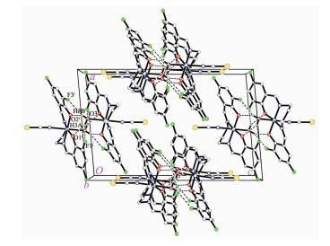
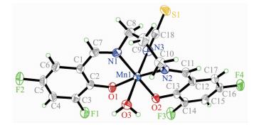
Molecular structure of complex 1 is shown in Fig. 1. The Schiff base ligand coordinates to the Mn atom through the phenolate O and imino N atoms. The Mn atom is in an octahedral coordination, with the four donor atoms of the Schiff base ligand defining the equatorial plane, and with the water O atom and thiocyanate N atom occupying the axial positions. The coordination bond lengths in the complex are comparable to those observed in manganese(Ⅲ) complexes with Schiff base ligands[27-28]. The displacement of the Mn atom from the equatorial plane towards the axial thiocyanate ligand is 0.008 8(2) nm. The dihedral angle between the two benzene rings of the Schiff base ligand is 19.2(5)°. In the crystal structure of the complex, molecules are linked through intermolecular O-H…O and O-H…F hydrogen bonds to form dimers (Fig. 2).
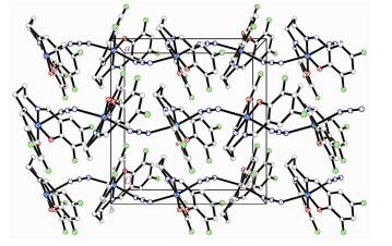
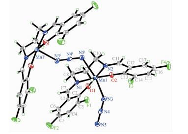
Molecular structure of complex 2 is shown in Fig. 3. The complex is an end-to-end azido-bridged poly-meric manganese(Ⅲ) compound. The Schiff base ligand coordinates to the Mn atom through the phenolate O and imino N atoms. The Mn atom is in an octahedral coordination, with the four donor atoms of the Schiff base ligand defining the equatorial plane, and with two azido N atoms occupying the axial positions. The coordination bond lengths in the complex are similar to complex 1, and also comparable to those observed in manganese(Ⅲ) complexes with Schiff base ligands[27-28]. The displacement of the Mn atom from the equatorial plane is 0.000 9(2) nm. The dihedral angle between the two benzene rings of the Schiff base ligand is 35.5(4)°. In the crystal structure of the complex, molecules are linked through end-to-end azido ligands, to form 1D chains along c-axis direction (Fig. 4).
The weak and broad absorptions in the range of 3 300~3 500 cm-1 in the spectra of H2L and complex 1 substantiate the presence of O-H groups, which are absent in complex 2. The strong absorption band at 1 637 cm-1 for H2L is assigned to the azomethine ν(C=N), which is shifted to lower wavenumber in the spectra of the complexes, 1 615 cm-1. This indicates the coordination through the two imino N atoms. The typical intense absorptions at 2 054 cm-1 for complex 1 and 2 035 cm-1 for complex 2 are assigned to the thiocyanate and azide groups, respectively.
Results of the urease inhibition are summarized in Table 4. Acetohydroxamic acid and manganese acetate were used as references. The free Schiff base and complex 2 showed poor inhibition, while complex 1 showed effective inhibition, with IC50 value of (81.0±3.7) μmol·L-1. The urease inhibition of the manganese complex is weaker than Schiff base copper(Ⅱ) comp-lexes, but higher than Schiff base zinc(Ⅱ) complexes reported previously[10, 13, 29-30].
下载:
导出CSV
| D-H…A | d(D-H) / nm | d(H…A) / nm | d(D…A) / nm | ∠D-H…A / (°) |
| O3-H3B…O2i | 0.085(1) | 0.201(4) | 0.277 9(4) | 149(6) |
| O3-H3B…F3i | 0.085(1) | 0.254(5) | 0.235(4) | 140(6) |
| O3-H3A…F1i | 0.085(1) | 0.214(2) | 0.944(5) | 156(5) |
| O3-H3A…O1i | 0.085(1) | 0.237(5) | 0.291 9(4) | 123(5) |
| Symmetry code: i 1-x, 1-y, -z. | ||||
下载:
导出CSV
| Tested material | Inhibition ratea / % | IC50 / (μmol·L-1) |
| H2L | 12.3±2.0 | — |
| Complex 1 | 52.0±3.1 | 81.0±3.7 |
| Complex 2 | 31.6±2.5 | — |
| Manganese acetate | 18.7±1.2 | — |
| Acetohydroxamic acid | 87.5±4.0 | 39.8±3.4 |
| a Concentration of the tested material is 100 μmol·L-1. | ||
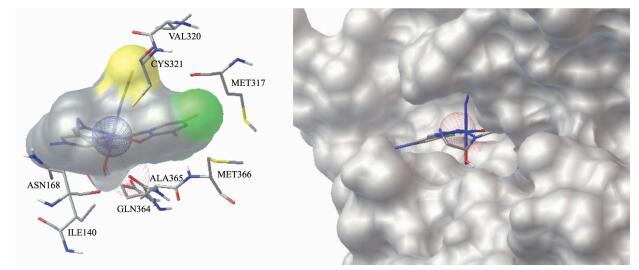
Molecular docking study was performed to investigate the binding effects between the molecule of complex 1 and the active site of the Jack bean urease. The binding model is depicted in Fig. 5. The results revealed that the molecule of complex 1 fit well with the active pocket of the urease. Additional interactions have been established in a variety of conformations because of the flexibility of the complex molecule and the amino acid residues of the enzyme. The optimized clusters (100 occurrences each) were ranked by energy level in the best conformation of inhibitor-urease modeled structures, where the docking score is -6.24. The negative value indicates that the molecule bind well with the urease. As a comparison, the docking score for the AHA inhibited model is -5.01.
The mechanism of urease inhibition was considered to be blockage of the entrance of the urease active pocket and the interaction of residues. The results endorse that complex 1 may serve as a structural template for the design and development of novel urease inhibitors.
The present study reports syntheses, characterization and crystal structures of two Schiff base manganese(Ⅲ) complexes with bis-Schiff bases. The Schiff base ligand coordinates to the Mn atoms through the phenolate oxygen and imino nitrogen. The other sites of the octahedral coordination are occupied by water, thiocyanate or azide ligands. The thiocyanato-coordinated mononuclear complex has effective inhibitory activity against Jack bean urease, while the azido-bridged polymeric complex has no activity. Considering that Schiff base manganese complexes have interesting biological activities, complex 1 described here may be used as a precursor for the design of novel urease inhibitors.
Myrach T, Zhu A T, Witte C P. J. Biol. Chem., 2017, 292:14556-14565 doi: 10.1074/jbc.M117.780403
Arshad T, Khan K M, Rasool N, et al. Bioorg. Chem., 2017, 72:21-31 doi: 10.1016/j.bioorg.2017.03.007
Mira A B, Cantarella H, Souza-Netto G J M, et al. Agric. Ecosyst. Environ., 2017, 248:105-112 doi: 10.1016/j.agee.2017.07.032
Dempsey R J, Slaton N A, Norman R J, et al. Agron. J., 2017, 109:363-377 doi: 10.2134/agronj2016.06.0374
Taha M, Ismail N H, Imran S, et al. J. Bioorg. Med. Chem., 2015, 23:7211-7218 doi: 10.1016/j.bmc.2015.10.017
Amtul Z, Siddiqui R A, Choudhary M I. Curr. Med. Chem., 2002, 9:1323-1348 doi: 10.2174/0929867023369853
Aslam M A S, Mahmood S U, Shahid M, et al. Eur. J. Med. Chem., 2011, 46:5473-5479 doi: 10.1016/j.ejmech.2011.09.009
Saeed A, Khan M S, Rafique H. Bioorg. Chem., 2014, 52:1-7 doi: 10.1016/j.bioorg.2013.10.001
Jing C L, Wang C F, Yan K, et al. Bioorg. Med. Chem., 2016, 24:270-276 doi: 10.1016/j.bmc.2015.12.013
You Z L, Liu M Y, Wang C F, et al. RSC Adv., 2016, 6:16679-16690 doi: 10.1039/C6RA00500D
Qu D, Niu F, Zhao X L, et al. Bioorg. Med. Chem., 2015, 23:1944-1949 doi: 10.1016/j.bmc.2015.03.036
You Z L, Yu H Y, Zheng B Y, et al. Inorg. Chim. Acta, 2018, 469:44-50 doi: 10.1016/j.ica.2017.09.011
Pan L, Wang C F, Yan K, et al. J. Inorg. Biochem., 2016, 159:22-28 doi: 10.1016/j.jinorgbio.2016.02.017
Abdullah M A A, Abuo-Rahma G E A A, Abdelhafez E M N, et al. Bioorg. Chem., 2017, 70:1-11 doi: 10.1016/j.bioorg.2016.11.002
Mazzei L, Cianci M, Contaldo U, et al. Biochemistry, 2017, 56:5391-5404 doi: 10.1021/acs.biochem.7b00750
Saeed A, Ur-Rehman S, Channar P A, et al. Drug Res., 2017, 67:596-605 doi: 10.1055/s-0043-113832
解庆范, 郭妙莉, 陈延民.无机化学学报, 2018, 34:309-316 doi: 10.11862/CJIC.2018.028XIE Qing-Fan, GUO Miao-Li, CHEN Yan-Min. Chinese J. Inorg. Chem., 2018, 34:309-316 doi: 10.11862/CJIC.2018.028
史娟, 李江, 葛红光.无机化学学报, 2017, 33:463-470 doi: 10.11862/CJIC.2017.050SHI Juan, LI Jiang, GE Hong-Guang. Chinese J. Inorg. Chem., 2017, 33:463-470 doi: 10.11862/CJIC.2017.050
Khan K M, Rahim F, Khan A, et al. J. Chem. Soc. Pak., 2015, 37:479-483
Qin Q P, Meng T, Tan M X, et al. Eur. J. Med. Chem., 2018, 143:1597-1603 doi: 10.1016/j.ejmech.2017.10.058
Oliveri V, Lanza V, Milardi D, et al. Mettallomics, 2017, 9:1439-1446 doi: 10.1039/C7MT00156H
SMART Ver. 5. 628 and SAINT Ver. 6. 02, Bruker AXS, Madison, Wisconsin, USA, 1998.
Sheldrick G M. SADABS, Program for Empirical Absorption Correction of Area Detector, University of Göttingen, Germany, 1996.
Sheldrick G M. Acta Crystallogr. Sect. A:Found. Crystallogr., 2008, A64:112-122
Mao W J, Lv P C, Shi L, et al. Bioorg. Med. Chem., 2009, 17:7531-7536 doi: 10.1016/j.bmc.2009.09.018
Krajewska B, Zaborska W. Bioorg. Chem., 2007, 35:355-365 doi: 10.1016/j.bioorg.2007.02.002
You Z L, Liu T, Zhang N, et al. Inorg. Chem. Commun., 2012, 19:47-50 doi: 10.1016/j.inoche.2012.01.034
Zhang N, Huang C Y, Shi D H, et al. Inorg. Chem. Commun., 2011, 14:1636-1639 doi: 10.1016/j.inoche.2011.06.027
Wang J, Qu D, Lei J X, et al. J. Coord. Chem., 2017, 70:544-555 doi: 10.1080/00958972.2016.1262538
Lu Y, Shi D H, You Z L, et al. J. Coord. Chem., 2012, 65:339-352 doi: 10.1080/00958972.2011.653785

Figure 1 Perspective view of the molecular structure of 1 with the atom labeling scheme
Thermal ellipsoids are drawn at 30% probability level

Figure 2 Molecular packing diagram of 1
Hydrogen bonds are shown as dashed lines; Symmetry codes: i 1-x, 1-y, -z

Figure 3 Perspective view of the molecular structure of 2 with the atom labeling scheme
Thermal ellipsoids are drawn at 30% probability level; Symmetry codes: i 1-x, 1-y, -z

Figure 5 Binding mode of complex 1 with the urease
Left: Only the interacting residues are displayed and the interactions are shown as dashed spheres; Right: Enzyme is shown as surface and the complex is shown as sticks
Table 1. Crystallographic and refinement parameters for the complexes
| 1 | 2 | |
| Formula | C18H14F4MnN3O3S | C17H12F4MnN5O2 |
| Formula weight | 483.3 | 449.3 |
| Crystal system | Monoclinic | Orthorhombic |
| Space group | P21/n | Pna21 |
| a / nm | 1.291 9(5) | 1.170 2(1) |
| b / nm | 0.724 92(3) | 1.293 1(2) |
| c / nm | 2.033 3(1) | 1.101 0(2) |
| β / (°) | 94.858(2) | |
| V / nm3 | 1.897 4(2) | 1.665 9(4) |
| Z | 4 | 4 |
| Dc / (g·cm-3) | 1.692 | 1.791 |
| μ(Mo Kα) / mm-1 | 7.299 | 0.862 |
| F(000) | 976 | 904 |
| Independent reflection | 3 368 | 3 031 |
| Observed reflection [I≥2σ(I)] | 2 787 | 2 407 |
| Parameter | 277 | 262 |
| Restraint | 3 | 1 |
| Final R indices [I≥2σ(I)] | R1=0.067 0, wR2=0.179 7 | R1=0.038 4, wR2=0.078 9 |
| R indices (all data) | R1=0.077 0, wR2=0.196 8 | R1=0.051 5, wR2=0.083 8 |
| GOF on F2 | 1.045 | 0.939 |
 下载: 导出CSV
Table 2. Selected bond lengths (nm) and angles (°) for the complexes
| 1 | |||||
| Mn1-O1 | 0.190 0(3) | Mn1-O2 | 0.189 2(3) | Mn1-N1 | 0.205(4) |
| Mn1-N2 | 0.204 0(4) | Mn1-N3 | 0.218 8(4) | Mn1-O3 | 0.228 6(3) |
| O2-Mn1-O1 | 89.79(13) | O2-Mn1-N1 | 178.82(15) | O1-Mn1-N1 | 91.28(14) |
| O2-Mn1-N2 | 88.88(14) | O1-Mn1-N2 | 170.07(14) | N1-Mn1-N2 | 89.98(15) |
| O2-Mn1-N3 | 91.12(16) | O1-Mn1-N3 | 98.12(16) | N1-Mn1-N3 | 89.20(17) |
| N2-Mn1-N3 | 91.75(16) | O2-Mn1-O3 | 90.53(13) | O1-Mn1-O3 | 86.65(13) |
| N1-Mn1-O3 | 89.06(13) | N2-Mn1-O3 | 83.52(13) | N3-Mn1-O3 | 174.96(15) |
| 2 | |||||
| Mn1-O1 | 0.187 4(3) | Mn1-O2 | 0.187 4(3) | Mn1-N1 | 0.205 8(3) |
| Mn1-N2 | 0.202 0(3) | Mn1-N3 | 0.228 5(3) | Mn1-N5A | 0.235 1(4) |
| O2-Mn1-O1 | 85.01(11) | O2-Mn1-N2 | 89.07(11) | O1-Mn1-N2 | 172.59(12) |
| O2-Mn1-N1 | 174.75(13) | O1-Mn1-N1 | 90.94(12) | N2-Mn1-N1 | 95.22(13) |
| O2-Mn1-N3 | 93.62(13) | O1-Mn1-N3 | 94.70(13) | N2-Mn1-N3 | 90.10(12) |
| N1-Mn1-N3 | 83.37(12) | O2-Mn1-N5A | 102.34(12) | O1-Mn1-N5A | 88.04(13) |
| N2-Mn1-N5A | 88.89(13) | N1-Mn1-N5A | 80.81(12) | N3-Mn1-N5A | 163.99(13) |
下载: 导出CSV
Table 3. Hydrogen bond parameters for complex 1
| D-H…A | d(D-H) / nm | d(H…A) / nm | d(D…A) / nm | ∠D-H…A / (°) |
| O3-H3B…O2i | 0.085(1) | 0.201(4) | 0.277 9(4) | 149(6) |
| O3-H3B…F3i | 0.085(1) | 0.254(5) | 0.235(4) | 140(6) |
| O3-H3A…F1i | 0.085(1) | 0.214(2) | 0.944(5) | 156(5) |
| O3-H3A…O1i | 0.085(1) | 0.237(5) | 0.291 9(4) | 123(5) |
| Symmetry code: i 1-x, 1-y, -z. | ||||
下载: 导出CSV
Table 4. Inhibition of urease by the tested materials
| Tested material | Inhibition ratea / % | IC50 / (μmol·L-1) |
| H2L | 12.3±2.0 | — |
| Complex 1 | 52.0±3.1 | 81.0±3.7 |
| Complex 2 | 31.6±2.5 | — |
| Manganese acetate | 18.7±1.2 | — |
| Acetohydroxamic acid | 87.5±4.0 | 39.8±3.4 |
| a Concentration of the tested material is 100 μmol·L-1. | ||
下载: 导出CSV

 扫一扫看文章


扫一扫关注我们