

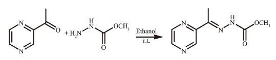
 Figure Scheme 1.
Synthesis route of HL
Figure Scheme 1.
Synthesis route of HL
Citation:
MAO Pan-Dong, HAN Xue-Feng, WU Wei-Na, WANG Guan-Jie, WANG Zhen, HOU Ying. Ni (Ⅱ) and Cd (Ⅱ) Complexes with a Schiff Base Ligand Derived from 2-Acetyl Pyrazine and Methyl Hydrazinocarboxylate: Crystal Structures and Fluorescence Properties[J]. Chinese Journal of Inorganic Chemistry,
2016, 32(1): 161-166.
doi:
10.11862/CJIC.2016.008

2-乙酰吡嗪缩肼基甲酸甲酯的镍和镉配合物的晶体结构及荧光性质
English
Ni (Ⅱ) and Cd (Ⅱ) Complexes with a Schiff Base Ligand Derived from 2-Acetyl Pyrazine and Methyl Hydrazinocarboxylate: Crystal Structures and Fluorescence Properties
Abstract:
Two complexes, namely [Ni (L)2] (1) and [Cd (HL)(CH3OH)(NO3)2] (2) (HL is derived from 2-acetyl pyrazine and methyl hydrazinocarboxylate) have been synthesized and characterized by single-crystal X-ray diffraction, elemental analysis and IR spectroscopy. X-ray diffraction analysis results show that the central Ni (Ⅱ) ion in complex 1 is surrounded by two independent anionic ligands with N2O donor set, thus possesses a distorted octahedral coordination geometry. However, in complex 2, the Cd (Ⅱ) ion with bicapped-triangular prism geometry is coordinated with one neutral ligand HL, two bidentate nitrate anions and one methanol molecule. In addition, the thermal stability and luminescent properties of the complexes are also studied in detail. CCDC: 1424257, 1; 1424258, 2.
-
Key words:
- methyl hydrazinocarboxylate
- / pyrazine
- / fluorescence
- / crystal structure
- / thermostability
-
Schiff bases as an important class of ligands play a crucial role in coordination chemistry and have been widely applied in different fields mainly due to their wide-spectrum biological applications [1-7]. In recent years, there are many reports about the acylhydrazones, thiosemicarbazones derived from actyl-pyridine/pyrazine, and their transition metal complexes because of high biological and pharmaceutical activities [3, 8-10]. By contrast, as their structurally analogous, carbazates (R-O-CO-NH-NH2) and their metal complexes have been paid much less attention [6].
Generally, Cd (Ⅱ) ion is closely related to biochemistry, clinical diagnostics as well as environ-mental pollution [11]. Furthermore, a large amount of Cd (Ⅱ) complexes have been reported for their fluore-scence properties [12-13]. Therefore, in this paper, Ni (Ⅱ) and Cd (Ⅱ) complexes with a Schiff base ligand derived from 2-acetyl pyrazine and methyl hydrazinocarbox-ylate have been synthesized and structural determined by single-crystal X-ray diffraction. In addition, the thermal stability and luminescent properties of the complexes are also investigated.
1 Experimental
1.1 Materials and measurements
Solvents and starting materials for synthesis were purchased commercially and used as received. Elemental analysis was carried out on an Elemental Vario EL analyzer. The IR spectra (ν=4 000~400 cm-1) were determined by the KBr pressed disc method on a Bruker V70 FT-IR spectrophotometer. 1H NMR spectra of L was acquired with Bruker AV400 NMR instrument in DMSO-d6 solution with TMS as internal standard. The UV spectra were recorded on a Purkinje General TU-1800 spectrophotometer. Fluorescence spectra were determined on a Varian CARY Eclipse spectrophotometer.
1.2 Preparations of the ligand, complexes 1 and 2
As shown in Scheme 1, the ligand HL was produced by condension of 2-acetyl pyrazine (1.22 g, 0.01 mol) and methyl hydrazinocarboxylate (0.90 g, 0.01 mol) in anhydrous methanol solution (30 mL) with continuous stirring at room temperature for 3 h. The white solid was filtered and washed three times by cold methanol. Yield: 1.13 g (58%). m.p. 183~185 ℃. Elemental analysis Calcd. for C8H10N4O2(%): C: 49.48; H: 5.19; N: 28.85; Found (%): C: 49.26; H: 5.34; N: 29.00. FT-IR (cm-1): ν(C=O) 1 748, ν(C=N) 1 613, ν(C=N)pyrazine 1 558. 1H NMR (400 MHz): δ 10.52(1H, s, NH), 9.09(1H)/8.55~8.58(2H) for pyrazine-H, 3.70 (3H, s, CH3), 2.24(3H, s, CH3).
Figure Scheme 1.
Synthesis route of HL
The complexes 1 and 2 were generated by reaction of the ligand HL (5 mmol) with equimolar of Ni (NO3)2 ·6H2O or Cd (NO3)2·6H2O in methanol solution (10 mL), respectively. Crystals suitable for X-ray diffraction analysis were obtained by evaporating the correspon-ding reaction solutions at room temperature.
1: Brown blocks. Anal. Calcd. for C16H18N8O4Ni (%): C: 43.18; H: 4.08; N: 25.18. Found (%): C: 43.05; H: 4.00; N: 25.33. FT-IR (cm-1): ν(N=C-O) 1 606, ν(C=N) 1 594, ν(C=N)pyrazine 1 535.
2: Colorless blocks. Anal. Calcd. for C9H14N6O9Cd (%): C: 23.36; H: 3.05; N: 18.16. Found (%): C: 23.22; H: 3.17; N: 18.27. FT-IR (cm-1): ν(O-H) 3 532, ν(C=O) 1 720, ν(C=N) 1 579, ν(C=N)pyrazine 1 523. ν1(NO3) 1 494, ν4(NO3) 1 311.
1.3 X-ray crystallography
The X-ray diffraction measurement for complexes 1 and 2 were performed on a Bruker SMART APEX Ⅱ CCD diffractometer equipped with a graphite monochromatized Mo Kα radiation (λ=0.071 073 nm) by using φ-ω scan mode. Semi-empirical absorption correction was applied to the intensity data using the SADABS program [14]. The structures were solved by direct methods and refined by full matrix least-square on F2 using the SHELXTL-97 program [15]. All non-hydrogen atoms were refined anisotropically. All the H atoms were positioned geometrically and refined using a riding model. Details of the crystal parameters, data collection and refinements for complexes 1 and 2 are summarized in Table 1.
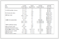 Table 1.
Crystal data and structure refinement for complexes 1 and 2
Table 1.
Crystal data and structure refinement for complexes 1 and 2
 Table 1. Crystal data and structure refinement for complexes 1 and 2
CCDC: 1424257, 1; 1424258, 2.
2 Results and discussion
2.1 Crystal structures description
Selected bond distances and angles, hydrogen bonds information for both complexes are listed in Table 2 and 3, respectively. As shown in Fig. 1a, the central Ni (Ⅱ) ion in complex 1 is surrounded by two independent anionic ligands with N2O donor set, thus possesses a distorted octahedral coordination geometry. The enolization of C=O bond of the ligand can be confirmed by the bond lengths of C-O being 0.125 3(4) and 0.124 1(4) nm, which are in excellent agreement with previously known semicarbazone complexes in the literature [6-7]. The distances of Ni-N/O bonds were in the range of 0.197 1(3)~0.209 5(2) nm, comparable with those in some reported complexes with similar donor set [3]. As expected, there exist none classic hydrogen bonds in the crystal of 1. However, certain C-H…N and C-H…O interactions are helpful to construct three dimensional net work.
Table 2.
Selected bond lengths (nm) and angles (°) in complexes 1 and 2
 Table 2. Selected bond lengths (nm) and angles (°) in complexes 1 and 2
Table 3.
Hydrogen bonds information in complexes 1 and 2
Table 2. Selected bond lengths (nm) and angles (°) in complexes 1 and 2
Table 3.
Hydrogen bonds information in complexes 1 and 2
 Table 3. Hydrogen bonds information in complexes 1 and 2
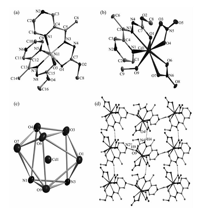 Figure 1.
Diamond drawing of 1 (a) and 2 (b) with 30% thermal ellipsoids; (c) Coordination enviorment of Cd (Ⅱ) ion in complex 2 (coordination atoms shown with 30% thermal ellipsoids); (d) Extend 2D supromolecular structure along c axis in complex 2
Figure 1.
Diamond drawing of 1 (a) and 2 (b) with 30% thermal ellipsoids; (c) Coordination enviorment of Cd (Ⅱ) ion in complex 2 (coordination atoms shown with 30% thermal ellipsoids); (d) Extend 2D supromolecular structure along c axis in complex 2
By contrast, the molar ratio of the ligand HL and metal is 1:1, and the ligand is neutral tridentate in complex 2 (Fig. 1b) with C-O bond length being 0.121 1(5) nm. The Cd (Ⅱ) ion is also coordinated with two bidentate nitrate anions and one methanol molecule, giving bicapped-triangular prism geometry (Fig. 1c). In the crystal, intermolecular O9-H9…N2iv and N4-H4…O4v (Symmetry codes: iv x-1/2, -y+1, z+1/2; v x, y-1, z) hydrogen bonds link the complexes into extended 2D supromolecular structure (Fig. 1d).
2.2 IR spectra
The ν(C=O) of the free ligand is 1 748 cm-1, it shifts to lower frequency value in complex 2, confir-ming the coordination of the carbonyl group [3]. How-ever, such absorption is disappeared in complex 1, meanwhile, new (N=C-O) stretching vibration absorp-tion is observed at 1 606 cm-1, revealing that the C=O in O=C-N moiety has enolizated and the oxygen atom coordinates to the Ni (Ⅱ) ion [6-7]. The ν(C=N) bands of the imine group and pyrazine ring in the ligand HL shift to lower frequency values in the complexes, indicating that the N atoms of both units take part in the coordination [8]. Meanwhile, the bands at 1 494 and 1 311 cm-1 in complex 2 could be assigned to the two split bands ν1 and ν4 of the coordinated nitrate group, respectively, showing that the nitrate group is bidentate [16]. It is in accordance with the crystal struc-ture study.
2.3 Thermal decomposition process of complexes 1 and 2
For detecting the thermal stabilities of complexes 1 and 2, thermal gravimetric (TG) analyses were carried out from the room temperature to 800 ℃ with the linear heating rate of 10 ℃·min-1 under argon atmosphere. Complex 1 (Fig. 2a) is thermally stable up to about 300 ℃, indicating there exist no solvent molecules in the complex. A sharp weight loss for complex 1 could be observed from 300 to 330 ℃, then gradually smooth until to about 700 ℃, corre-sponding to the decomposition of two organic ligands. However, the first stage occurs with weight loss of 3.62% below 150 ℃ for complex 2, contributing to the loss of one coordinated methanol molecule (Calcd. 6.89%). The second process of weight loss appears between 150 to 780 ℃, considered as the decomposi-tion of the ligand L and two nitrate anions. The remainders of the complexes 1 and 2 might be the metal oxides because the residue weights (11.53% and 15.08%) are agreement with the calculated values of 10.12%, and 15.72 %, respectively.
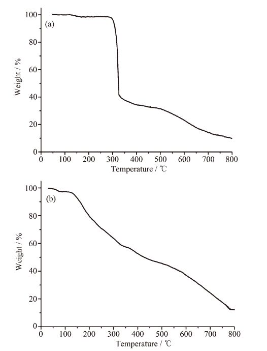 Figure 2.
TG curves for complexes 1 (a) and 2 (b)
Figure 2.
TG curves for complexes 1 (a) and 2 (b)
2.4 UV spectra
The UV spectra of HL, complexes 1 and 2 in CH3OH solution (concentration: 1×10⁻⁵ mol·L-1) were measured at room temperature (Fig. 3). The spectra of HL features only one main band located around 290 nm (ε=10 674 L·mol-1·cm-1), which could be assigned to characteristic π-π* transition of pyrazine unit [3]. Similar bands are observed at 289 nm (ε=12 422 L·mol-1·cm-1) in the complex 2. However, there are three bonds in spectra of 1 at 252 (ε=7914 L·mol-1·cm-1), 296 (ε=16 464 L·mol-1·cm-1) and 385 nm (ε=12 538 L·mol-1·cm-1). The former two could be contributed to the characteristic π-π* transition of pyrazine and imine unit, respectively [16], while the final one is probably due to the ligand-to-metal charge transfer (LMCT) [3]. This indicates that an extended conjugation is formed in anionic ligand after complexation in complex 1.
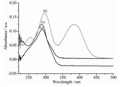 Figure 3.
UV spectra of the ligand HL (a), 1 (b) and 2 (c) in CH3OH solution at room temperature
Figure 3.
UV spectra of the ligand HL (a), 1 (b) and 2 (c) in CH3OH solution at room temperature
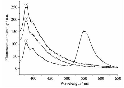 Figure 4.
Fluorescence emission spectra of the ligand L (a), 1 (b) and 2 (c) in CH3OH solution at room temperature
Figure 4.
Fluorescence emission spectra of the ligand L (a), 1 (b) and 2 (c) in CH3OH solution at room temperature
2.4 Fluorescence spectra
The fluorescence spectra of the ligand HL, complexes 1 and 2 have been studied in CH3OH solution (concentration: 1×10⁻⁵ mol·L-1) at room temperature. The results show that the emission spectra of all three compounds exhibit one emission peak at 380 nm when excited at 340 nm, while complex 2 shows another peak at 550 nm. The behavior of Cd (Ⅱ) ion coordinated to the ligand is regarded as that of emissive species resulted in a CHEF (chelation enhancement of the fluorescence emission) effect [17].
-
-
[1]
Alagesan L, Bhuvanesh N S P, Dharmaraj N. Dalton Trans., 2013, 42:7210-7223 doi: 10.1039/c3dt50371b
-
[2]
Ye X P, Zhu T F, Wu W N, et al. Inorg. Chem. Commun., 2014, 47:60-62 doi: 10.1016/j.inoche.2014.07.022
-
[3]
陈延民, 解庆范, 刘金花.无机化学学报, 2015, 31(1):74-80 http://www.wjhxxb.cn/wjhxxbcn/ch/reader/view_abstract.aspx?file_no=20150111&flag=1CHEN Yan-Min, XIE Qing-Fan, LIU Jin-Hua, et al. Chinese J. Inorg. Chem., 2015, 31(1):74-80 http://www.wjhxxb.cn/wjhxxbcn/ch/reader/view_abstract.aspx?file_no=20150111&flag=1
-
[4]
Singh P, Singh D P, Singh V P. Polyhedron, 2014, 81:56-65 doi: 10.1016/j.poly.2014.05.066
-
[5]
Nath M, Vats M, Roy P. Eur. J. Med. Chem., 2013, 59:310-321 doi: 10.1016/j.ejmech.2012.11.023
-
[6]
Milenković M, Cantoni G, Bacchi A, et al. Polyhedron, 2014, 80:47-52 doi: 10.1016/j.poly.2014.01.022
-
[7]
Milenković M, Bacchi A, Cantoni G, et al. Inorg. Chim. Acta, 2013, 395:33-43 doi: 10.1016/j.ica.2012.09.043
-
[8]
Chang H Q, Jia L, Xu Z Q, et al. Inorg. Chem. Commun., 2015, 57:8-10 doi: 10.1016/j.inoche.2015.04.010
-
[9]
Li M X, Zhang L Z, Yang M, et al. Bioorg. Med. Chem. Lett., 2012, 22:2418-2433 doi: 10.1016/j.bmcl.2012.02.024
-
[10]
Li M X, Zhang L Z, Zhang D, et al. Eur. J. Med. Chem., 2011, 46:4383-4390 doi: 10.1016/j.ejmech.2011.07.009
-
[11]
王召璐, 冯慧云, 李艳.无机化学学报, 2015, 31(10):2-7 http://www.wjhxxb.cn/wjhxxbcn/ch/reader/view_abstract.aspx?file_no=20151004&flag=1WANG Zhao-Lu, FENG Hui-Yun, LI Yan, et al. Chinese J. Inorg. Chem., 2015, 31(10):2-7 http://www.wjhxxb.cn/wjhxxbcn/ch/reader/view_abstract.aspx?file_no=20151004&flag=1
-
[12]
王桂仙, 张启伟, 温一航.无机化学学报, 2014, 30(11):2571-2576 http://www.wjhxxb.cn/wjhxxbcn/ch/reader/view_abstract.aspx?file_no=20141117&flag=1WANG Gui-Xian, ZHANG Qi-Wei, WEN Yi-Hang. Chinese J. Inorg. Chem., 2014, 30(11):2571-2576 http://www.wjhxxb.cn/wjhxxbcn/ch/reader/view_abstract.aspx?file_no=20141117&flag=1
-
[13]
李可, 李书静, 张小鹏.无机化学学报, 2014, 30 (7):1647-1652 http://www.wjhxxb.cn/wjhxxbcn/ch/reader/view_abstract.aspx?file_no=20140723&flag=1LI Ke, LI Shu-Jing, ZHANG Xiao-Peng, et al. Chinese J. Inorg. Chem., 2014, 30 (7):1647-1652 http://www.wjhxxb.cn/wjhxxbcn/ch/reader/view_abstract.aspx?file_no=20140723&flag=1
-
[14]
Sheldrick G M. SADABS, University of Göttingen, Germany, 1996.
-
[15]
Sheldrick G M. SHELX-97, Program for the Solution and the Refinement of Crystal Structures, University of Göttingen, Germany, 1997.
-
[16]
Song X Q, Zang Z P, Liu W S, et al. J. Solid State Chem., 2009, 182:841-848 doi: 10.1016/j.jssc.2008.12.025
-
[17]
Vicente M, Bastida R, Lodeiro C, et al. Inorg. Chem., 2003, 42:6768-6779 doi: 10.1021/ic034245z
-
[1]
-

Figure 1 Diamond drawing of 1 (a) and 2 (b) with 30% thermal ellipsoids; (c) Coordination enviorment of Cd (Ⅱ) ion in complex 2 (coordination atoms shown with 30% thermal ellipsoids); (d) Extend 2D supromolecular structure along c axis in complex 2
In (a)~(c): H atoms are omitted for clarity; In (d): H atoms of C-H bonds are omitted for clarity, Symmetry codes: iv x-1/2, -y+1, z+1/2; v x, y-1, z

Figure 3 UV spectra of the ligand HL (a), 1 (b) and 2 (c) in CH3OH solution at room temperature

Figure 4 Fluorescence emission spectra of the ligand L (a), 1 (b) and 2 (c) in CH3OH solution at room temperature
-


 下载:
下载:


 扫一扫看文章
计量
- PDF下载量: 0
- 文章访问数: 919
- HTML全文浏览量: 94
 下载:
下载:

